Evaluar la concordancia de la resonancia magnética (RM) de mama con la histología en la valoración del tamaño y extensión del carcinoma ductal in situ puro (CDIS), y compararla con la de las técnicas convencionales (mamografía y ecografía).
Material y métodosEstudio retrospectivo de pacientes consecutivas con biopsia percutánea con resultado de CDIS. Se estimó el coeficiente de concordancia de correlación de Lin para cada uno de las 3 técnicas de imagen con la histología. Además, se revisó la concordancia con gráficos de Bland-Altman. Se evaluaron los cambios de conducta quirúrgica generados por la RM.
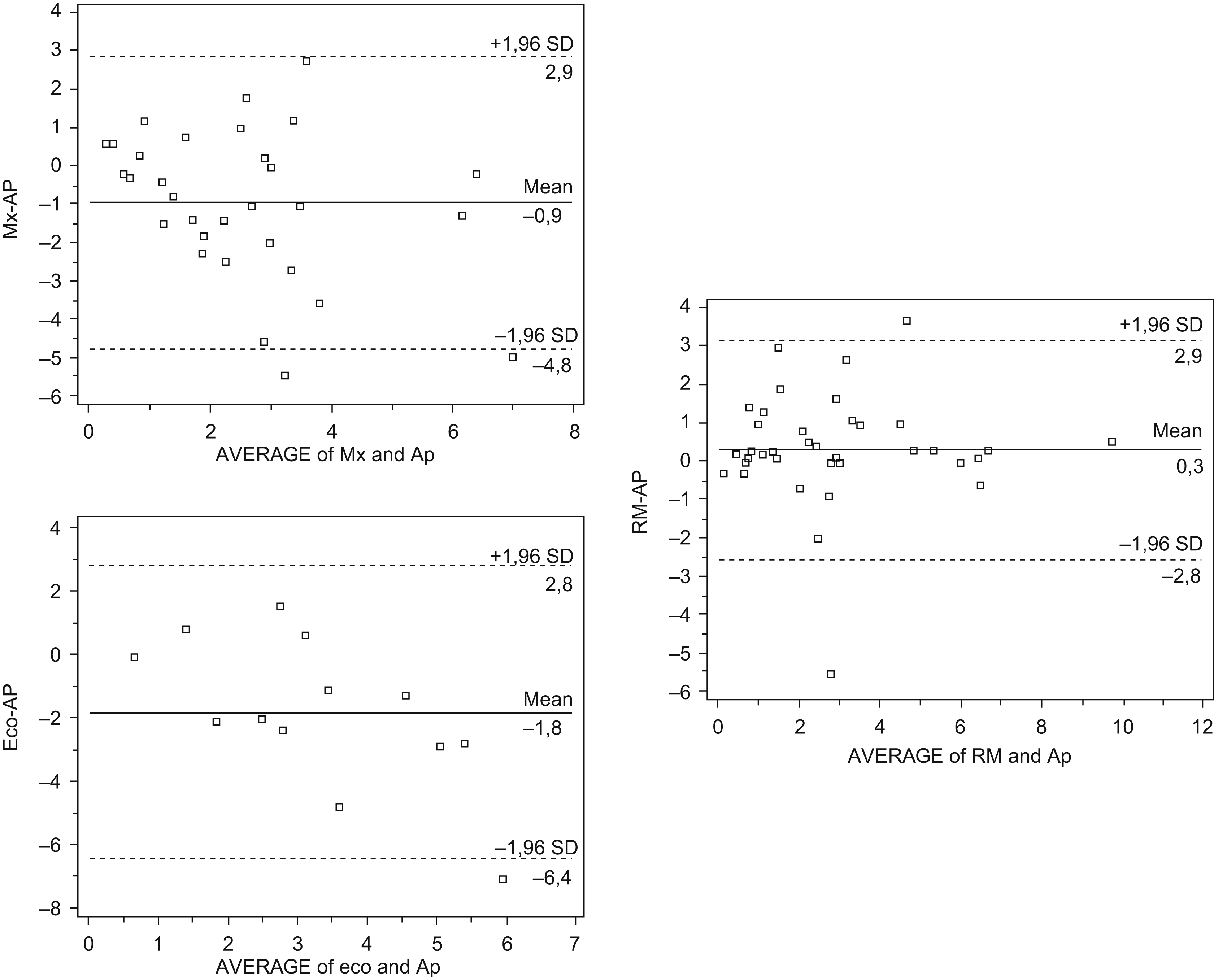
ResultadosEl grupo estudiado fue de 32 pacientes. En cuanto al tamaño tumoral, la concordancia fue superior en la RM (0,78; intervalo de confianza [IC] de 95%, 0,62–0,87) que en la mamografía (0,43; IC del 95%, 0,19–0,62) o en la ecografía (0,27; IC del 95%, 0,09–0,43). La RM sobrestimó el tamaño con un promedio de 3mm, mientras que la mamografía y la ecografía lo subestimaron en 9 y 18mm, respectivamente. La RM fue superior para la detección del multifocalidad o multicentricidad (7 casos) frente a la mamografía (3 casos) y a la ecografía (0 casos). Hubo 6 cambios de conducta quirúrgica correctos basados en los hallazgos de la RM.
ConclusiónLa RM de mama es superior a las técnicas convencionales en la valoración del tamaño de los CDIS. Además, detecta más casos de multifocalidad y multicentricidad, por lo que aconsejamos su utilización prequirúrgica en pacientes diagnosticadas de CDIS, especialmente en mamas densas.
To evaluate the concordance between the breast MRI findings and the histologic findings for the size and extension of pure ductal carcinoma in situ (DCIS) and to compare this concordance with that of conventional techniques (mammography and ultrasonography).
Material and methodsThis is a retrospective study of consecutive patients diagnosed with DCIS after percutaneous biopsy. We estimated Lin's coefficient of concordance for the histologic findings with each of the three techniques. We also assessed concordance using Bland-Altman graphs. Finally, we determined the impact of the MRI findings on the surgical management of patients with DCIS.
ResultsA total of 32 patients were included in the study. Concordance between imaging and histology on tumor size was higher for MRI (0.78; 95%CI, 0.62–0.87) than for mammography (0.43; 95%CI, 0.19–0.62) or for ultrasonography (0.27; 95%CI, 0.09–0.43). MRI overestimated the size of DCIS by a mean of 3mm, whereas mammography and ultrasonography underestimated it by 9mm and 18mm, respectively. MRI detected multifocality and multicentricity (7 cases) better than mammography (3) or ultrasonography (0). The MRI findings correctly changed the surgical management in six patients.
ConclusionBreast MRI is better than conventional techniques for the evaluation of the size of DCIS. Breast MRI also detects more cases of multifocality and multicentricity. We recommend that all patients diagnosed with DCIS (especially those with dense breasts) undergo breast MRI prior to surgery.
Artículo
Comprando el artículo el PDF del mismo podrá ser descargado
Precio 19,34 €
Comprar ahora


