Describir los hallazgos ecográficos de los abscesos hepáticos tras la administración de contraste de segunda generación. Realizar el diagnóstico diferencial con otras lesiones focales hepáticas.
Material y métodosSe valoraron 28 abscesos hepáticos en 15 pacientes mediante ecografía basal y tras la administración de SonoVue. Se valoraron las lesiones hepáticas de 6 pacientes en las que se planteó el diagnóstico diferencial con absceso en la ecografía basal.
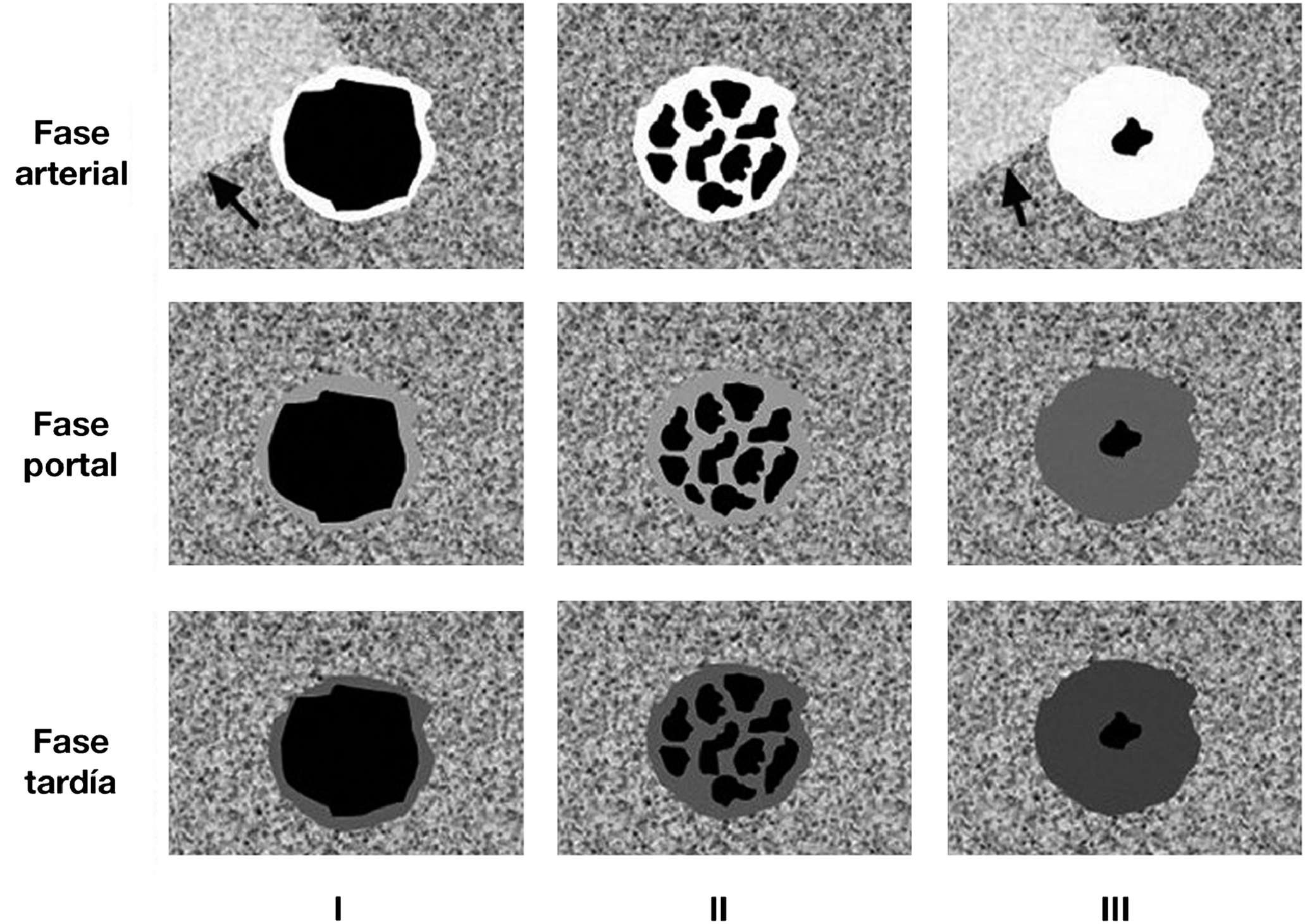
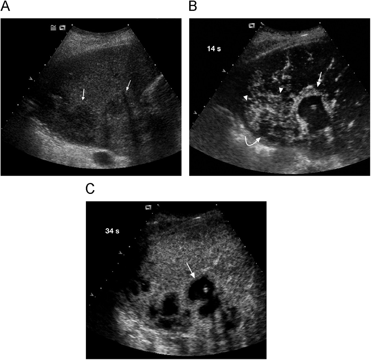
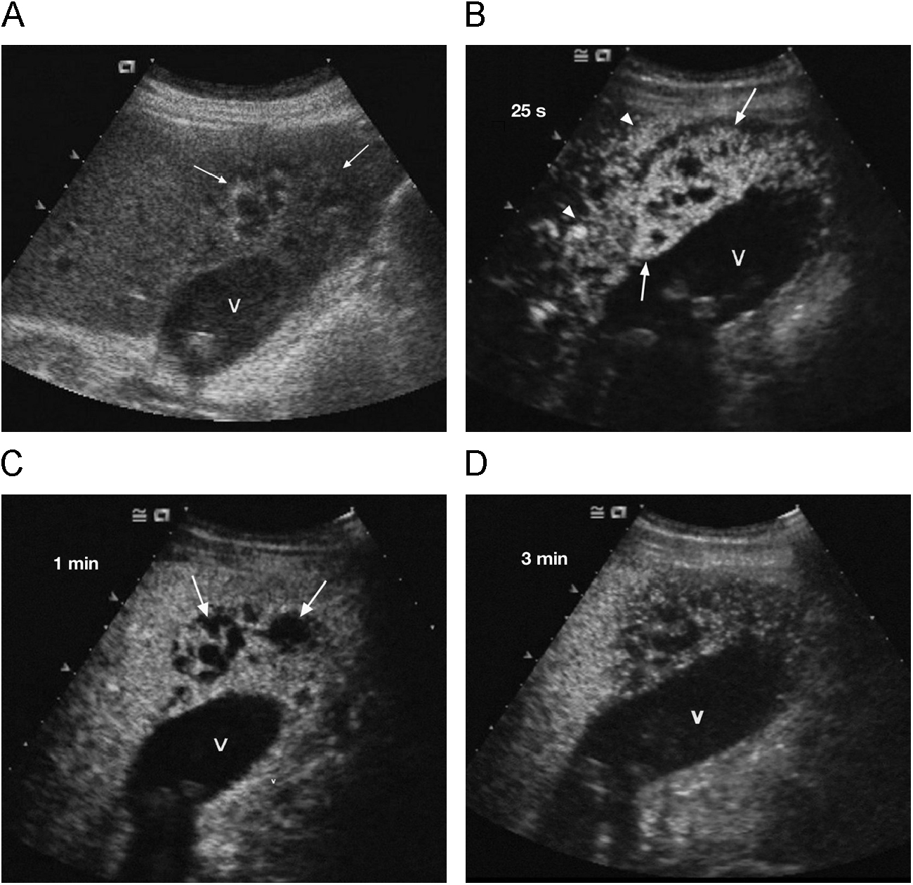
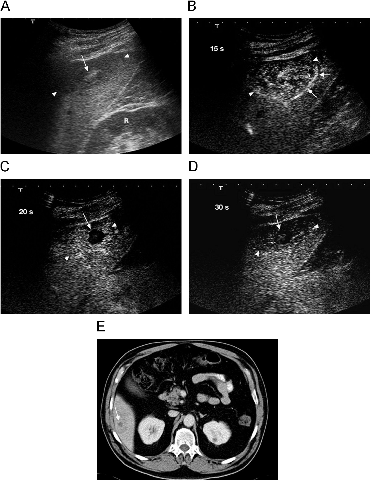
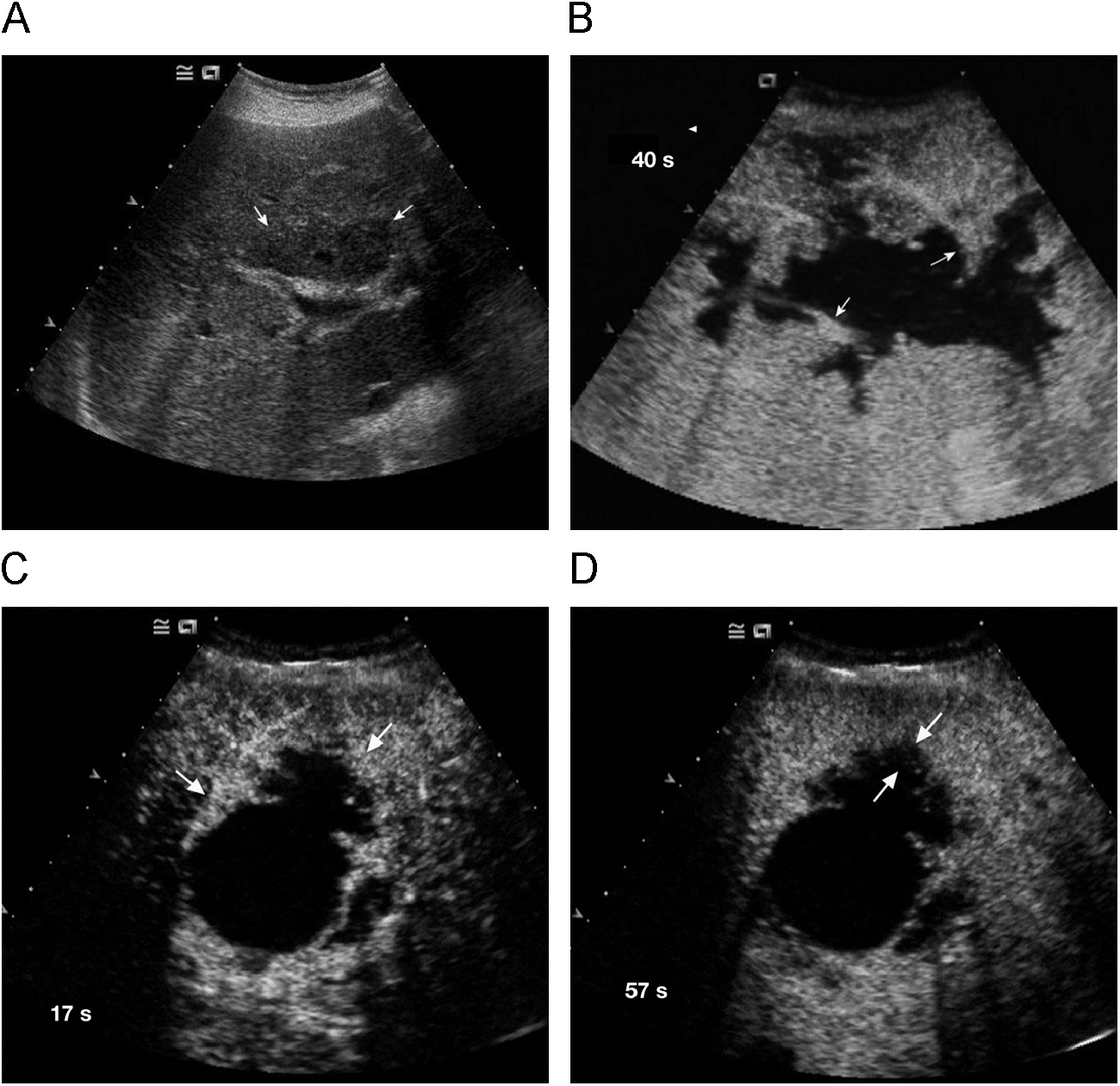
ResultadosEn 21 abscesos (75%) se vio un patrón de realce típico (realce anular periférico arterial y ausencia de realce central). En otros 6 (21,4%), el realce arterial se vio en gran parte de la lesión, con zonas de ausencia de captación. Otro caso (3,6%) mostró un patrón de realce multiseptado. En 6 de los abscesos se apreció un realce segmentario hepático. En cuanto a las lesiones con las que se planteó el diagnóstico diferencial, 5 de las 6 no presentaron realce en ninguna de las fases. La otra lesión, una metástasis quística, presentó un realce periférico arterial irregular. Ninguna de estas lesiones presentó un realce arterial segmentario hepático.
ConclusionesLa ecografía con contraste mejora el rendimiento de la ecografía en el diagnóstico de los abscesos hepáticos, observándose 3 patrones de realce, superponibles a los hallazgos de la tomografía computarizada y resonancia magnética. Es muy útil para definir la arquitectura interna del absceso, lo cual es importante en la elección del tipo de tratamiento. Permite hacer el diagnóstico diferencial con otras lesiones focales hepáticas.
To describe the ultrasonographic findings in liver abscesses after the administration of a second generation agent. To perform the differential diagnosis of liver abscesses with other focal liver lesions.
Material and methodsWe evaluated 28 liver abscesses in 5 patients before and after the administration of SonoVue. We also evaluated liver lesions in six patients in whom the differential diagnosis with liver abscess was considered in the baseline ultrasonographic examination.
ResultsA typical enhancement pattern consisting of peripheral ring enhancement in the arterial phase and absence of central enhancement was observed in 21 (75%) abscesses. In another 6 (21.4%) abscesses, arterial enhancement was seen in large areas of the lesion, while other areas showed no uptake. One case (3.6%) had a multiseptated pattern of enhancement. Segmental hepatic enhancement was observed in 6 abscesses. In the liver lesions in which the differential diagnosis with abscess was carried out, 5 of the 6 showed no enhancement in any phase. The other lesion, a cystic metastasis, had irregular peripheral enhancement in the arterial phase. None of these lesions had segmental hepatic enhancement in the arterial phase.
ConclusionsContrast administration improves the performance of ultrasonography in the diagnosis of liver abscesses. There are three patterns of enhancement and these correlate well with the findings at CT and MRI. Contrast-enhanced ultrasonography is very useful for defining the internal architecture of the abscess, which is important for choosing the type of treatment. Contrast-enhanced ultrasonography also enables the differential diagnosis with other focal liver lesions.
Artículo
Comprando el artículo el PDF del mismo podrá ser descargado
Precio 19,34 €
Comprar ahora


