


Información del artículo
Resumen
Texto completo
Bibliografía
Descargar PDF
Estadísticas
Tablas (2)
TABLA 1. Características de los pacientes
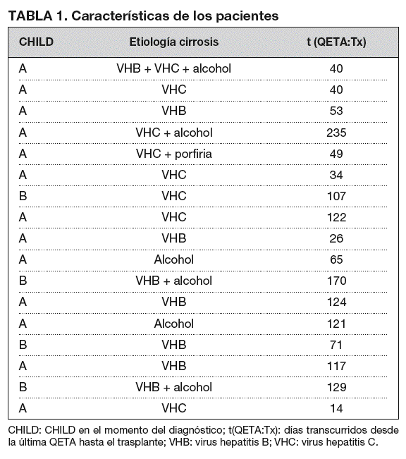
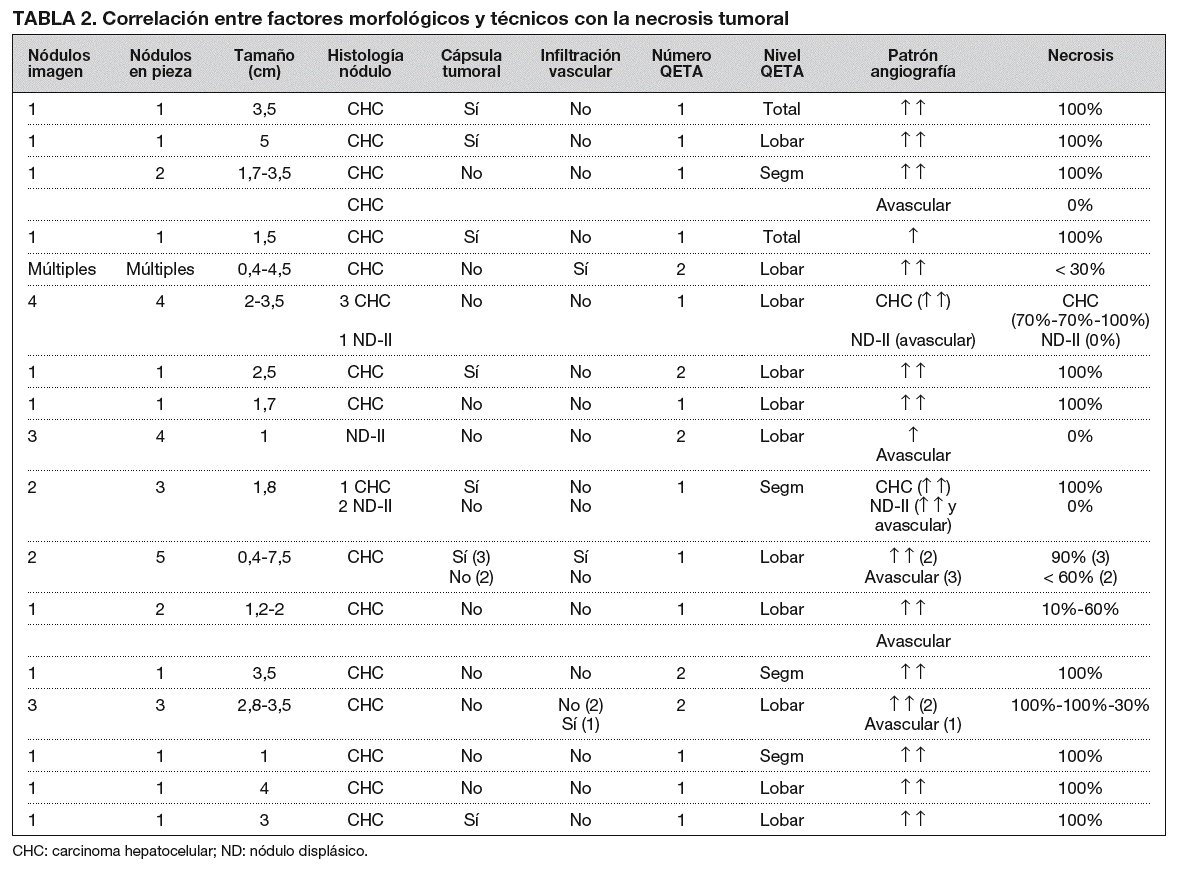
TABLA 2. Correlación entre factores morfológicos y técnicos con la necrosis tumoral
Mostrar másMostrar menos
Figuras (10)


Mostrar másMostrar menos
Objective. To retrospectively evaluate the degree of necrosis brought about by chemoembolization of hepatocellular carcinoma by correlating the histological study of livers explanted in liver transplantations with morphological (number, size, histological type, encapsulation, and vascular invasion) and technical (angiographic patterns, number of sessions, and level of chemoembolization) factors of the tumors. Material and methods. Seventeen cirrhotic patients that underwent chemoembolization of hepatocellular carcinoma prior to liver transplantation were studied. Chemoembolization consisted of introducing adriamycin, lipiodol, and particles of polyvinyl alcohol into the hepatic artery. The explanted livers were studied macroscopically and microscopically, evaluating the degree of necrosis achieved in each of the nodules found. Results. A total of 32 nodules (26 hepatocellular carcinomas and 7 type II dysplastic nodules) were detected in 16 patients; the remaining patient had multiple hepatocellular carcinomas. The mean size of the hepatocellular carcinomas was 3.2 cms (range 0.4-7.5) and the mean size of the dysplastic nodules was 1.2 cm (range 1-1.7). Greater than 90% necrosis was achieved in 17 (68%) of the 25 hepatocellular carcinomas; 6 of these lesions were encapsulated and none had vascular infiltration. In the patient with multiple hepatocellular carcinomas (all were hypervascularized and had no capsule), the degree of necrosis achieved was < 30% in each lesion. In three patients with nodular vascular infiltration, multiple noduleswere present; the necrosis achieved in these lesions ranged from 0%-70%, and extrahepatic metastases after transplantation. Eighteen of the hepatocellular carcinomas had a hypervascularized angiographic pattern and seven were hypovascular or avascular. In the seven type II dysplastic nodules (none of which was encapsulated), no necrosis was achieved. These nodules were found in three patients; all but one were avascular and coincided with simultaneous hepatocellular carcinomas in two patients. Conclusion. The greatest degree of necrosis was achieved in single, encapsulated, hypervascularized hepatocellular carcinomas without vascular infiltration. Chemoembolization fails to achieve necrosis in dysplastic nodules.
Keywords:
liver neoplasm, hepatic arteries, therapeutic chemoembolization, transplant, liver
Artículo
Opciones para acceder a los textos completos de la publicación Radiología
Suscriptor
Suscribirse
Comprar
Contactar
Teléfono para suscripciones e incidencias
De lunes a viernes de 9h a 18h (GMT+1) excepto los meses de julio y agosto que será de 9 a 15h
Llamadas desde España
932 415 960
Llamadas desde fuera de España
+34 932 415 960
E-mail

