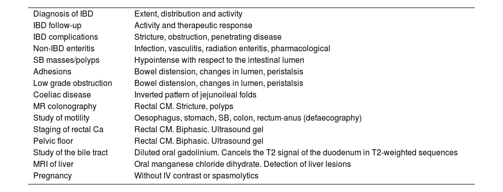
Magnetic resonance enterography is primarily indicated for inflammatory bowel diseases. The study of the gastrointestinal tract using MRI has become feasible due to the emergence of ultrafast sequences with higher spatial resolution and phased-array coils enabling wider fields of view. However, to ensure the quality of the examination, it is essential to have prior preparation with oral or rectal contrast to distend the lumen and improve the definition of the intestinal wall. These contrast agents can be positive, negative or biphasic, depending on the signal intensity they induce in the intestinal lumen. Most commonly used biphasic contrasts agents behave as hyperintense in T2 and hypointense in T1. Achieving a “black” intestinal lumen in 3D T1-weighted sequences with intravenous contrast injection is crucial for mucosal assessment and parietal enhancement. Although more cost-effective and accessible, biphasic agents like PEG and mannitol are relatively discomforting for patients. While negative agents are preferred, they are currently unavailable. The purpose of this article is to review the different types of contrast agents mentioned in the literature and their application in intestinal resonance, analyzing the effects they generate on the image, their possible indications and associated limitations.
La enterografía por Resonancia Magnética está principalmente indicada en la enfermedad inflamatoria intestinal. El estudio del tracto gastrointestinal mediante resonancia hoy día es posible gracias a la aparición de secuencias ultrarrápidas con mayor resolución espacial y bobinas phased-array que permiten campos de visión de todo el abdomen. Sin embargo, para garantizar la calidad de la exploración es fundamental una preparación previa con contraste oral o rectal para distender la luz y mejorar la definición de la pared intestinal. Estos agentes de contraste pueden ser positivos, negativos o bifásicos, dependiendo de la señal que generen en la luz intestinal. Los contrastes bifásicos son los más empleados en la práctica diaria y se comportan como hiperintensos en T2 e hipointensos en T1. Lograr una luz intestinal "negra" en secuencias 3D ponderadas en T1 con inyección intravenosa de contraste es crucial para la evaluación de la mucosa y el realce parietal. Aunque son más económicos y accesibles, los agentes bifásicos como el PEG y el manitol son relativamente molestos para los pacientes. Los agentes negativos son los más deseados, pero no están disponibles de manera rutinaria. El propósito de este artículo es revisar los diferentes tipos de contrastes mencionados en la literatura para su aplicación en resonancia intestinal, analizando los efectos que generan en la imagen, sus posibles indicaciones y limitaciones asociadas.
Artículo
Comprando el artículo el PDF del mismo podrá ser descargado
Precio 19,34 €
Comprar ahora


