metricas
covid
Buscar en
Revista Española de Anestesiología y Reanimación (English Edition)
Toda la web
Inicio Revista Española de Anestesiología y Reanimación (English Edition) Spanish Society of Anesthesiology, Reanimation and Pain Therapy (SEDAR), Spanish...
Journal Information
Vol. 71. Issue 3.
Pages 207-247 (March 2024)
Visits
625
Vol. 71. Issue 3.
Pages 207-247 (March 2024)
CONSENSUS STATEMENT
Full text access
Spanish Society of Anesthesiology, Reanimation and Pain Therapy (SEDAR), Spanish Society of Emergency and Emergency Medicine (SEMES) and Spanish Society of Otolaryngology, Head and Neck Surgery (SEORL-CCC) Guideline for difficult airway management. Part II
Guía de la Sociedad Española de Anestesiología, Reanimación y Terapéutica del Dolor (SEDAR), Sociedad Española de Medicina de Urgencias y Emergencias (SEMES) y Sociedad Española de Otorrinolaringología y Cirugía de Cabeza y Cuello (SEORL-CCC) para el manejo de la vía aérea difícil. Parte II
Visits
625
M.Á. Gómez-Ríosa,
Corresponding author
magoris@hotmail.com

Corresponding author.
, J.A. Sastreb, X. Onrubia-Fuertesc, T. Lópezb, A. Abad-Gurumetad, R. Casans-Francése, D. Gómez-Ríosf, J.C. Garzónb, V. Martínez-Ponsg, M. Casalderrey-Rivash, M.Á. Fernández-Vaqueroi, E. Martínez-Hurtadod, R. Martín-Larraurij, L. Reviriego-Agudok, U. Gutierrez-Coutol, J. García-Fernándezm,n, A. Serrano-Morazao, L.J. Rodríguez Martínp, C. Camacho Leisp, S. Espinosa Ramírezo..., J.M. Fandiño Orgeiraq, M.J. Vázquez Limar,s, M. Mayo-Yáñezt, P. Parente-Ariast, J.A. Sistiaga-Suárezu, M. Bernal-Sprekelsenv,w, P. Charco-MoragVer más
a Anesthesiology and Perioperative Medicine, Complejo Hospitalario Universitario de A Coruña, A Coruña, Spain
b Anesthesiology and Perioperative Medicine, Complejo Asistencial Universitario de Salamanca, Salamanca, Spain
c Department of Anesthesiology, Hospital Universitary Dr Peset, Valencia, Spain
d Department of Anesthesiology, Hospital Universitario Infanta Leonor, Madrid, Spain
e Department of Anesthesiology, Hospital Universitario Infanta Elena, Valdemoro, Madrid, Spain
f Hospital de Barbanza, Ribeira, A Coruña, Spain
g Department of Anesthesiology, Hospital Universitari i Politècnic La Fe, Valencia, Spain
h Department of Anesthesiology. Complejo Hospitalario Universitario de Ourense, Ourense, Spain
i Department of Anesthesiology, Hospital Clínica Universitaria de Navarra, Madrid, Spain
j Department of Anesthesiology, Hospital Infanta Elena, Málaga, Spain
k Department of Anesthesiology, Hospital Clínico Universitario, Valencia, Spain
l Biblioteca, Complejo Hospitalario Universitario de Ferrol (CHUF), Ferrol, A Coruña, Spain
m Department of Anesthesiology, Hospital Universitario Puerta de Hierro-Majadahonda, Majadahonda, Madrid, Spain
n President of the Spanish Society of Anesthesiology, Resuscitation and Pain Therapy (SEDAR), Spain
o SUMMA 112
p Emergencias SAMUR Protección Civil, Madrid, Spain
q Emergency Department, Complejo Hospitalario Universitario de A Coruña, A Coruña, Spain
r Emergency Department, Hospital do Salnes, Vilagarcía de Arousa, Pontevedra, Spain
s President of the Spanish Emergency Medicine Society (SEMES), Spain
t Department of Otorhinolaryngology/Head Neck Surgery, Complexo Hospitalario Universitario A Coruña, A Coruña, Spain
u Department of Otorhinolaryngology, Hospital Universitario Donostia, Donostia, Gipuzkoa, Spain
v Department of Otorhinolaryngology, Hospital Clínic Barcelona, University of Barcelona, Barcelona, Spain
w President of the Spanish Society for Otorhinolaryngology Head & Neck Surgery (SEORL-CCC), Spain
Ver más
This item has received
Article information
Abstract
Full Text
Bibliography
Download PDF
Statistics
Figures (6)
Show moreShow less
Tables (7)
Table 1. Main drugs used for sedation during awake tracheal intubation.
Table 2. Main indications for extracorporeal membrane oxygenation (ECMO).
Table 3. Maneuvers to overcome difficulties in videolaryngoscopy-guided tracheal intubation.
Table 4. Requirements of an ideal position of a SGA, causes of malpositioning and how to correct them.
Table 5. Characteristics of pre-hospital airway management.
Show moreShow less
Additional material (5)
Abstract

The Airway Management section of the Spanish Society of Anesthesiology, Resuscitation, and Pain Therapy (SEDAR), the Spanish Society of Emergency Medicine (SEMES), and the Spanish Society of Otorhinolaryngology and Head and Neck Surgery (SEORL-CCC) present the Guide for the comprehensive management of difficult airway in adult patients. Its principles are focused on the human factors, cognitive processes for decision-making in critical situations, and optimization in the progression of strategies application to preserve adequate alveolar oxygenation in order to enhance safety and the quality of care. The document provides evidence-based recommendations, theoretical-educational tools, and implementation tools, mainly cognitive aids, applicable to airway management in the fields of anesthesiology, critical care, emergencies, and prehospital medicine. For this purpose, an extensive literature search was conducted following PRISMA-R guidelines and was analyzed using the GRADE methodology. Recommendations were formulated according to the GRADE methodology. Recommendations for sections with low-quality evidence were based on expert opinion through consensus reached via a Delphi questionnaire.

Keywords:
Airway management
Practice guideline
Conscious sedation
General anesthesia
Endotracheal intubation
Laryngeal mask
Tracheostomy
Airway obstruction
Monitoring
Resumen

La sección de Vía Aérea de la Sociedad Española de Anestesiología, Reanimación y Terapéutica del Dolor (SEDAR), la Sociedad Española de Medicina de Urgencias y Emergencias (SEMES) y la Sociedad Española de Otorrinolaringología y Cirugía de Cabeza y Cuello (SEORLCCC) presentan la Guía para el manejo integral de la vía aérea difícil en el paciente adulto. Sus principios están focalizados en el factor humano, los procesos cognitivos para la toma de decisiones en situaciones críticas y la optimización en la progresión de la aplicación de estrategias para preservar una adecuada oxigenación alveolar con el objeto de mejorar la seguridad y la calidad asistencial. El documento proporciona recomendaciones basadas en la evidencia científica actual, herramientas teórico-educativas y de implementación, fundamentalmente ayudas cognitivas, aplicables al tratamiento de la vía aérea (VA) en el campo de la anestesiología, cuidados críticos, urgencias y medicina prehospitalaria. Para ello, se realizó una amplia búsqueda bibliográfica según las directrices PRISMA-R y se analizó utilizando esta metodología. Las recomendaciones se formularon de acuerdo con esta metodología. Las recomendaciones de aquellas secciones con evidencia de baja calidad se basaron en la opinión de expertos mediante el consenso alcanzado a través de un cuestionario Delphi.

Palabras clave:
Manejo de la vía aérea
Guía clínica
Sedación consciente
Anestesia general
Intubación endotraqueal
Mascarilla laríngea
Traqueostomía
Obstrucción de la vía aérea
Monitorización
Full Text
Known or anticipated difficult airway

Awake airway management is the technique of choice in patients with known or anticipated difficult airway (EO 85.7%)1 since it (1) preserves airway patency and spontaneous ventilation, increases respiratory reserve, and protects against aspiration by preserving laryngeal reflexes1,2; (2) facilitates a gradual transition to positive pressure ventilation (PPV) and slow induction of general anaesthesia (GA) in patients at risk of haemodynamic collapse3,4; (3) facilitates tracheal intubation by preventing soft tissue collapse, dilating peritracheal structures, improving the glottic view by preventing the larynx from adopting a more anterior position, and allowing the operator to locate a distorted glottis by the presence of air bubbles; (4) can be performed in a sitting position in a collaborative patient, which also helps evaluate the patient’s neurological status; (5) keeps all treatment options open and allows for decision-making based on findings.1,3,5–8

A known or anticipated DA requires formulation of team strategies with a comprehensive multidisciplinary discussion beforehand about sequential plans (primary and alternative) to achieve oxygenation and ventilation and prevent aspiration.1,7,9,10

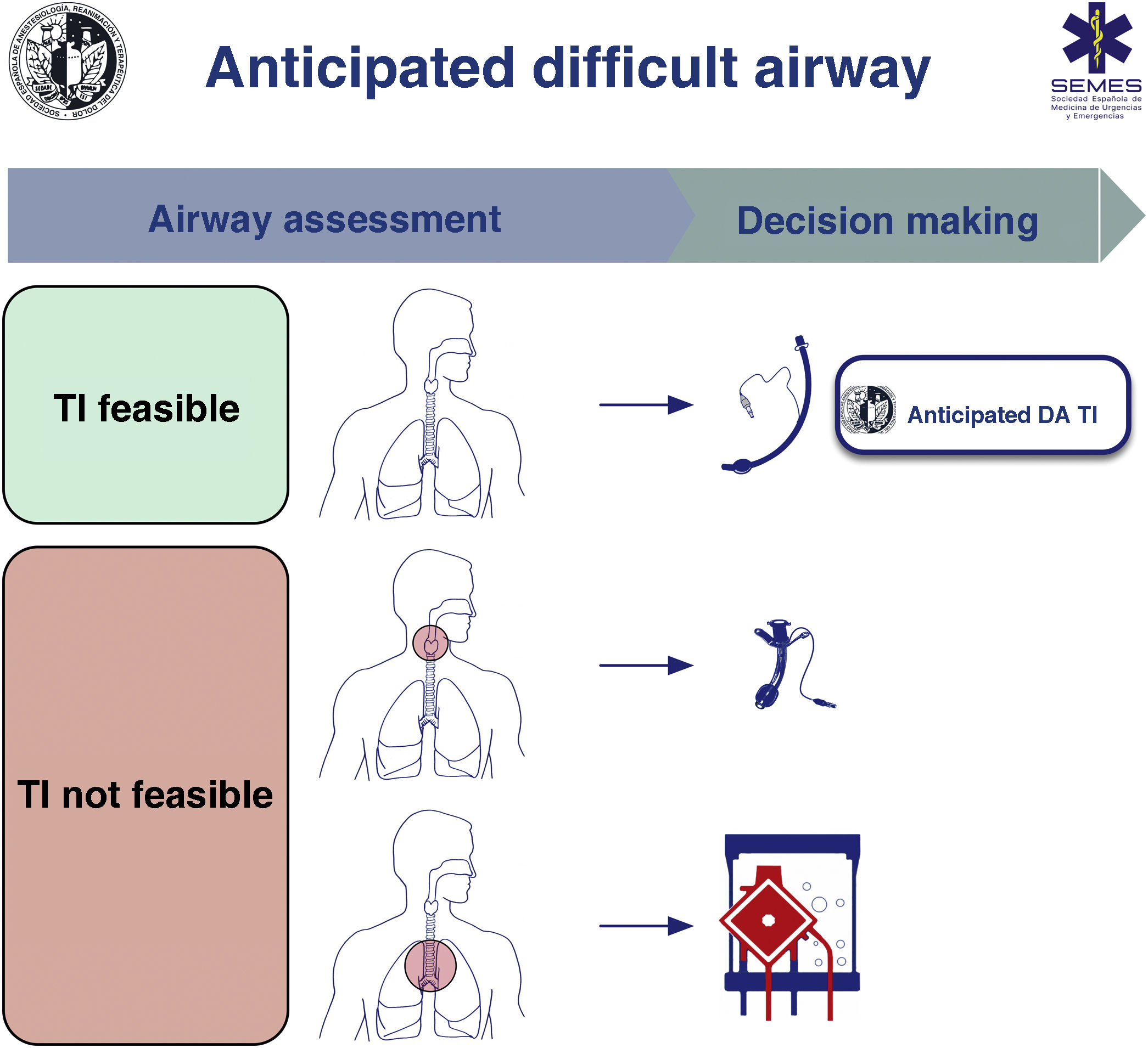
When the DA is secondary to an obstructive pathology, the team will need to evaluate the patient’s respiratory status and the cause, location, and degree of obstruction (greater or less than 50%) using clinical signs and symptoms, imaging tests, and flexible nasolaryngoscopy (FNL).1,7,10,11 The risks and benefits of each approach must be carefully considered and the strategy must be agreed by the entire medical-surgical team.1Fig. 1 shows a cognitive aid for decision-making in the event of an anticipated DA. Lower-tier plans act as rescue strategies in the event of failure of the upper-tier plan if selected as primary. In all cases, it is advisable to perform awake airway management. (1) Supraglottic lesions causing mild obstruction that can be overcome with an ETT allow for TI, usually fibreoptic intubation (FOI). (2) In the case of obstructive supraglottic lesions causing stenosis greater than 50% (or with inspiratory stridor at rest),7 non-obstructive supraglottic lesions that impede TI (or would cause unacceptable morbidity), and glottic or subglottic lesions, it is advisable to perform tracheotomy or cricothyrotomy as a primary approach.10 (3) Lower tracheal obstructive lesions not salvageable with an ETT or tracheal cannula require the application of extracorporeal membrane oxygenation (ECMO).10

Fig. 1.

Cognitive aid to facilitate decision-making in the anticipated difficult airway. DA: difficult airway; TI: tracheal intubation.

(0.28MB).

Active oxygenation strategies should be implemented throughout the procedure. High-flow nasal oxygen therapy (HFNO), although requiring further validation in this context, may be the technique of choice. HFNO is recommended over conventional low-flow cannulas (EO 91.4%).
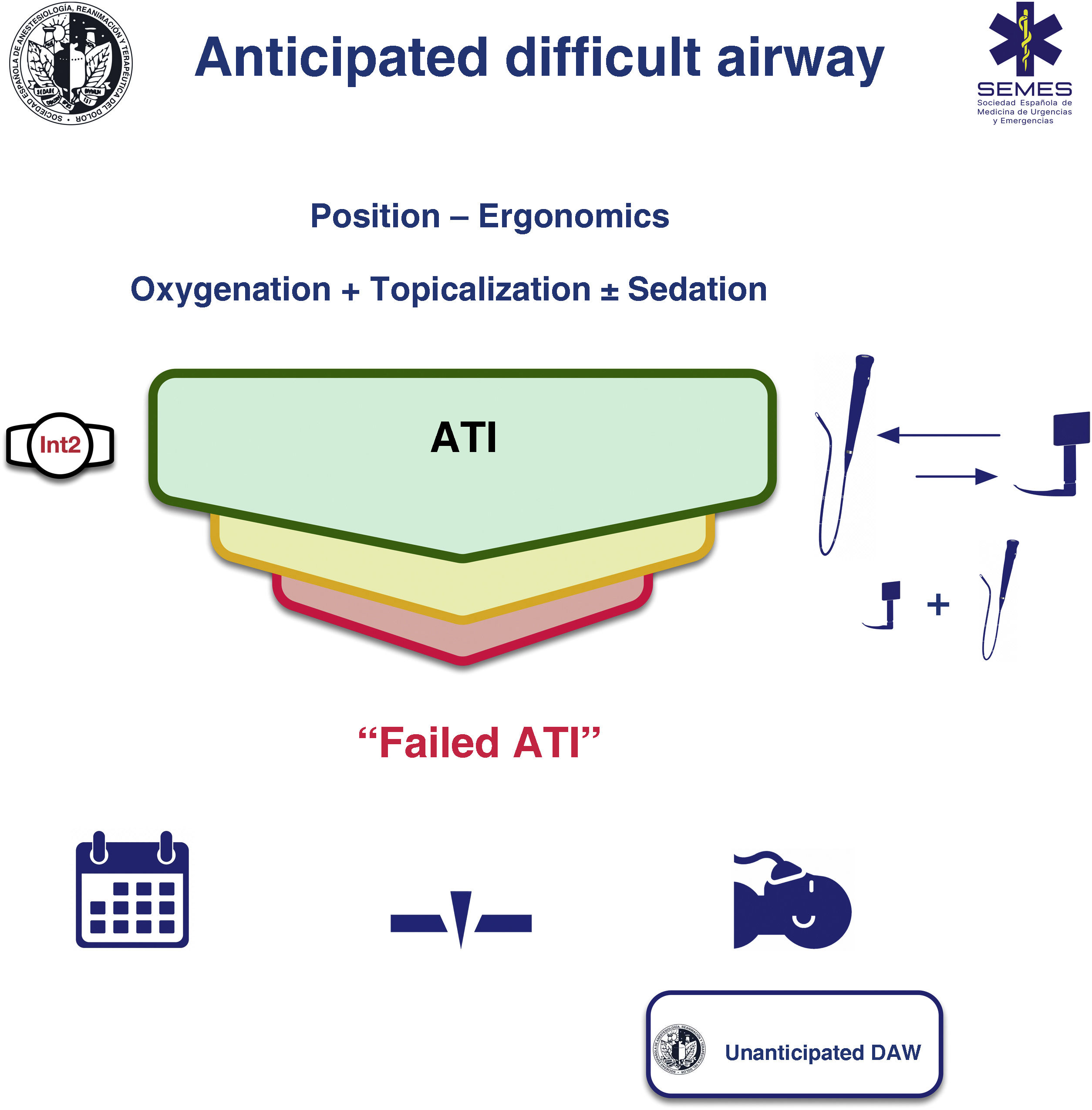
Awake tracheal intubation

When the airway can be secured noninvasively, awake tracheal intubation (ATI) remains the gold standard technique3,12,13 due to its safety and reliability.14,15 The cornerstones of successful ATI are: continuous oxygenation, topicalization of the airway, sedation (optional), and selection, experience, and use of an appropriate TI device and technique. The ideal protocol in terms of efficacy and safety is unknown, so the most appropriate one should be chosen based on the clinical context and the individual characteristics of each patient, as well as the experience and preferences of the operator.16–18Fig. 2 shows the SEDAR SEMES SEORL-CCC cognitive aid for TI of the anticipated DA.

Fig. 2.

Cognitive aid for tracheal intubation of an anticipated difficult airway. ATI: awake tracheal intubation; DA: difficult airway; Int2: second operator.

(0.34MB).
Oxygenation

Continuous oxygenation improves safety by preventing or minimizing hypoxaemia.12,19 Conventional methods may be insufficient to avoid desaturation.20 HFNO increases tolerance of an airway obstruction, hypoventilation, and longer apnoea time,20–23 and despite the scant evidence, it is becoming the method of choice.13,24

NIV through an endoscopy face mask could be useful when intubating critically ill patients with severe hypoxaemia (EO: 82.9%).25

Topicalization

Applying topical anaesthesia to the airway is a key step in successful ATI.1 Lidocaine (2%–4%), which has a favourable cardiovascular and systemic toxicity risk profile, is the most widely used local anaesthetic.12,19,26–28 The maximum dose should not exceed 9 mg/kg,13,29 and only the minimum necessary dose should be used.

The “spray as you go” (SAYGO) technique with an epidural catheter or an atomizer, and regional nerve blocks (glossopharyngeal, superior laryngeal, or transtracheal injection) are the most employed methods for topicalization of the respiratory mucosa.2,12,27 These techniques are often used in combination.28 There is no evidence of the superiority of any particular technique,16 although nerve blocks are more invasive, require multiple bilateral injections, demand experience, and are associated with a higher incidence of complications.2,13,16 Transtracheal injection (4ml of 4% lidocaine) is perhaps the most useful invasive method as it: (1) anaesthetises the infraglottic larynx, upper trachea, and even the supraglottic structures30; (2) has a success rate of over 95%12; and (3) is seldom associated with complications (1:10,000), although they can be important.28,31

The SAYGO technique can minimize the risk of aspiration, because laryngeal reflexes are maintained until just before inserting the ETT.2 Premedication with an antisialogogue is recommended to maximise the local anaesthetic effect and improve the field of vision (EO 80%). The antisialogogue of choice is glycopyrrolate (3 μg/kg), because it is fast-acting, has no effect on the central nervous system, and has a mild vagolytic action.2,12,32 When administered 15−20 min before reduces the dilution and esophageal elimination of the local anesthetic by secretions.1

Regardless of the method used, topicalization should include the oral cavity, nasal cavity if nasotracheal intubation is planned, oropharynx, periglottic area, larynx and trachea.1,30 Otherwise, the insertion of the device and the ETT can provoke reflex responses from the airway, such as coughing or laryngospasm, as well as a cardiovascular response mediated by the sympathetic nervous system.33

Sedation

Sedation is an optional complement to adequate topical anaesthesia in ATI16 (EO 88.6%), as awake tracheal intubation with prior psychological preparation can be performed safely and effectively without sedation.19,28,34 It should never compensate for inadequate topicalization. Although very high levels of anxiety can increase the physiological response to stress and reduce tolerance, oversedation can cause loss of cooperation, respiratory depression, hypoxia, hypercapnia, airway obstruction, aspiration, and cardiovascular instability,19,35,36 so sedation should only be administered after a detailed risk-benefit analysis. The goals of sedation are (EO 94.3%)1,19,35: (1) effective anxiolysis and amnesia while maintaining patient cooperation (“conscious sedation”, sedation level 2–3 on the Ramsay scale)28,37; (2) analgesia to suppress the cough and gag reflexes and reduce the haemodynamic response while preserving airway patency, spontaneous ventilation, and avoiding aspiration. Sedation must be carefully monitored to avoid oversedation, so ideally a second operator should be exclusively responsible for its administration and monitoring.13,19

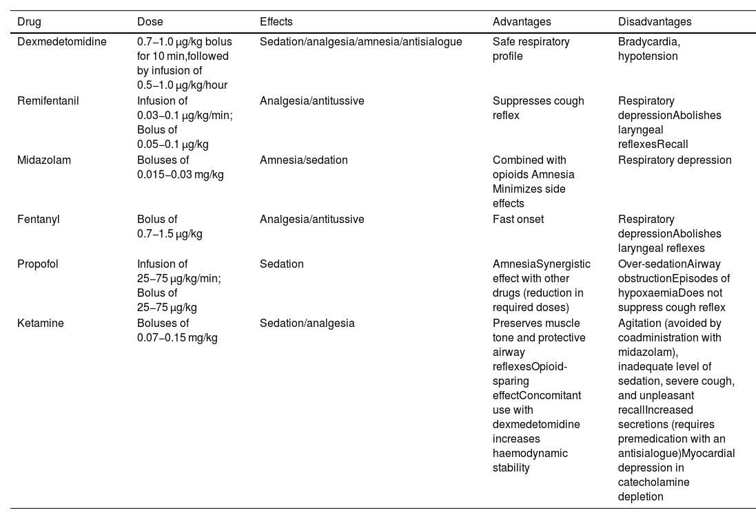
All sedation regimens used have shown a satisfactory level of efficacy and safety16; however, dexmedetomidine, with its anxiolytic, sedative, analgesic, and sympatholytic action, may be the safest and most effective option because it is less likely to cause apnoea and desaturation, is well tolerated, and is associated with better intubation conditions and less recall compared to other drugs16,38–40; however, it can produce episodes of severe bradycardia and hypotension.35 Its ability to maintain respiratory function integrity, even with deep levels of sedation, makes it a good choice for patients at risk of airway obstruction and/or respiratory failure.35 Opioids, particularly remifentanil, attenuate the cough and gag reflexes, although they may increase the risk of chest rigidity and laryngospasm.6,13,19 They are also associated with a high incidence of recall when used alone, and are therefore usually combined with a benzodiazepine such as midazolam.19,28 Monotherapy is usually more predictable and reliable, although the availability of specific opioid and benzodiazepine antagonists improves their safety profile. Remifentanil is a good option when topical anaesthesia has been ruled out.35,41 Propofol is associated with a low incidence of recall, although it can increase the risk of over sedation, airway obstruction, and coughing.13,35,42Table 1 shows the main drugs used for sedation.

Table 1.

Main drugs used for sedation during awake tracheal intubation.

Drug  Dose  Effects  Advantages  Disadvantages 
Dexmedetomidine  0.7−1.0 μg/kg bolus for 10 min,followed by infusion of 0.5−1.0 μg/kg/hour  Sedation/analgesia/amnesia/antisialogue  Safe respiratory profile  Bradycardia, hypotension 
Remifentanil  Infusion of 0.03−0.1 μg/kg/min; Bolus of 0.05−0.1 μg/kg  Analgesia/antitussive  Suppresses cough reflex  Respiratory depressionAbolishes laryngeal reflexesRecall 
Midazolam  Boluses of 0.015−0.03 mg/kg  Amnesia/sedation  Combined with opioids Amnesia Minimizes side effects  Respiratory depression 
Fentanyl  Bolus of 0.7−1.5 μg/kg  Analgesia/antitussive  Fast onset  Respiratory depressionAbolishes laryngeal reflexes 
Propofol  Infusion of 25−75 μg/kg/min; Bolus of 25−75 μg/kg  Sedation  AmnesiaSynergistic effect with other drugs (reduction in required doses)  Over-sedationAirway obstructionEpisodes of hypoxaemiaDoes not suppress cough reflex 
Ketamine  Boluses of 0.07−0.15 mg/kg  Sedation/analgesia  Preserves muscle tone and protective airway reflexesOpioid-sparing effectConcomitant use with dexmedetomidine increases haemodynamic stability  Agitation (avoided by coadministration with midazolam), inadequate level of sedation, severe cough, and unpleasant recallIncreased secretions (requires premedication with an antisialogue)Myocardial depression in catecholamine depletion 

Doses extracted from Gil K, et al.6

Tracheal intubation device

FOI is classically considered the method of choice for ATI,2,17,37,43 due to its versatility and unique ability to combine with other devices in any treatment plan,44 as well as its efficacy and safety.16,45 However, FOI has a steep learning curve and skills must be maintained with regular practice; it is also fallible and not available in all settings.37,46

VL-guided ATI may be faster than FOI – a factor that could reduce the risk of aspiration - and the skills are easier to learn and maintain.46,47 It has a similar success rate and safety profile as FOI, and equally high patient and operator satisfaction rates,17,43,47,48 making it a valid alternative for a first-line technique.13,17,46–49 Videolaryngoscopes with a hyperangulated blade, with or without a guide channel, are indicated for ATI. In certain circumstances, VL offers additional advantages over FOI.17,37,43 It can accommodate an ETT of any diameter37 and allows the operator to change the ETT without removing the videolaryngoscope,47 and unlike FOI, which is performed blind, it allows the operator to observe the passage of the ETT through the vocal cords, thus reducing the risk of ETT impingement and the resulting trauma.37,50,51 However, unlike FOI, VL-guided ATI is not feasible in patients with a mouth opening of less than 18−20 mm or a space-occupying lesion in the oral cavity,37 and in patients with cervical spine instability without manual stabilization it may cause greater cervical spine movement,52 although the outcomes appear to be similar.53,54 VL, therefore, cannot completely replace FOI.37,43

There is insufficient evidence to recommend any particular technique, so the choice should be context-oriented.43,48 Both approaches are equivalent and complementary43; a failed attempt with one can be rescued with the other, and they can be used in combination - mainly in highly complex cases.2,17,37

Single-use flexible FBs are as safe as reusable scopes,55,56 although there is evidence to suggest that they minimise the risk of cross-contamination infection and are more resource-saving and cost-efficient.57,58

Procedure

Conditions of the 4 components of ATI must be optimal from the first attempt in order to maximize the likelihood of success and minimize the number of attempts. If the first-line technique (FOI or VL) fails, the alternative technique should be used (EO 80%). A third attempt may benefit from a multimodal approach, namely VL + FOI (EO 100%), which combines the advantages of both devices.59 This combination can improve the first attempt success rate, reduce TI time, TI-related morbidity,60,61 and could be the approach of choice in patients with severely distorted airways.

The combination of an intubating SGA and FOI can be a useful rescue technique to maintain oxygenation and airway patency and be used as a conduit to facilitate TI (EO 100%). SGA acts as a conduit for FOI, and protects the periglottic structures from secretions or blood.62–64 It also facilitates the localization of the glottis and reduces the difficulty of railroading the ETT over the FB,51 while the FB aids correct SGA placement. This technique can be further simplified by using the latest video laryngeal masks.65,66

No more than 3 attempts at TI should be made (EO 88.6%), because repeated attempts increase the risk of oedema, laryngeal bleeding, and total airway obstruction.51 This is particularly important in the case of preexisting airway obstruction, which can rapidly progress to a CICO situation.9,67 Each failed attempt should be followed by an evaluation of each component of ATI to determine if optimization is feasible.

Factors such as surgical access, anatomical characteristics, extubation plan, and operator preference determine the choice of TI technique.68

The main cause of TI failure19,51 and glottic or subglottic injury50,69 is difficulty railroading the ETT over the FB, stylet or exchanger and through the vocal cords due to impingement on periglottic structures, mainly the right arytenoid cartilage.2,50 Silicone ETTs with a Murphy eye, such as the LMA Fastrach™ ETT (Teleflex Medical, Dublin, Ireland),70 those with a central orifice and backward-facing bevel such as the Parker Flex-Tip™ ETT (Parker Medical, Highlands Ranch, CO 80126, USA),71–74 LMA Fastrach™ ETT75 and BlockBuster ETT (Tuoren Medical Instrument co, Ltd, Changyuan city, China),69 and those made of flexible material with or without a slight anterior curvature, such as the LMA Fastrach™ wire-reinforced ETT69,76 reduce the incidence of this complication.50,69,72,76,77 The Parker Flex-Tip ETT is recommended over conventional ETTs for FOI in the general population (1B). Inserting the tube with the bevel facing backwards, or rotating the tube 90º counter-clockwise after insertion facilitates advancement.78–80 Other useful manoeuvres are neck flexion and releasing mandibular traction or cricoid pressure.51,80 There should be less difference between the external diameter of the FB and the internal diameter of the ETT in order to facilitate FOI (EO 85.7%),2,51,81 or an intubation catheter (e.g., Aintree catheter [AIC; Cook Critical Care, Bloomington, IN, USA]) can be used between the fiberscope and the ETT. Similarly, using the specific ETT for each device will facilitate VL-guided TI.82 The Parker Flex-Tip ETT should be considered instead of conventional ETTs to reduce the risk of FOI- and laryngoscopy-related complications in the general population (1C).

Narrow-bore ETTs give better laryngeal vision during laryngoscopy and facilitate TI by reducing the risk of impingement on periglottic structures.51,83,84 Large-bore ETTs are associated with greater morbidity.83,85–90 ETTs with up to 6.0 mm internal diameter allow the passage of intubation devices, suction devices, and narrow-bore FBs. PPV can be performed without increasing the risk of ventilator-induced lung injury or air trapping,91,92 even when high minute volumes (Vm) are required. The use of narrow-bore ETTs does not increase the risk of injury from aspiration or high cuff pressure, and might even provide a better seal than large bore ETTs.93 Narrow-bore ETTs may not be safe in all cases, such as in patients with abundant secretions or impaired airflow. Therefore, a smaller than usual ETT is recommended when performing FB and VL (EO 85.7%), provided a context-oriented risk-benefit analysis has been performed.83,84,90,94

After TI has been confirmed visually (visualisation of passage of the ETT through the glottis on VL and identification of the carina and advancement of the ETT up 2 or 3 tracheal rings above the carina on FB), GA should be induced after inflating the cuff and confirming TI with capnography (EO 94.3%).2

Fallback techniques and approaches should be planned in advance and implemented without delay after failure of the primary technique (EO 100%).12,67 If a failed ATI is declared, the team has 3 options (1) postpone the procedure, if feasible; (2) perform awake FONA or emergency FONA in the case of airway obstruction and loss of control of the airway; (3) induce GA with complete NMB and follow the algorithm for unanticipated DA. This is a high risk option. Deep sedation or GA induced with ketamine under spontaneous ventilation could be a more favorable step than establishing apnea95 (Fig. 2).

A FONA plan must be prepared before attempting ATI if it is likely to fail and result in complete airway obstruction (EO 88.6%)96 (the “double setup”, see below): the CTM should be marked on the neck to guide the incision,3 and the neck and FONA kit must be prepared with the surgical team present.9 Multidisciplinary management and coordination with an ENT specialist is essential.1,97

Recommendation

The use of the Parker Flex-Tip ETT is recommended over conventional ETTs for FOI in the general population.

Strong recommendation; moderate level of evidence (⊕⊕⊕⊝)

The Parker Flex-Tip ETT is suggested instead of conventional ETTs for FOI and laryngoscopy to reduce the risk of complications in the general population.

Strong recommendation; low level of evidence (⊕⊕⊝⊝)

Awake alternative techniquesFront of neck access

Awake tracheostomy under local anaesthesia should be performed in patients with pre-existing critical airway compromise (EO 82.9%). If the upper airway is severely distorted or obstructed due to a tumour, haematoma, severe oedema, bilateral vocal cord paralysis, or haemorrhage, awake FONA under local anaesthesia could be the safest option to secure the airway as the primary plan67,98–102 because: (1) instrumentation of the upper airway can cause bleeding or oedema, can aggravate occlusion, and even cause distal tumour seeding7,9; (2) topical anaesthesia may exacerbate a pre-existing occlusion1,103 or trigger laryngospasm1,9; (3) FB can cause a “cork in a bottle” effect7 and complete airway collapse. Multidisciplinary DA teams work faster and have a higher first-attempt success rate.104

The recommended technique is tracheostomy performed by a trained operator.105 Sedation should be avoided if possible. HFNO appears to be effective in extending safe apnoea time,106 although full precautions should be taken and no electrical instruments should be used during the tracheal opening.107 The procedure requires patient collaboration, since the supine position and neck extension are often poorly tolerated.106 Anaesthesia can be induced once ventilation has been confirmed with capnography.108 The full moon view of the tracheal lumen on FB confirms that the cannula is correctly positioned. A crescent moon image indicates the need to reposition or change the cannula.

Awake cricothyrotomy secures the airway more rapidly, and is therefore indicated in patients presenting critical airway compromise (EO 91.4%).109
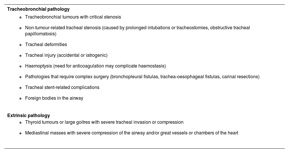
Extracorporeal membrane oxygenation

Awake ECMO under local anaesthesia may be the safest option when all 4 conventional plans are likely to be impossible, to fail, or to be ineffective and the patient is at risk of complete airway obstruction (EO 90.6%). Recent advances in technology have made it possible to include ECMO as a means of securing adequate gas exchange in these situations,110,111 particularly when difficulty is due to tracheobronchial disease or extrinsic compression that causes critical central airway obstruction or makes FONA unfeasible.67,110 In these cases, elective awake ECMO under local anaesthesia may be the safest option.67,112,113 Once the situation has been brought under control, the airway is secured to prevent aspiration. Table 2 shows the main indications for ECMO.

Table 2.

Main indications for extracorporeal membrane oxygenation (ECMO).

Tracheobronchial pathology 
  • Tracheobronchial tumours with critical stenosis

  • Non-tumour-related tracheal stenosis (caused by prolonged intubations or tracheostomies, obstructive tracheal papillomatosis)

  • Tracheal deformities

  • Tracheal injury (accidental or iatrogenic)

  • Haemoptysis (need for anticoagulation may complicate haemostasis)

  • Pathologies that require complex surgery (bronchopleural fistulas, trachea-oesophageal fistulas, carinal resections)

  • Tracheal stent-related complications

  • Foreign bodies in the airway

 
Extrinsic pathology 
  • Thyroid tumours or large goitres with severe tracheal invasion or compression

  • Mediastinal masses with severe compression of the airway and/or great vessels or chambers of the heart


ECMO, however, is costly and can lead to complications,111,114–116 so the decision to use it must be taken by a multidisciplinary team.116 If the indication is uncertain, the ECMO system can be put on standby with the vessels cannulated and a perfusionist present before proceeding with airway management.116

Veno-venous ECMO is associated with fewer complications, does not require therapeutic levels of anticoagulation, requires only a single double-lumen cannula, and could therefore be the technique of choice in these situations.110,111 In haemodynamically unstable patients requiring cardiopulmonary support due to obstructive pathology such as a large mediastinal mass, veno-arterial ECMO or even a conventional extracorporeal circulation circuit may be needed.110

In extreme cases, such as massive haemoptysis or central foreign bodies, ECMO may be the last resort.64,117 Establishing ECMO, however, can be complicated and time-consuming, so it is currently not indicated to rescue a CICO situation after induction of GA, even though several authors have described its use in this context.

Unanticipated difficult airwayPeriprocedure oxygenation

Discussed in the corresponding section

Airway management

TI is associated with more complications than other non-invasive plans, so it should be carefully weighed up and only performed when indicated.118

The primary goal in airway management should be to minimize the number of attempts in order to avoid a CICO situation and the need for FONA. Planning and optimization are essential.119

Given the unreliability of predictors,120,121 planning should be geared towards a potentially DA.122 Decision-making should be “context-oriented” rather than focused on specific devices and techniques.8,123

The first attempt must be made under optimal conditions (EO 100%) in order to maximize the likelihood of success (“make the first attempt the best attempt”).119,122,124–126 Additional attempts are only justified when there is room for improvement and a new approach will optimise the foregoing technique or substantially improve the probability of success (for example, changing the size or type of device, using a new adjunct, or changing the operator, as required).119,127

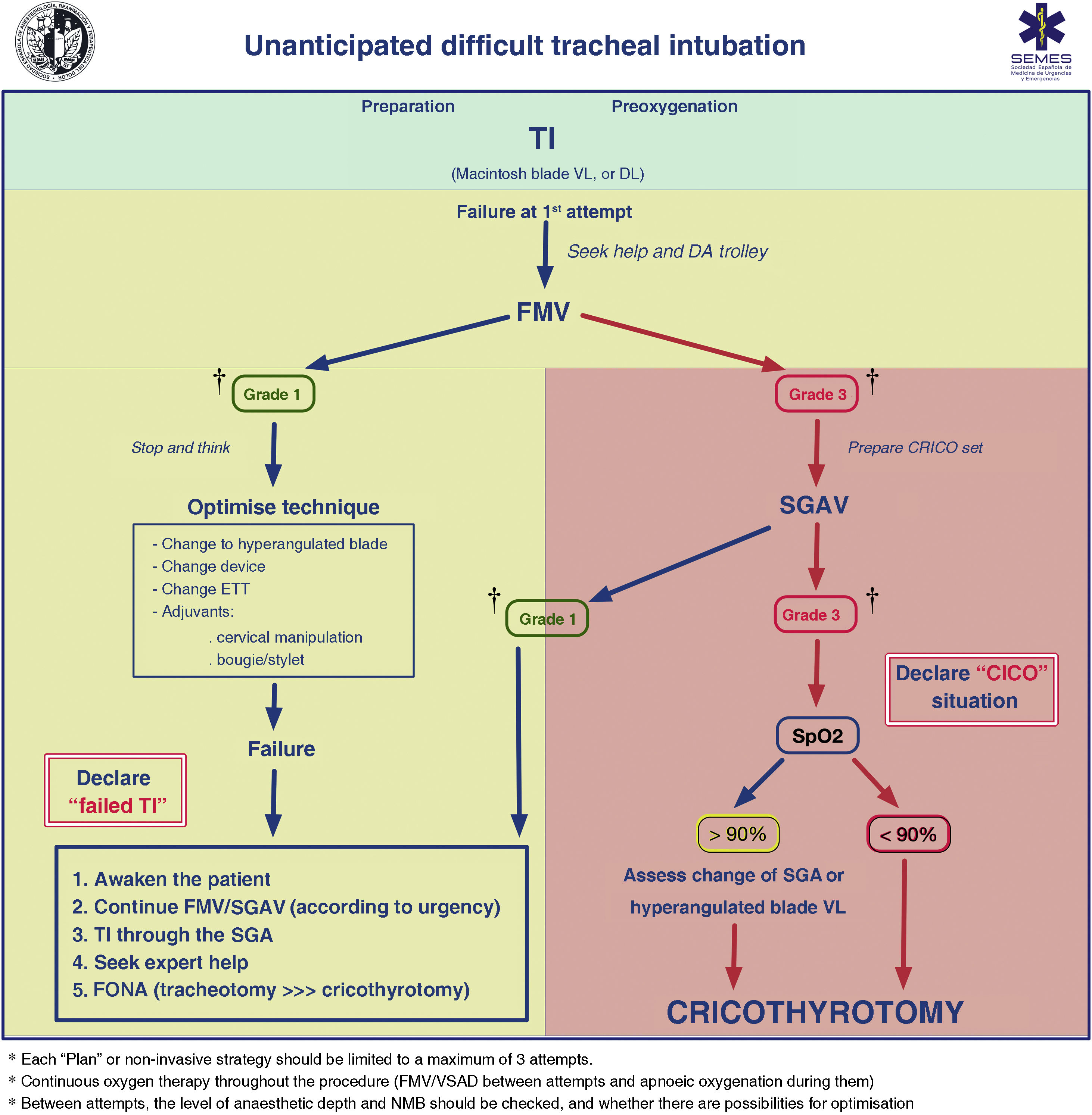
FMV and the depth of anaesthesia and NMB should be verified between attempts. Adequate ventilation between attempts using a face mask or second-generation supraglottic airway device (2GSGA) provides the opportunity to “stop and think”, reformulate the strategy, or bring in new resources while maintaining the initial principles. Fig. 3 shows the unanticipated DA management algorithm.

Fig. 3.

Unanticipated difficult tracheal intubation algorithm. † Ventilation status on capnography waveform. CICO: Can’t intubate, can’t oxygenate; CRICO: cricothyrotomy; DA: difficult airway; DL: direct laryngoscopy; ETT: endotracheal tube; FMV: face mask ventilation; FONA: front of neck access; SGA: supraglottic airway; SGAV: supraglottic airway ventilation; SpO2: peripheral oxygen saturation; TI: Tracheal intubation; VL: videolaryngoscope.

(0.74MB).

Failure of non-invasive plans necessitates declaring a CICO situation, ensuring adequate NMB, and immediately performing FONA regardless of SpO2.

Tracheal intubationVideolaryngoscopy

Failure of the first TI attempt implies a lower probability of success in subsequent attempts.60,119,128 Multiple attempts can lead to trauma, oesophageal intubation, hypoxaemia, cardiovascular events, a CICO situation, unplanned admission to a critical care unit, or death.129–134 Up to 93% of difficult TIs are unanticipated,121 so the most appropriate first-line technique should be the one that gives the greatest likelihood of achieving success on the first attempt (EO 94.3%).119,122,124–126

Most meta-analyses, despite their heterogeneity,135,136 suggest the superiority of VL over direct laryngoscopy (DL) (Supplementary data 4). Overall, VL compared to DL increases first-attempt success,137–150 improves glottis visualization,140,141,143,145–148,150–158 and reduces the incidence of complications - mainly trauma and oesophageal intubation - 141,142,144–147,149,152,155–157,159–162 by up to 50%.150

The outbreak of the COVID-19 pandemic,135,163–167 cost reduction,18 and the widespread availability of videolaryngoscopes coupled with emerging evidence of their cost-effectiveness168,169 and capacity to improve quality of care,129 teaching, documentation and teamwork (by promoting shared mental models and improving human factors), 122,167,170–173 and mastery of the technique with regular practice129 have overcome resistance to change among clinicians,174,175 and VL is now practically the gold standard for laryngoscopy and DA management.171

Based on all the aforementioned factors, SEDAR SEMES SEORL-CCC recommend the routine use of VL over DL as first-choice TI device (1B). Standard Macintosh blade devices (allowing both direct and indirect laryngoscopy) are appropriate for managing the airway without predictors of difficulty, while hyperangulated blade devices (with or without a guiding channel) are indicated for known or anticipated difficult airway (EO 94.3%).120,135,176,177 Hyperangulated blade devices are first-choice to rescue a failed first attempt.14,178–180 Consequently, it is recommended to have a videolaryngoscope available and the necessary expertise in every location where airway management is performed.

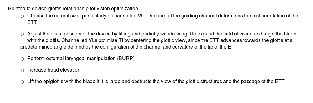
Failure to pass the ETT through the glottis is the most common cause of failed VL-guided TI166,172,173,181,182; however, a skilled operator is highly likely to overcome this difficulty.171 Recommended maneuvers to address this difficulty are listed in Table 3.182–186

Table 3.

Maneuvers to overcome difficulties in videolaryngoscopy-guided tracheal intubation.

Related to device-glottis relationship for vision optimization 
  • o

    Choose the correct size, particularly a channelled VL. The bore of the guiding channel determines the exit orientation of the ETT

  • o

    Adjust the distal position of the device by lifting and partially withdrawing it to expand the field of vision and align the blade with the glottis. Channelled VLs optimise TI by centering the glottic view, since the ETT advances towards the glottis at a predetermined angle defined by the configuration of the channel and curvature of the tip of the ETT

  • o

    Perform external laryngeal manipulation (BURP)

  • o

    Increase head elevation

  • o

    Lift the epiglottis with the blade if it is large and obstructs the view of the glottic structures and the passage of the ETT

 
Related to the use of adjuncts 
  • o

    Semi-rigid stylet, either a malleable stylet that can be pre-shaped like a “hockey stick” to match the ETT with the same angulation as the device blade, or the manufacturer's own stylet designed for use with a specific VL. Stylets are mandatory when using VLs with a hyperangulated blade without a guiding channel. As soon as the ETT has passed through the vocal cords, the stylet should be withdrawn to avoid airway injury

  • o

    Static introducer or “bougie” with a curved tip. Rigid bougies are of limited utility because they do not follow the curvature of the blade or the guiding channel. They are more useful with videolaryngoscopes with a Macintosh blade

  • o

    Dynamic bougie or flexible FB that can “negotiate” the acute angle between the glottis and the tip of the ETT

 
Related to the endotracheal tube 
  • o

    Type of ETT. Flexible tubes with a distal silicone end and conical tip

  • o

    Correct ETT size. A smaller size than usual is generally recommended to facilitate passage through the glottis, although excessively small ETTs in channelled VLs can hamper intubation because the large gap between the ETT and the channel means that the tip of the ETT will adopt an excessively posterior position as it emerges from the channel

  • o

    Change the curvature of the ETT

  • o

    Use ETTs with a flexible articulated tip that allows articulation at different angles

  • o

    Rotate the ETT 90º clockwise so that the bevel faces forward, thus reducing the angle of incidence of the ETT when not using intubation adjuncts, or when the stylet is removed before advancing the ETT into the trachea. If using a dynamic bougie or a flexible FB, the ETT should be rotated 90º counterclockwise to position the bevel backwards, overcoming advancement difficulties


BURP: backward, upward, right lateral position; FB: Fibreoptic bronchoscope; TI: tracheal intubation; ETT: endotracheal tube; VL: Videolaryngoscope.

Introducers are associated with a higher first-attempt success rate in elective and emergency TI, particularly in patients with predictors of DA or impaired glottic views.187–190 The availability of a tracheal tube introducer is recommended at all locations where airway management is performed (EO 97.1%)191 and its use should be considered at the first attempt.120,185,188,192 SEDAR SEMES SEORL-CCC recommend using an articulating introducer instead of a conventional stylet for TI in patients with DA (1C), as it improves the rate of first-attempt success and expedites TI, thus reducing instrumentation and the use of alternative adjuncts.60,69,186,193,194

Recommendation

The routine use of VL is recommended over DL as the first-choice TI device.

Strong recommendation; moderate level of evidence (⊕⊕⊕⊝)

A dynamic or articulating introducer (Flex-tip or FB) is recommended instead of a conventional stylet for TI in patients with difficult airway.

Strong recommendation; low level of evidence (⊕⊕⊝⊝)

Flexible fibreoptic bronchoscopy

FOI in unconscious or under GA patients can be highly effective in expert hands195–198; however, it is technically more challenging than in awake patients, it is not fail-safe, leading to episodes of desaturation or complete obstruction of the airway.105 The presence of blood, emesis, or secretions in the airway of patients requiring emergency TI further reduces the likelihood of success.

Manoeuvres such as tongue pulling and jaw thrusting open the pharynx and larynx, respectively, improving vision and the likelihood of success.197,199 Oral cannulas, anterior displacement of the tongue base with laryngoscopy, or placing the patient in the left semilateral position with left head rotation are strategies that facilitate passage of the FB and improve view.200 When ETT encounters resistance, jaw thrust and counterclockwise rotation of the ETT can facilitate passage through the glottis.198

In the NAP4 Audit, TI failed and an emergency FONA was necessary in all patients undergoing FOI after induction of GA as the primary technique or after failure of DL.

FB, either alone or as part of a multimodal strategy, serves as a highly effective rescue approach following failure of most devices.6 However, its availability, preparation, and execution are more labor-intensive and time-consuming than VL in an emergency,43,47 so its use as a rescue device is less widespread.201

Confirmation of tracheal intubation

After successful TI, it is crucial to immediately rule out oesophageal intubation - a common complication that can have devastating consequences.167,202–204

None of the available techniques for confirming TI are 100% reliable in all circumstances, so they should be used in combination.205,206

Waveform capnography is the most accurate (sensitivity of 98%–100% and specificity of 100%) and rapid method of confirming TI,4,120,205,207–212 and is therefore the gold standard (1B), Although penetration of waveform capnography as a standard of care remains low,204 it is mandatory to use this technique to confirm ETT placement, and it should be available wherever intubation is performed or may be required.167,203,211,213–216 A flat capnography trace (grade 3 ventilation) indicates failed TI until proven otherwise (EO 80%)211,217 and active steps should be taken to exclude oesophageal intubation.218 The capnography trace remains present, though attenuated (not flat), even during cardiac arrest.211,212,217–219 EtCO2 has a lower predictive value when perfusion is low or absent,212 and can give false positive readings when the tip of the ETT is in the hypopharynx.205 Confirming TI using point-of-care ultrasound is a valid alternative in these cases, because it has a sensitivity of 99% and a specificity of 97%, is not affected by pulmonary blood flow, takes only 13 seconds on average, and even allows real time visualization of insertion of the ETT into the trachea or oesophagus.220,221 The “double trachea sign” allows oesophageal intubation to be detected before starting ventilation.216,222

Colorimetric capnography should only be used when waveform capnography is not available, such as in pre-hospital or emergency settings.205

Clinical examination alone has an unacceptably high false positive rate,223 particularly in emergency situations,224 and confirmation bias can lead to seeing and hearing what one expects,225 although clinical examination combined with capnography can be useful. Ultrasound or FB examination through the ETT are alternative methods of confirming TI. Other methods include the oesophageal detector device, transtracheal illumination, pulse oximetry, and chest x-ray.205

Capnography waveform monitoring during mechanical ventilation is highly recommended in all settings (EO 100%),207,209,211,226–230 as it allows continuous monitoring of the ETT position, confirms airway patency, and provides early diagnosis of accidental extubation or partial displacement of an artificial airway.211,218,227,229,231–233 The NAP4 audit found that absence of monitoring might have contributed to more than 70% of airway-related deaths in the ICU,234 so the widespread adoption of capnography in critical care units and training medical and nursing staff in its use211,217,218 could be the single most impactful change in preventing morbidity and mortality arising from TI or the use of other artificial airways outside the operating room.211,234

Recommendation

Capnography waveform is the gold standard for confirming alveolar ventilation.

Strong recommendation; moderate level of evidence (⊕⊕⊕⊝)

Ventilation with a supraglottic airway

SGAs, in addition to their use as a primary technique in elective surgical procedures or cardiopulmonary resuscitation,235–237 are essential for rescuing a difficult or failed TI, since they enable ventilation and oxygenation, maintain airway patency with a certain degree of protection against aspiration, and act as conduits for FOI.238–243 Insertion and function of an SGA is not usually affected by the anatomical and/or technical factors that prevent effective FMV and TI.239 Therefore, an SGA should be inserted without delay to preserve alveolar oxygenation in the event of difficult or failed TI (EO 85.7%).

2GSGAs possess most of the ideal characteristics: they are easy to insert, provide high oropharyngeal sealing pressures, facilitate gastric decompression, and can be used as a conduit for TI.49,239,244 Thus, they have demonstrated superior performance compared to first-generation devices and are more suitable for advanced uses and as rescue devices.244–246 Therefore, the immediate availability of a 2GSGA and the necessary competence for its use are recommended in all locations where airway management is performed (EO 100%).

The selection of an SGA for rescuing a DA should be made prior to the procedure. Devices with a high first-attempt ventilation success rate that can be used as a conduit for TI are the first choice238,247: i-gel™ (Intersurgical Ltd., Wokingham, United Kingdom), Ambu® AuraGain™ (Ambu A/S, Ballerup, Denmark), LMA® Protector™ (The Laryngeal Mask Company Limited, Mahé, Seychelles) and iLTS-D (VBM Medizintechnik GmbH, Sulz, Germany).

An optimal attempt or a maximum of 3 attempts is recommended before declaring a failed plan, since the success rate significantly decreases with successive attempts,248,249 and these increase trauma and delay the transition between plans. If cricoid pressure has been applied, it must be released during placement of an SGA (EO 80%). Each new attempt must include a change that improves the chances of success. Periprocedural oxygenation methods must continue both between and during attempts.

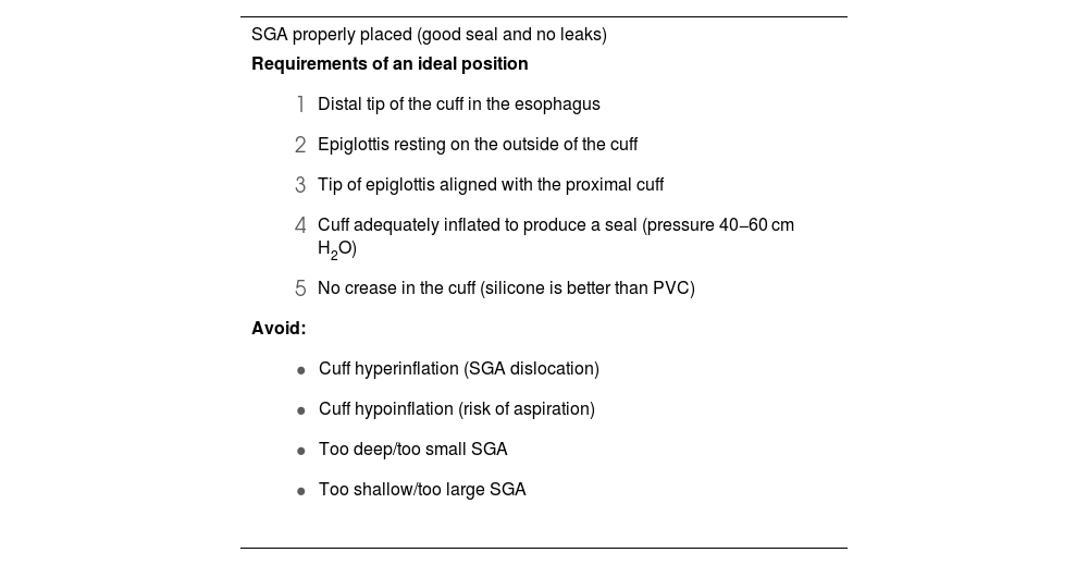
Rapid insertion and correct placement of the SGA is essential for securing the airway.250 Ninety degree rotation, jaw thrust, and the use of DL or VL (first choice) with the “insert-detect-correct as you go” technique facilitate SGA placement, increase the first-attempt success rate while reducing pharyngeal trauma (EO 82.9%),246,250–257 and can prevent malpositioning.257,258 Blind insertion, however, results in malpositioning in 50%–80% of cases,246,256,257 leading to suboptimal airway control, leaks or obstruction, increased risk of further displacement, and increased morbidity.251,255,259 Malpositioned SGAs are 26 times more likely to cause gastric insufflation followed by aspiration.255Table 4 shows the requirements for an ideal SGA position, the causes of malpositioning, and how to correct them.246,255 The recently developed video laryngeal mask (VLM)259 can be placed under direct vision, and malpositioning can be immediately corrected without the need for additional devices,66 although more evidence on their use is required.259–264

Table 4.

Requirements of an ideal position of a SGA, causes of malpositioning and how to correct them.

SGA properly placed (good seal and no leaks) 
Requirements of an ideal position
  • 1

    Distal tip of the cuff in the esophagus

  • 2

    Epiglottis resting on the outside of the cuff

  • 3

    Tip of epiglottis aligned with the proximal cuff

  • 4

    Cuff adequately inflated to produce a seal (pressure 40−60 cm H2O)

  • 5

    No crease in the cuff (silicone is better than PVC)

Avoid:
  • Cuff hyperinflation (SGA dislocation)

  • Cuff hypoinflation (risk of aspiration)

  • Too deep/too small SGA

  • Too shallow/too large SGA

 
Malpositioned SGA confirmed with video laryngoscopy255 (leaks and airway obstruction) 
Causes
  • 1

    Tip of distal cuff

    • a.

      folding over

    • b.

      folding backward

    • c.

      located between the vocal cords

  • 2

    Epiglottis in bowl of SGA

    • a.

      without downfolding

    • b.

      downfolding epiglottis

    • c.

      epiglottis folding double

Correction:
  • Jaw thrust to open the oropharyngeal space (increase the distance between the epiglottis and the posterior wall of the oropharynx) in order to reposition the SGA268

  • Use a different size or type of SGA (change from PVC cuff to silicone cuff or reinforced tip)

  • Railroad the SGA over an introducer or orogastric tube269

  • Use Magill forceps270


SGA: suplaglottic airway.

Adapted from Van Zundert AA, et al.246

Correct placement of an SGA is confirmed clinically by a normal capnography waveform (grade 1 ventilation) and the maintenance of adequate alveolar oxygenation with peak inspiratory pressures of 20 cmH2O and an oropharyngeal leak pressure >25 cm H2O. Lung auscultation and successful passage of a tube through the gastric canal are complementary signs.259

Achieving effective ventilation and oxygenation provides time to stop and think about the next step, depending on the degree of urgency and type of procedure:

  • 1

    If the situation is not urgent (e.g., elective surgical procedure), the safest option is to wake the patient and perform the surgery under regional anaesthesia or postpone the intervention in order to perform ATI.

  • 2

    If the situation is urgent due to: (a) emergency surgery: the team may decide to go ahead with surgery with the 2GSGA, although this is a high-risk option due to the potential compromise of airway patency during surgery; (b) critically ill patient: a definitive airway is likely to be required, so a FONA (tracheostomy) can be performed to prevent potentially fatal hypoxaemia.

  • 3

    In intermediate cases, FOI through the SGA can be performed if the situation is stable, NMB is adequate, and the operator has the required skills (EO 97.1%, success rates close to 100%238). Blind TI is not recommended due to its low success rate (10%–20%), the need for repeated attempts, and the potential for causing greater trauma and deterioration of oxygenation.265,266 A VLM allows the operator to perform TI without FB guidance,66 so it can shorten intubation time and may be particularly advantageous when FB is not available, such as in the prehospital and emergency setting.267

Grade 2–3 ventilation or ineffective oxygenation after exhausting all attempts requires immediate progression to a new plan.

Face mask ventilation

FMV is an essential transition technique during anaesthesia induction and management of the emergent airway, and an indispensable rescue plan when other techniques fail.271

The presence of predictors of difficult FMV, as well as its use during an emergent airway or as a rescue from failed plans, makes it especially advisable to use the optimal technique from the outset (the triple airway manoeuvre of head extension, jaw thrust and mouth opening, placement of a nasopharyngeal or oropharyngeal cannula272 and the two-handed “VE” technique 271,273 performed either by 2 operators or with pressure-controlled ventilation from a ventilator or other device274,275 in a patient with optimal positioning under intense NMB271,276–280) (EO 80%) in order to limit airway obstruction and optimize mask sealing to achieve grade 1 alveolar ventilation without causing gastric insufflation.126,275,281 This speeds up the transition between plans and reduces the maximum ventilation pressure.282 Face mask ventilation with a modified triple airway manoeuvre is recommended over the “CE” technique in the general population (1C).

Successful FMV must be defined as stable or improved SpO2, acceptable tidal volume and airway pressure (4−5 ml/kg and <15–20 cmH2O, respectively273,275,283), and the presence of a plateau phase on capnography.284 It is advisable to use scales to objectively stratify the difficulty of FMV, such as the instrument developed by the Japanese Society of Anaesthesiologists (Fig. 2, Part I). This allows the declaration of a failed FMV attempt before desaturation occurs (late sign).275

FMV is a dynamic procedure that must be continuously monitored until the airway has been secured.285 The declaration of failed FMV necessitates an immediate transition to SGAV (EO 85.7%). Clinical deterioration and worsening oxygenation should prompt the declaration of CICO and an immediate transition to FONA if SGAV has also failed.

A CICO situation should be declared using clear language that is easily understood by all and creates a shared model that will facilitate effective transition to FONA.119,286

Recommendation

Face mask ventilation with a modified triple airway manoeuvre is recommended over the “CE” technique in the general population.

Strong recommendation; low level of evidence (⊕⊕⊝⊝)

Front of neck access techniques

Failure of all 3 non-invasive supraglottic plans (primary and rescue) in an apneic patient necessitates the verbalization of CICO and the prompt execution of FONA regardless of the SpO2 value (EO 90.6%), because in these circumstances deterioration is either imminent or already evident. In in-hospital CICO situations, the SEDAR SEMES SEORL-CCC recommend asking for an ENT specialist (or surgeon trained in tracheostomy) as soon as CICO has been declared, although no procedure should be delayed while waiting for their arrival.

Delayed FONA is a leading cause of morbidity and mortality.99,234,247,287,288 Situational awareness and shared decision-making, together with good training in technical skills and human factors management will eliminate the psychological barriers to relinquishing further attempts at non-invasive techniques.99,119,287,289–291 Cognitive aids can help alleviate stress and speed up the transition to FONA292 (Fig. 1, Part I).

Teams should be prepared both psychologically and technically before declaring a CICO situation, and for this purpose the “double setup” may help293: one team performs conventional airway management while a second team stands by ready to perform FONA if needed. Multidisciplinary approach is more likely to secure the airway faster and at the first attempt.

It is recommended to continue providing 100% oxygen via a supraglottic route, ensure intense NMB127,294 and the neck must be extended to expose the larynx.295

There are 3 emergency FONA techniques: percutaneous cricothyrotomy, surgical cricothyrotomy, and surgical tracheotomy.

Cricothyrotomy is the technique of choice in a CICO situation (EO 91.4%) because it is relatively simple, fast, and has a high success rate with few complications.100,296 However, the presence of an experienced ENT specialist or surgeon allows for a tracheostomy to be performed with the same efficacy and safety as a scalpel cricothyrotomy.297,298 Therefore, if an ENT specialist is present, he or she should perform FONA using the technique they consider most appropriate.

Cricothyrotomy can be either surgical or percutaneous. The evidence to support any single FONA technique is week. The few comparison studies published have been performed in simulation or animal models,99 therefore, it is impossible to recommend one technique over the other.99,101,299–302 However, surgical cricothyrotomy is successful in practically 100% of cases.234,287,303

The SEDAR SEMES SEORL-CCC recommend performing surgical cricothyrotomy using the scalpel-bougie-tube technique (EO 91.4%) for the following reasons98–100,234,247,287,291,294,301,304,305: (1) the equipment needed is available in all settings; (2) it can be performed by a single operator; (3) it has a short learning curve; (4) although it is psychologically more challenging,300,306,307 it is a simple, rapid technique; (5) it permits a large bore ETT or cuffed cannula to be inserted into the trachea to definitively secure the airway, protect against aspiration, and achieve effective gas exchange with conventional PPV that can be confirmed with capnography; (6) it is both safe and effective. The equipment includes a scalpel with a number 10, 20 or 21 blade, an ETT or cannula with a bore of up to 6 mm, and a bougie.

Other cricothyrotomy techniques are also valid, provided they are performed by an experienced operator using the correct equipment. Percutaneous cricothyrotomy to re-establish oxygenation and adequate ventilation involves inserting either a ≥4 mm tube with a 15 mm connector, or a wide-bore cannula techniques.247,291,294,301,305,308 The SEDAR SEMES SEORL-CCC leave it to the operator to decide whether to use percutaneous cricothyrotomy as their first-choice or rescue technique,309 and therefore recommend acquiring skills in more than one technique.300,302,310 The operator’s experience, the availability of the material, and the characteristics of the patient will determine the technique used.99,101,294,297,309,311 The percutaneous technique, being more familiar and less intimidating, could be initiated earlier.300

The success of the technique relies on identification of the cricothyroid membrane (CTM). Identification by palpation is prone to error. Ultrasound is recommended over palpation to identify the CTM (1C), so the SEDAR SEMES SEORL-CCC recommend acquiring the necessary skills. The laryngeal handshake technique (by palpation), though slightly more time-consuming than conventional techniques, is associated with a higher success rate.312,313 When the anatomy of the neck makes it difficult to identify the CTM by palpation, the operator should perform a vertical cutaneous incision measuring more than 4 cm in the midline of the neck in a distal-proximal direction above the sternal notch to identify the relevant anatomy.98,247,314 Ultrasound, though superior to palpation in identifying the CTM,315–319 can be time consuming, so in a CICO situation it should only be used if it is immediately available and the operator has the required training.

After securing the airway with FONA, adequate ventilation and alveolar oxygenation should be confirmed by capnography waveform, clinical evaluation, pulse oximetry and arterial blood gases when indicated.294 The examination can be complemented by a fibrobronchoscopic and radiological study. Emergency cricothyrotomy should be converted to an ETT or tracheostomy in a timely fashion, as there is insufficient evidence to support its long term use (EO 85.7%).296,320 The cricothyrotomy tube should be replaced with a tracheostomy within 72 h to avoid subglottic stenosis.301,321

Failure of a cricothyrotomy to secure the airway makes it advisable to perform a tracheostomy by an experienced operator (EO 94.3%).100

According to the NAP4 audit, CICO situations account for 39% of critical events and 25% of all anaesthesia-related deaths.287 FONA is the last resort when non-invasive plans have failed, so it is vitally important.322 Each institution should protocolise the technique used, and a commercial or pre-assembled cricothyrotomy kit should be available wherever airway management is performed.99,247,311,323 The kit should be placed in an easily accessible transparent bag, ideally in the DA trolley, so that the team can become familiar with its contents and mentally rehearse the procedure. All clinicians involved in airway management must acquire and maintain the skills needed to perform a surgical or percutaneous cricothyrotomy using the Seldinger technique (EO 100%).99,101,119,247,291,298,301 The performance of FONA should be feasible at any location where airway management is conducted (EO 100%).324
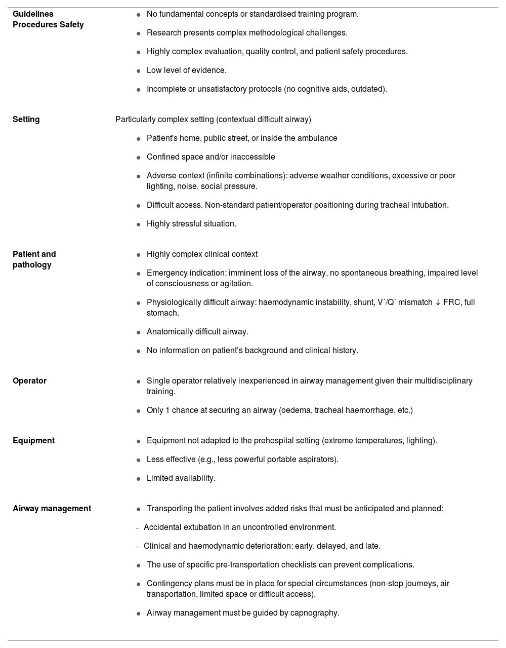
Recommendation

The use of ultrasound is recommended over palpation to identify the cricothyroid membrane

Strong recommendation; low level of evidence (⊕⊕⊝⊝)

The prehospital setting

Prehospital airway management is essential for patient survival, and is a major challenge for Emergency Medical Services (EMS).325,326 Different factors and limitations come together in each scenario to create a high level of uncertainty that transforms any ordinary airway into a DA,327 and it is not always possible for patient and operator to be positioned as described in intubation guidelines (patient trapped, confined, crushed, buried, or inaccessible). Survival depends on effective coordination of the entire emergency care chain. Table 5 shows the distinguishing characteristics of prehospital airway management.328,329

Table 5.

Characteristics of pre-hospital airway management.

Guidelines Procedures Safety 
  • No fundamental concepts or standardised training program.

  • Research presents complex methodological challenges.

  • Highly complex evaluation, quality control, and patient safety procedures.

  • Low level of evidence.

  • Incomplete or unsatisfactory protocols (no cognitive aids, outdated).

 
Setting  Particularly complex setting (contextual difficult airway)
  • Patient's home, public street, or inside the ambulance

  • Confined space and/or inaccessible

  • Adverse context (infinite combinations): adverse weather conditions, excessive or poor lighting, noise, social pressure.

  • Difficult access. Non-standard patient/operator positioning during tracheal intubation.

  • Highly stressful situation.

 
Patient and pathology 
  • Highly complex clinical context

  • Emergency indication: imminent loss of the airway, no spontaneous breathing, impaired level of consciousness or agitation.

  • Physiologically difficult airway: haemodynamic instability, shunt, V˙/Q˙ mismatch ↓ FRC, full stomach.

  • Anatomically difficult airway.

  • No information on patient’s background and clinical history.

 
Operator 
  • Single operator relatively inexperienced in airway management given their multidisciplinary training.

  • Only 1 chance at securing an airway (oedema, tracheal haemorrhage, etc.)

 
Equipment 
  • Equipment not adapted to the prehospital setting (extreme temperatures, lighting).

  • Less effective (e.g., less powerful portable aspirators).

  • Limited availability.

 
Airway management 
  • Transporting the patient involves added risks that must be anticipated and planned:

  • -

    Accidental extubation in an uncontrolled environment.

  • -

    Clinical and haemodynamic deterioration: early, delayed, and late.

  • The use of specific pre-transportation checklists can prevent complications.

  • Contingency plans must be in place for special circumstances (non-stop journeys, air transportation, limited space or difficult access).

  • Airway management must be guided by capnography.


FRC: Functional residual capacity; V˙/Q˙ mismatch: ventilation/perfusion mismatch.

Human and ergonomic factors

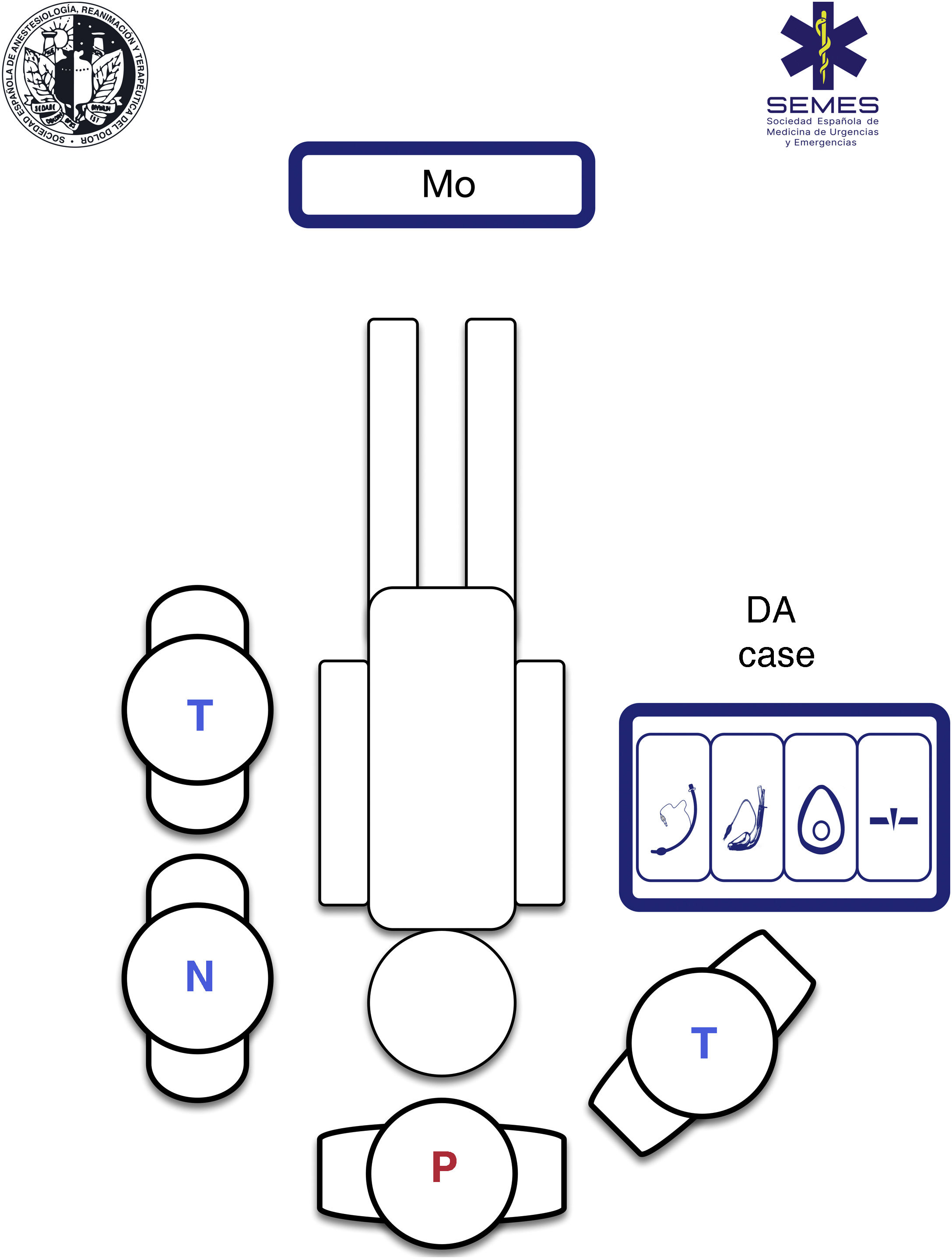
Using a standardised RSI protocol will optimize outcomes, reduce the cognitive burden in high-stress situations, and improve technical and non-technical performance. Therefore, it is advisable to standardize the contents and layout of the airway management bag or “kit dump”, and have a checklist on hand.330Fig. 4 shows our suggested contents and ergonomic layout of the airway management bag suggested by SEDAR SEMES SEORL-CCC for the prehospital setting.

Fig. 4.

Placement of team members and materials in an Ideal Prehospital Setting. DA: difficult airway; N: nurse; Mo: monitor; P: physician; T: technician.

(0.28MB).
Airway management bag

All the airway material is stowed in the bag using a standardised, modular design so that it is always visible and accessible in less than a minute. The material needed for each of the four treatment plans is stowed in a separate compartment separated by Velcro® or zippers, and marked with easily recognizable pictograms.331 Each compartment must contain at least 1 airway device for each plan (see Fig. 4).

For field use, bags, cases, or backpacks must be easy to carry (reasonable size and weight), made of officially approved flexible, water- and corrosion-proof material of an easily identifiable colour that is clearly labelled and can be easily washed and sterilize. They must have both handles and straps to enable them to be carried on the back to keep the hands free for care procedures. They should ideally be checked daily, and also at each change of shift, following a checklist.

Ergonomics of positions during airway management

Pre-hospital care of critically ill patients relies on teamwork, usually made up of a physician, a nursing component, and one or more technicians. Therefore, there is a single operator.

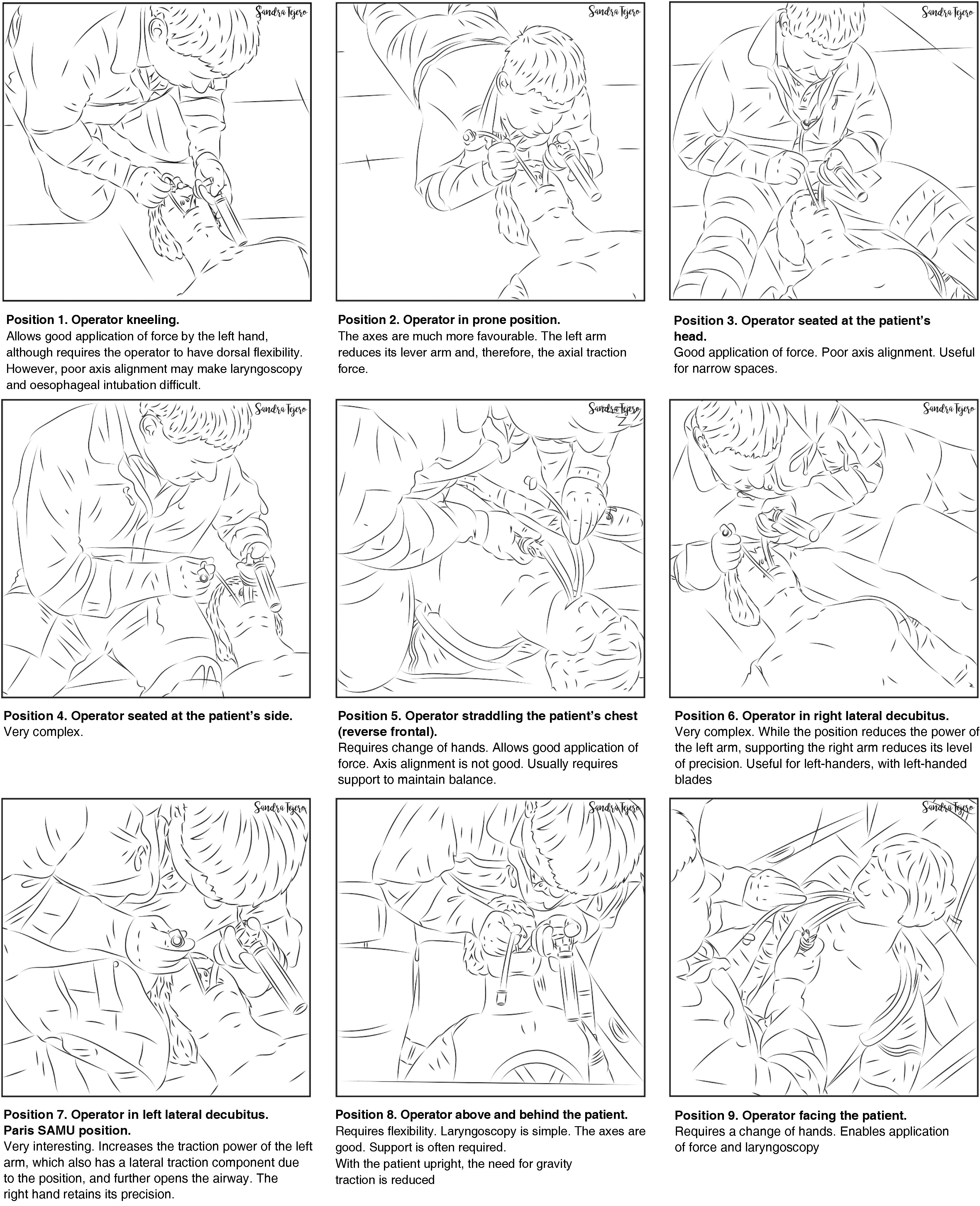
The airway approach will vary greatly, depending on the many different scenarios, risks, patients, positions, and obstacles encountered. Each intervention is unique and unrepeatable. The emergency team must access and control the scene, identify the most seriously ill patient, determine their priorities, and establish more than one approach plan. The patient will usually be lying on the ground allowing for a safety roll, although this is not always possible. Tracheal intubation on the ground is a predictor of DA.332 Non-standard positions create varying degrees of airway access difficulties, so it is important to bear in mind the anatomical changes and the pathophysiology associated with each position (Fig. 5). However, in an ideal scenario, the SEDAR SEMES SEORL-CCC suggest arranging team members as shown in Fig. 4. The physician is situated at the head of the patient and controls ventilation and airway. A technician is situated on his or her right to assist with the technique (Sellick, BURP, jaw thrust, handing over material, etc.). The nursing member and the second technician are positioned at the patient's left shoulder/arm, while the airway management bag is placed on the patient's right, and the monitor is placed at the patient’s feet where it can be clearly seen by all team members. If the patient is in cardiac arrest, the monitor should be placed by the patient's left shoulder to facilitate manoeuvres.

Fig. 5.

Strategies for intubating difficult access airway.

(1.48MB).
Pre-procedure assessment and planning

A DA encountered in the pre-hospital setting is, by definition, unanticipated. However, it is still essential to evaluate the airway in order to anticipate possible difficulties and plan the treatment.331 Predictive tests can be challenging to use, and often there is only time for a “one second look”. Although this quick evaluation can be useful, it should be combined with other tests as far as possible.333

The following independent predictors of DA in the pre-hospital setting have been described in the literature: (1) airway obstruction, patient lying on the ground, and thyromental distance of less than 3 fingers,332 (2) cramped conditions, short neck, obesity, facial and neck injuries, mouth opening less than 3 cm, and ankylosing spondylitis,334 (3) Glasgow scale >3, limited neck movement, trismus/jaw clenched, inability to palpate landmarks on the neck, presence of blood or vomit in the airway.335

Teams dealing with an unanticipated DA should also have experience in ATI, although the indications are more limited in emergency TI.336,337

Periprocedure oxygenation

Apnoeic oxygenation significantly reduces hypoxaemia during emergency TI,338,339 so this, together with preoxygenation, is essential in prehospital airway management.340 Patients should be oxygenated using a standard nasal cannula at 15 l/min before induction and until the ETT is in place, except in the case of epistaxis, severe head trauma with possible skull base fracture, or complex facial fractures, because it could worsen intubating conditions and potentially cause pneumocephalus.331

Any factor that may affect airway management must be optimized before attempting TI to make sure that the first attempt will be the best.

Rapid sequence induction

RSI is the most widely used induction technique in the pre-hospital setting. However, alternative techniques should also be available.95 It is advisable to follow a standardized checklist-based RSI protocol that includes drugs and dose calculation, and all the material required.341

Cardiopulmonary resuscitation

Any abnormal situation increases the delay in achieving ventilation and alveolar oxygenation, interrupts chest compressions, and delays the return of spontaneous circulation (ROSC).342

According to current evidence, there are no significant differences between non-invasive airway management plans.343–345 Outcomes are determined by TI success rates. Therefore, if the desired level of TI efficacy is not achieved, preference should be given to establishing ventilation and alveolar oxygenation over a specific treatment plan, and airway management should not interfere with resuscitation interventions, such as chest compressions, defibrillation, and treatment of the reversible causes of the cardiac arrest.328

Severe trauma

Severe traumatic brain injury carries a high risk of airway obstruction, pulmonary aspiration, hypoxia, brain injury, and death.346 Pre-hospital TI is beneficial when performed by experienced practitioners following standardized protocols.346,347 Pre-hospital TI together with air transport could reduce overall mortality by 47%.326

Various factors can complicate airway management,348 including: (1) possibility of an unstable cervical spine injury requiring bimanual alignment; (2) contamination or flooding of the airway by tissue, vomit, secretions, blood, etc. This requires aggressive management with manual removal of fragments and aspiration of secretions using the SALAD (suction assisted laryngoscopy and airway decontamination) technique349; (3) uncooperative or agitated patient; (4) facial bone fractures; (5) Direct Airway Trauma: Both penetrating and blunt trauma (burnt airway and/or inhalation syndrome). The presence of penetrating cervical trauma is the most common indication for ATI, while the highest incidence of FONA is reported in patients with maxillofacial trauma.348,350

In these conditions, RSI and VL-guided TI with a hyperangulated blade and preconfigured stylet are recommended348,351 The procedure can be affected by sunlight reflecting off the screen or contamination of the airway by blood or vomit.352 If a VL is not available, an SGA or DL with reduced traction can be used.329 The use of DL may increase the risk of cervical spine injury.348,350

Patient trapped, buried, crushed, or inaccessible

This represents the paradigm of contextual difficult airway. To deal with this setting, teams need to acquire additional skills in order to take part in rescue support manoeuvres (8 modalities) and work in hostile environments.353

Cuff pressure monitoring

A large percentage of laryngotracheal morbidity is related to improper establishment of cuff pressure.354 Underinflation can cause hypoventilation or increase the risk of aspiration, while excessive pressure, even for short periods, can cause hoarseness, sore throat, impaired ciliary motility, and injuries such as mucosal inflammation and ischaemia, laryngeal oedema, ulcers, stenosis, tracheoesophageal fistula, tracheomalacia, tracheal rupture, vocal cord paralysis, or nerve injury.241,355–357 The incidence of these complications has declined since the introduction of low-pressure, high-volume cuffs358; however, these devices still cause preventable injuries.94,359

Intermittent monitoring with manometry of cuff pressure is desirable after establishment and periodically during maintenance360–364 (not applicable in crisis situations). The use of continuous monitoring to constantly maintain cuff pressure within a desired range in critically ill patients could reduce the risk of ventilator-associated pneumonia.365–367 A manometer should be used for continuous monitoring of cuff pressure (1C).

Cuff pressure must be established with the minimum pressure needed to guarantee a safe, effective seal. Cuff pressure should remain between 20−30 cm H2O (ideally lower than 25 cm H2O) in ETTs and tracheostomy and cricothyrotomy cannulas, and <60 cm H2O in the case of SGAs (EO 94.3%)241,354,358,368–371 from insertion to extraction. If the seal is inadequate even at these pressures, it may be necessary to reposition or re-size the device.372

Nitrous oxide spreads into the cuff,359 so pressure should be re-measured after 20 min, at which time the pressure will have stabilized, and again if nitrous oxide concentration increases.373
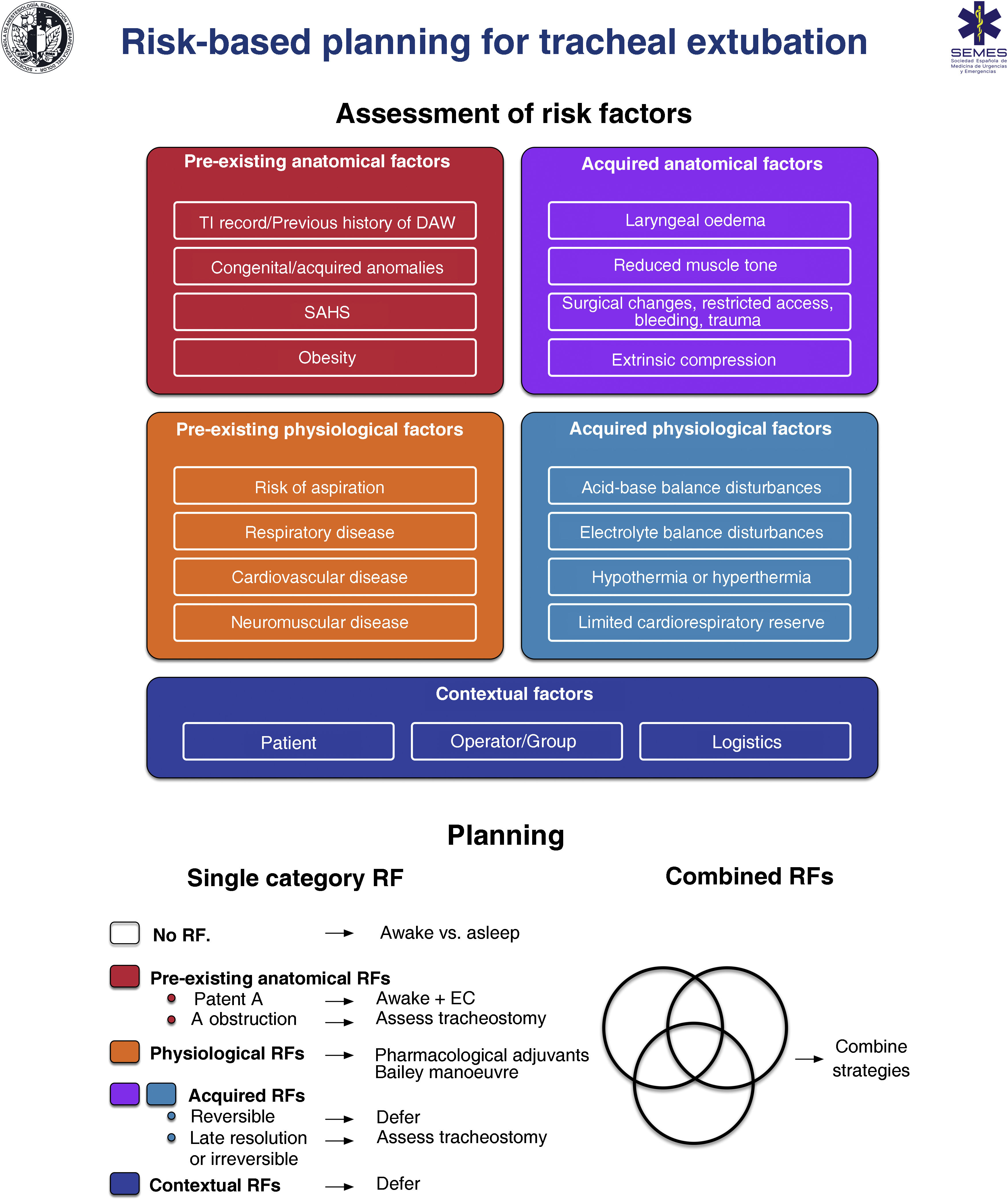
Recommendation

A manometer should be used for continuous monitoring of cuff pressure

Strong recommendation; low level of evidence (⊕⊕⊝⊝)

Extubation

Tracheal extubation is a high-risk procedure that has a major physiological impact.374 It may trigger a haemodynamic stress response, coughing, laryngospasm or agitation which may in turn increase intracranial or intraocular pressure.375–378 Laryngotracheal protective reflexes remain impaired for several hours after extubation, facilitating aspiration. Extubation failure occurs when a patient is unable to maintain adequate oxygenation and ventilation, clear secretions, or maintain airway patency, and can have catastrophic consequences, particularly in patients with a DA.376 Airway obstruction is the main cause, and is usually associated with post-obstructive pulmonary oedema and severe hypoxia.378 Failed extubation, generally defined as the need for reintubation within 24−72 hours, occurs in approximately 0.1%–0.45% of patients undergoing general anaesthesia.379 Prevalence increases 10-fold in patients with sleep apnoea-hypopnea syndrome (SAHS) or in those undergoing airway surgery, and another 10-fold in patients treated in the ICU.380 Failed extubation can potentially lead to severe adverse outcomes.381–383 One third of all cases of anaesthesia-related deaths or permanent brain damage are due to failed extubation.288,384 Poor anticipation and planning for “at risk” extubation are core issues.288,384 Extubation is an elective procedure that must be prepared, executed a stepwise strategy, and meticulously followed up.375,376,378,385Fig. 6 shows a cognitive aid for planning extubation based on risk assessment.

Fig. 6.

Cognitive aid for extubation: planning, risk stratification and decision making.

A. airway; Adjs: adjuvants; DA: difficult airway; EC: exchange catheter; RF: risk factors; SAHS: sleep apnoea-hypopnea syndrome; TI: tracheal intubation.

(1.17MB).

The main objective in tracheal extubation is to preserve oxygenation and avoid reintubation. Reintubation should always be considered potentially difficult due to the additional complexity involved (EO 97.1%, distorted anatomy with restricted access, secretions, blood or oedema, time constraints, and highly stressful setting).375,376,385 Anatomical, physiological and contextual risk stratification376,383,386,387 determines the likelihood of successful extubation on the basis of tolerance to extubation and the feasibility of reintubation, and allows to establish an individualized strategy and optimise the patient’s status in anticipation of airway-related and physiological factors, such as hypoxaemia, hypercapnia, residual NMB, hypothermia, or airway oedema.355,383,387 The leak test,388 preferably quantitative (leak volume <110 ml or <12%–24% of delivered tidal volume determines diminished airway patency and risk for postextubation stridor due to laryngeal oedema387,389), ultrasound evaluation,390,391 and laryngeal visualization with VL or FB can be used to evaluate airway patency and identify periglottic oedema or bleeding before extubation, thereby facilitating decision making (EO 97.1%),355 although none of these are specific predictors of successful extubation.392

Once the decision has been made to proceed with extubation, the first step is to review the original TI and re-evaluate the airway and overall risk factors.383 Careful advance preparation, notifying the team of potential problems and early warning signs, and establishing appropriate rescue plans for oxygenation and reintubation if the primary strategy fails will help ensure safe, successful extubation.378,383 Important considerations before extubation include378: (1) preoxygenate until extubation; (2) aspirate visible airway secretions or blood, ideally using a laryngoscope to avoid soft tissue trauma; (3) place a bite bloc386; (4) properly positioning the patient; (5) reverse NMB. Neuromuscular monitoring in combination with reversal agents to achieve a train-of-four ratio of ≥0.90 is essential to avoid residual NMB393–396; (6) minimise head and neck movements and reduce noxious stimuli to avoid laryngospasm; (7) emergence to an awake state.377,378,385–387 Deep extubation is inappropriate in patients with DA or risk of aspiration377,386; (8) apply positive pressure, deflate the cuff, and remove the ETT; (9) administer 100% oxygen, confirm airway patency and adequate spontaneous breathing; (10) maintain continuous oxygen therapy until full recovery, along with surveillance, monitoring, and qualified assistance to handle potential emergent tracheal reintubation.385 DA equipment must be prepared and immediately accessible.

Prophylactic administration of corticosteroids before elective extubation significantly reduces the incidence of airway-related post-extubation adverse events and reintubation, so patients at high risk of obstruction could benefit from this strategy.397–399 In patients with an absent or reduced cuff leak, administration of a corticosteroid at least 4 h before extubation is advised.376,387,389 Prophylactic administration of corticosteroids is recommended before extubation in patients at high risk of airway obstruction (1B).

Various advanced techniques have been developed for high-risk extubation, but these should only be performed by clinicians experienced in each technique.378 Administration of pharmacological adjuvants,400–405 such as infusion of remifentanil,406 or performing the Bailey manoeuvre, which involves the placement of a SGA over the ETT followed by the removal of the ETT,407 may be considered when smooth emergence and attenuation of undesirable cardiovascular or respiratory responses are required.408–410 The use of VLM for TI could play a significant role in making this manoeuvre safer and simpler, as it allow for the easy withdrawal of the ETT through its ventilation channel under direct vision and reinsert it immediately if required.66 Both techniques require a deep level of anesthesia for their execution, so they may be inappropriate in patients at risk of aspiration and in whom reintubation may be difficult.1,378

Awake extubation and the use of advanced techniques is the most suitable approach in patients with a difficult airway (EO 94.3%), as the maintain airway patency and muscle tone, protective reflexes, and spontaneous breathing.410 Airway exchange catheters are usually used to rescue extubation in patients with anticipated or known DA.1 These devices can be left in place after extubation if reintubation is likely,376 in which case an ETT can be railroaded over the catheter under direct vision.185 They can also be used to deliver low-flow oxygen or jet ventilation in the event of life-threatening hypoxaemia, although this should be avoided as far as possible, since even low flows have been known to cause barotrauma.411 Reintubation with an airway exchange catheter is successful in 92% of cases, with a first attempt success rate of 87% vs 14%. They are associated with a low incidence of oesophageal intubation and desaturation, bradycardia, and hypotension.412 The main risks of the technique are airway stimulation, injury due to subcarinal insertion, and accidental dislodgement of the catheter. In adults, they should never be inserted deeper than 25 cm from the lips.378 Staged extubation sets, consisting of a guide wire and reintubation catheter,413,414 are associated with an overall success rate of 93%415 and appear to improve tolerance at the expense of increased risk of accidental dislodgement.416 If reintubation over an intubation catheter is necessary, reducing the gap between the ETT and the catheter, administering adequate NMB, and using a VL to visualise the advance of the ETT facilitate the procedure by preventing arytenoid and epiglottic impingement.383,417 Other recommendations to facilitate FOI are applicable to exchange catheter-guided TI. If re-intubation fails, the operator must follow the unanticipated DA algorithm.

Waveform capnography should be available in recovery units and should be used in high-risk patients (EO 97.1%). After extubation, oxygen therapy with nasal prongs and face mask with capnography waveform monitoring will promptly detect respiratory depression, hypoventilation, hypercapnia or post-extubation airway obstruction.379,387

Strategies to prevent extubation failure include head tilt and administration of supplemental oxygen. A good oxygenation strategy with NIV or HFNO can avoid reintubation in at-risk patients.374,418–421

Recommendation

Prophylactic administration of corticosteroids is recommended before extubation in patients at high risk of airway obstruction

Strong recommendation; moderate level of evidence (⊕⊕⊕⊝)

Documentation

The airway management process must be detailed in a report that can orient future intubations. In addition to the informed consent and the results of the preintubation pre-procedure airway evaluation, the report should detail the techniques used for periprocedure oxygenation, topicalization, awake sedation, induction, the devices, adjuncts and ETT used, the approach (right nasal, left nasal or oral), number of attempts, the extubation strategy, and any difficulties or complications encountered. Supplementary data 5 shows an airway management report template.

A history of failure in previous procedures is the most accurate predictor of failure in subsequent treatments (EO 97.1%).422 Reporting difficulty is one of the most important ways of preventing future complications. The report facilitates decision-making and forms the basis for a structured, targeted approach that ensures an efficient transition and reduces airway instrumentalization by steering away from plans that have been unsuccessful in the past.422,423 Using a variety of resources to report a DA will increase the likelihood of this critical information fulfilling its purpose.423,424 For example11,294,424: (1) a detailed description of the successful technique used together with a visual record with images and/or video of the anatomy and the technique can be added to the patient’s clinical history425; (2) the patient and their next of kin or caregiver can be notified verbally in person; (3) the patient can be given a written report. Supplementary data 6 shows our suggested DA notification template; (4) the patient can be issued with a medical alert bracelet, necklace or ID card with a QR code for access to clinical information. Supplementary data 7 shows our suggested DA alert wearables; (5) a DA alert can be programmed to pop up whenever the patient's electronic record is accessed; and (6) the patient can be registered in national or international DA databases.426 The registration form should be standardised, and should include mandatory fields describing the characteristics of the airway, the nature of the difficulty, the techniques that failed and succeeded, and a free text field for additional information.294,424 Using structured difficult airway notes in electronic records has been shown to reduce the number of adverse events.427,428

Organisational aspects of airway management

The implementation of airway strategies requires not only individual commitment, but also a proactive attitude on the ministerial, institutional and departmental levels.429,430 The action plan to improve the safety, cost-effectiveness and quality of airway management includes: (1) standardising the techniques and equipment used across all locations wherever airway management is performed, effective adherence to guidelines, standardising the DA trolley,431 eliminating barriers and promoting facilitators to encourage compliance with guidelines432–434; (2) coordinating all departments involved in airway management (anaesthesia, intensive care, emergency, otolaryngology, and prehospital care); (3) acquiring and ensuring accessibility of the right equipment; (4) providing staff with training and opportunities for continuous professional development; (5) performing periodic audits and introducing a human error prevention program: a) analysing airway-related incidents, b) identifying and addressing departmental and institutional factors that may have contributed to the event in order to learn from mistake and make improvements, c) reviewing cases and discuss alternative plans; (6) implementing a system for reporting and notifying airway-related incidents435; (7) introducing a system of coding, registering, identifying and warning of DAs; (8) drafting organizational guidelines on the use of resources and continuous improvement.436,437

Each institution should appoint an “Airway Lead” (EO 100%)11,429,430,438 - a clinician with experience in the field who liaises with hospital management to ensure local policies and organisation are implemented. The airway lead’s primary objective is to provide each member of staff with the tools they need.429,439 In order to improve safety, some hospitals have formed multidisciplinary specialist teams with clearly defined roles.440–442 The creation of a national network of airway leads and airway teams would further improve airway management and patient safety.438,439

Decisions to acquire airway devices must be supported by a formal, evidence-based evaluation.24,443,444

Education and training

Airway management is an essential skill in anaesthesiology, critical care, and prehospital and emergency medicine.445,446 Poor training is a common causal or contributing factor in complications234,384 and undermines the operator’s confidence in performing essential techniques, such as FOI and cricothyrotomy.447–449 Optimizing teaching and training are key to improving safety.322

There is scant evidence on airway management curricula, and training is not standardized,324,450 so strategies used in other specialist fields have been adapted to airway management training. Good training should include both cognitive skills based on theoretical principles, and technical and non-technical skills.322,451 Skills must be acquired gradually through a process of theoretical, simulation-based, and clinical training with problem solving until the learning curve has been completed. Instructors must give evaluation and feedback in each phase of learning (EO 100%).437,452,453 Competency-based training must be individualized.454 Training should follow a mastery learning approach that consists of achieving predefined standard learning objectives involving mastery of a particular skill before moving on to a higher level of difficulty and stress.455 Mastery learning is a student-centred, evidence-based method456 that could give better results.457,458

Education and training must be structured in such a way as to address all aspects of airway management,37,322,445,455 with emphasis on core, versatile skills, and skills to be used in critical emergency situations.445 Standardization of a training program is highly recommended.322 Simulation-based training is important for acquiring non-technical skills, such as teamwork, using guidelines and cognitive aids in clinical practice, and practising procedures and resolving life-threatening issues that would rarely be encountered in real-world practice.437,448,455,459–464 Guidelines and algorithms are tools for learning skills and strategies, and can be used as a reference during debriefing,322,465,466 while cognitive aids are useful when practising procedures in simulation-based training.127,322 Structured debriefing improves clinical knowledge and the acquisition and implementation of skills in clinical practice.455,467

More senior operators may be at risk of providing lower quality care,446,468 and therefore require a continuing education and training program, preferably annual, with refresher courses to acquire the new skills needed to use the latest devices or new techniques and to maintain their existing skills322,445,455,469,470 (EO 97.1%).471 Both hospital management and individual clinicians must take a proactive approach to training. It is highly recommended that each service appoint a local airway leader to organise top quality interprofessional, multidisciplinary training programs with defined learning goals, evaluations, and supervision, and to ensure that guidelines and cognitive aids are disseminated and followed.322,324,439,445,448,472 Training must be diversified to provide the operator with several alternatives.43Supplementary data 8 shows the skills that we believe should be included in all airway management training programs, as well as the teaching methods needed to achieve these objectives.

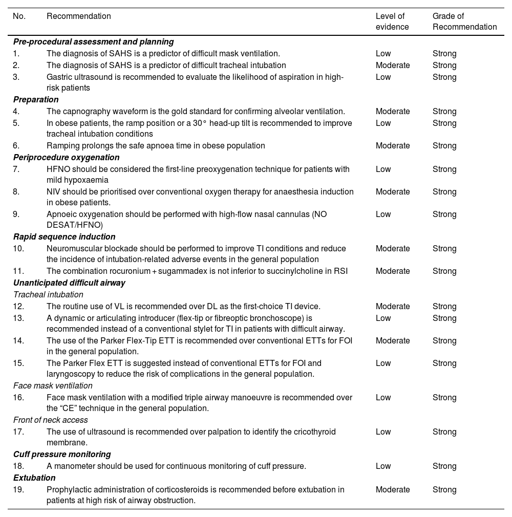
Summary of recommendations derived from the systematic literature search

Search strategies and GRADE tables are shown in supplementary data.

No.  Recommendation  Level of evidence  Grade of Recommendation 
Pre-procedural assessment and planning
1.  The diagnosis of SAHS is a predictor of difficult mask ventilation.  Low  Strong 
2.  The diagnosis of SAHS is a predictor of difficult tracheal intubation  Moderate  Strong 
3.  Gastric ultrasound is recommended to evaluate the likelihood of aspiration in high-risk patients  Low  Strong 
Preparation
4.  The capnography waveform is the gold standard for confirming alveolar ventilation.  Moderate  Strong 
5.  In obese patients, the ramp position or a 30° head-up tilt is recommended to improve tracheal intubation conditions  Low  Strong 
6.  Ramping prolongs the safe apnoea time in obese population  Moderate  Strong 
Periprocedure oxygenation
7.  HFNO should be considered the first-line preoxygenation technique for patients with mild hypoxaemia  Low  Strong 
8.  NIV should be prioritised over conventional oxygen therapy for anaesthesia induction in obese patients.  Moderate  Strong 
9.  Apnoeic oxygenation should be performed with high-flow nasal cannulas (NO DESAT/HFNO)  Low  Strong 
Rapid sequence induction
10.  Neuromuscular blockade should be performed to improve TI conditions and reduce the incidence of intubation-related adverse events in the general population  Moderate  Strong 
11.  The combination rocuronium + sugammadex is not inferior to succinylcholine in RSI  Moderate  Strong 
Unanticipated difficult airway
Tracheal intubation
12.  The routine use of VL is recommended over DL as the first-choice TI device.  Moderate  Strong 
13.  A dynamic or articulating introducer (flex-tip or fibreoptic bronchoscope) is recommended instead of a conventional stylet for TI in patients with difficult airway.  Low  Strong 
14.  The use of the Parker Flex-Tip ETT is recommended over conventional ETTs for FOI in the general population.  Moderate  Strong 
15.  The Parker Flex ETT is suggested instead of conventional ETTs for FOI and laryngoscopy to reduce the risk of complications in the general population.  Low  Strong 
Face mask ventilation
16.  Face mask ventilation with a modified triple airway manoeuvre is recommended over the “CE” technique in the general population.  Low  Strong 
Front of neck access
17.  The use of ultrasound is recommended over palpation to identify the cricothyroid membrane.  Low  Strong 
Cuff pressure monitoring
18.  A manometer should be used for continuous monitoring of cuff pressure.  Low  Strong 
Extubation
19.  Prophylactic administration of corticosteroids is recommended before extubation in patients at high risk of airway obstruction.  Moderate  Strong 

AW: airway; DL: direct laryngoscopy; ETT: endotracheal tube; FOB: fiberoptic bronchoscopy; HFNO: high flow nasal oxygen therapy; NIV: non-invasive ventilation; NOT DESETT: Nasal oxygen therapy during efforts to secure an ETT; RSI: Rapid sequence induction; SAHS: Sleep apnoea-hypopnoea syndrome; TI: tracheal intubation; VL: videolaryngoscopy.

Expert statement derived from the results of the Delphi questionnaire

No.  Statement  % in favour [for; neutral; against] 
Human factors
1.  No more than 3 attempts should be made in each non-invasive airway management plan  88.6 [31; 2; 2] 
2.  The first attempt must be made under optimal conditions  100 [35; 0; 0] 
3.  The most appropriate first-line technique should be the one that gives the greatest likelihood of achieving success on the first attempt.  94.3 [33; 1; 1] 
4.  Clinicians should use visual cognitive aids to deal with emergencies  97.1 [34; 1; 0] 
5.  A standardized difficult airway trolley should be placed near any room where airway management is performed  100 [35; 0; 0] 
6.  Checklists should be used to reduce human error, speed up tasks, and promote an airway management safety culture  100 [35; 0; 0] 
7.  Systems that promote ergonomics and communication models should be implemented  91.4 [32; 3; 0] 
Pre-procedural assessment and planning
8.  A pre-procedure evaluation should be performed in all patients who require airway management  100 [35; 0; 0] 
9.  Pre-procedural airway assessment should be multifactorial, structured, and aimed at detecting anatomical, physiological, and contextual difficult airway  97.1 [34; 1; 0] 
10.  The airway assessment can begin by detecting predictors of difficulty or failure for the primary plan and subsequently for the three alternative plans  97.1 [34; 1; 0] 
11.  Multivariate models could have greater predictive capacity  97.1 [34; 1; 0] 
12.  Decision-making must be individualized to the patient, operator, context, and time of day  97.1 [34; 1; 0] 
13.  Intake of food and liquids should be restricted according to preoperative fasting guidelines  97.1 [34; 1; 0] 
14.  A full stomach is an indication for TI to protect the airway  88.6 [31; 2; 2] 
Preparation
15.  Waveform capnography must be available at all locations where airway management is performed to confirm successful ventilation with any of the 4 plans used  97.1 [34; 1; 0] 
Difficult airway management: basic options
16.  A pre-intubation risk-benefit analysis must be performed to determine the suitability of the airway management technique  97.1 [34; 1; 0] 
17.  Awake airway management is recommended when TI is likely to be highly difficult or impossible, or when there are composite predictors of difficulty, physiological alterations, and negative contextual issues  82.9 [29; 5; 1] 
18.  General anaesthesia with preserved spontaneous breathing should be induced whenever awake tracheal intubation is advisable but general anaesthesia is unavoidable due to lack of cooperation or the urgency of the situation, and when there are no predictors of physiological or context-related difficulties or obstructive airway pathology  91.4 [32; 2; 1] 
19.  In patients with predictors of physiological or context- difficulties, postponement can be considered if it outweighs the risk of proceeding with airway management, or alternative anaesthesia strategies can be considered  85.7 [30; 5; 0] 
Known or anticipated difficult airway
20.  Awake tracheal intubation is the technique of choice in patients with known or anticipated difficult airway  85.7 [30; 4; 1] 
21.  High-flow nasal oxygen therapy is recommended over conventional low-flow cannulas  91.4 [32; 3; 0] 
22.  NIV through an endoscopy face mask could be useful when intubating critically ill patients with hypoxaemia  82.9 [29; 6; 0] 
23.  Premedication with an antisialogogue, preferably glycopyrrolate, is recommended to optimize the local anaesthetic effectiveness and improve the field of vision.  80 [28; 5; 2] 
24.  Sedation is an optional complement to adequate topical anaesthesia in awake tracheal intubation  88.6 [31; 2; 2] 
25.  The goals of conscious sedation in awake tracheal intubation are effective amnesia, patient satisfaction, and analgesia to reduce coughing, gagging, and haemodynamic reflexes while preserving airway patency, spontaneous breathing, and protective laryngeal reflexes.  94.3 [33; 2; 0] 
26.  If the first-line technique (FB or VL) fails, the alternative technique should be used.  80 [28; 6; 1] 
27.  A third attempt may benefit from a multimodal approach (VL + FB)  100 [35; 0; 0] 
28.  The combination of intubating SGA and FOI can be a useful rescue technique to maintain oxygenation and airway patency and be used as a conduit to facilitate TI  100 [35; 0; 0] 
29.  A smaller calibre ETT than usual is recommended when performing FB and VL.  85.7 [30; 4; 1] 
30.  There should be less difference between the external diameter of the FB and the internal diameter of the ETT in order to facilitate FOI.  85.7 [30; 3; 2] 
31.  Standard PVC ETTs are not recommended for FOI as they are more likely to impinge on the glottic structures.  71.9 [23; 4; 5] 
32.  After visual confirmation of tracheal intubation, general anaesthesia should be induced after inflating the cuff and confirming intubation with capnography.  94.3 [33; 2; 0] 
33.  Fallback techniques and approaches should be planned in advance and implemented without delay after failure of the primary technique.  100 [35; 0; 0] 
34.  A FONA plan must be prepared before attempting awake tracheal intubation if it is likely to fail and result in complete airway obstruction.  88.6 [31; 4; 0] 
35.  Awake tracheostomy under local anaesthesia should be performed in patients with pre-existing critical airway compromise.  82.9 [29; 6; 0] 
36.  Awake cricothyrotomy would be the most appropriate technique in the event of emergency critical airway compromise.  91.4 [32; 3; 0] 
37.  Awake ECMO under local anaesthesia may be the safest option when all 4 conventional plans are likely to be impossible, to fail, or to be ineffective and the patient is at risk of complete airway obstruction.  90.6 [29; 1; 2] 
Unanticipated difficult airway
Periprocedure oxygenation
38.  HFNO should be considered the first-line preoxygenation technique for patients with mild hypoxaemia (PaO2/FiO2 > 200 mmHg), while NIV is the technique of choice in those with severe hypoxaemia (PaO2/FiO2 ≤ 200 mmHg).  87.5 [28; 3; 1] 
39.  Preoxygenation with NIV + HFNO and apnoeic oxygenation with HFNO should be a priority in critically ill patients undergoing TI.  85.7 [30; 4; 1] 
Rapid sequence induction
40.  RSI is the recommended technique when there is a considerable risk of aspiration in an airway without predictors of difficulty  97.1 [34; 1; 0] 
41.  RSI, with or without the Sellick manoeuvre, should be performed in in all emergency TIs  84.4 [27; 1; 4] 
42.  An RSI checklist should be used to improve patient safety  97.1 [34; 1; 0] 
43.  Patients with high risk of aspiration should be premedicated with a nonparticulate antacid immediately before induction or an H2 receptor antagonist or a proton pump inhibitor 40−60 min before induction to increase pH and reduce the volume of gastric contents  82.9 [29; 5; 1] 
44.  Use of a nasogastric tube should be individualized  88.6 [31; 4; 0] 
45.  High-efficiency, large-calibre, multi-hole suction instruments must be readily available to treat possible regurgitation  100 [35; 0; 0] 
46.  A 20–30° head-up position is recommended to prevent passive regurgitation. Should this occur, the patient should be placed in the Trendelenburg position with the head turned to one side to aspirate fluid from the pharynx and trachea before starting PPV  94.3 [33; 2; 0] 
47.  The choice, dose, and rate of administration of hypnotic agent must be individualized  91.4 [32; 3; 0] 
48.  In agitated, non-cooperative patients, delayed sequence induction can be considered to achieve adequate preoxygenation.  71.9 [23; 3; 6] 
49.  The routine use of cricoid pressure cannot be recommended  81.3 [26; 2; 4] 
50.  A “modified RSI” can be used in patients at high risk of hypoxia who are not candidates for awake tracheal intubation  85.7 [30; 5; 0] 
Tracheal intubation
51.  Videolaryngoscopes with standard Macintosh blade (which allow for both direct and indirect laryngoscopy) are appropriate for tracheal intubation in patients without predictors of difficulty, while those with hyperangulated blade (with or without a guiding channel) are indicated in patients with known or anticipated difficult airway.  94.3 [33; 1; 1] 
52.  The availability of a tracheal tube introducer is recommended at all locations where airway management is performed.  97.1 [34; 1; 0] 
53.  A flat capnography trace (grade 3 ventilation) indicates failed TI until proven otherwise.  80 [28; 6; 1] 
54.  Capnography waveform monitoring during maintenance of mechanical ventilation is highly recommended in all settings  100 [35; 0; 0] 
Ventilation with a supraglottic airway
55.  An SGA should be inserted without delay to preserve alveolar oxygenation in the event of difficult or failed TI  85.7 [30; 3; 2] 
56.  Immediate availability of a 2GSGA is recommended, along with the necessary proficiency for its use in all locations where airway management is conducted.  100 [35; 0; 0] 
57.  If cricoid pressure has been applied, it must be released during placement of an SGA  80 [28; 5; 2] 
58.  A 90-degree rotation, jaw thrust, and the use of DL or VL (of choice) with the “insert-detect-correct-as-you-go” technique enhance the efficacy and safety of SGAs by facilitating insertion, increasing the success rate on the first attempt, and reducing pharyngeal trauma.  82.9 [29; 4; 2] 
59.  FOI can be performed through the SGA if the situation is stable, NMB is adequate, and if the operator is skilled in the technique  97.1 [34; 1; 0] 
Mask ventilation
60.  The optimal mask ventilation technique should be used as first-line the triple airway manoeuvre of head extension, jaw thrust and mouth opening, placement of a nasopharyngeal or oropharyngeal cannula and the two-handed “VE” technique in a patient with optimal positioning under intense NMB.  80 [28; 3; 4] 
61.  The declaration of failed mask ventilation necessitates an immediate transition to SGAV.  85.7 [30; 2; 3] 
Front of neck access
62.  The failure of the 3 non-invasive plans (primary and rescue), regardless of the SpO2 value, requires the verbalization of CICO and subsequent execution of FONA.  90.6 [29; 0; 3] 
63.  Cricothyrotomy is the technique of choice in a CICO situation  91.4 [32; 2; 1] 
64.  The scalpel-introducer-tube cricothyrotomy technique is recommended  91.4 [32; 2; 1] 
65.  The performance of FONA should be feasible at any location where airway management is conducted.  100 [35; 0; 0] 
66.  Emergency cricothyrotomy should be converted to ETT or tracheostomy, as there is insufficient evidence to support its use as long-term treatment  85.7 [30; 3; 2] 
67.  Failure of a cricothyrotomy to secure the airway makes it advisable to perform a tracheostomy by an experienced operator.  94.3 [33; 1; 1] 
68.  All clinicians involved in airway management must acquire and maintain the skills needed to perform a surgical or percutaneous cricothyrotomy using the Seldinger technique.  100 [35; 0; 0] 
Cuff pressure monitoring
69.  Cuff pressure should be established with the minimum pressure needed to guarantee a safe, effective seal. Cuff pressure should remain between 20−30 cm H2O in ETTs and tracheostomy and cricothyrotomy cannulas, and <60 cm H2O for SGAs  94.3 [33; 1; 1] 
Extubation
70.  Reintubation should always be considered potentially difficult due to the addition complexity involved  97.1 [34; 1; 0] 
71.  The cuff leak test, preferably using a quantitative evaluation technique, ultrasound evaluation and laryngeal visualization with VL or fibreoptic bronchoscope can facilitate decision-making.  97.1 [34; 1; 0] 
72.  Awake extubation and the use of advanced techniques is the most suitable approach in patients with difficult airway  94.3 [33; 2; 0] 
73.  Waveform capnography should be available in recovery units and should be used in high-risk patients  97.1 [34; 1; 0] 
Documentation
74.  A history of failure in previous procedures is the most accurate predictor of failure in subsequent treatments.  97.1 [34; 1; 0] 
Organisational aspects of airway management
75.  Each institution should appoint an “Airway Lead”  100 [35; 0; 0] 
Teaching and training
76.  Skill acquisition should be gradual, involving a cognitive phase, simulation, and clinical training with problem-solving until the learning curve is completed, with evaluation and feedback from the instructor at each stage.  100 [35; 0; 0] 
77.  Continuing education and regular training are required for the development of new skills or techniques and the maintenance of competencies, preferably on an annual basis.  97.1 [34; 1; 0] 

Authors’ contribution

  • Manuel Á. Gómez-Ríos: drafting of the manuscript, preparation of all cognitive aids and graphic material, tables and annexes, literature review, critical reading, levels of evidence, final revision of the document.

  • José Alfonso Sastre: Draft of RSI sections, SGA, and checklist, risk factor tables, information document models, literature review, critical reading, levels of evidence, final revision of the document.

  • Xavier Onrubia-Fuertes: FONA contribution, unanticipated difficult tracheal intubation, literature review, final revision of the document.

  • Teresa López: Draft of SGA and ECMO sections, literature review, critical reading, levels of evidence, final revision of the document.

  • Alfredo Abad-Gurumeta: literature review, critical reading, levels of evidence, final revision of the document.

  • Rubén Casans-Francés: literature review, critical reading, final revision of the document.

  • David Gómez-Ríos: literature review, critical reading, final revision of the document.

  • José Carlos Garzón: literature review, critical reading, final revision of the document.

  • Vicente Martínez-Pons: Algorithm for unanticipated difficult tracheal intubation, literature review, final revision of the document.

  • Marta Casalderrey-Rivas: literature review, critical reading, final revision of the document.

  • Miguel Ángel Fernández-Vaquero: Algorithm for unanticipated difficult tracheal intubation, literature review, and critical reading aimed at airway predictors and assessment, final revision of the document.

  • Eugenio Martínez-Hurtado: Algorithm for unanticipated difficult tracheal intubation, final revision of the document.

  • Ricardo Martín-Larrauri: Algorithm for unanticipated difficult tracheal intubation, final revision of the document.

  • Laura Reviriego-Agudo: Algorithm for unanticipated difficult tracheal intubation, literature review, final revision of the document.

  • Uxía Gutierrez-Couto: literature search strategies.

  • Javier García-Fernández: critical reading, final revision of the document.

  • Alfredo Serrano Moraza: prehospital setting section, literature review, critical reading, final revision of the document.

  • Luis Jesús Rodríguez Martín: prehospital setting section, final revision of the document.

  • Carmen Camacho Leis: prehospital setting, final revision of the document.

  • Salvador Espinosa Ramírez: prehospital setting, final revision of the document.

  • José Manuel Fandiño Orgeira: critical reading, final revision of the document.

  • Manuel José Vázquez Lima: critical reading, final revision of the document.

  • Miguel Mayo-Yáñez: FONA contribution, final revision of the document.

  • Pablo Parente-Arias: FONA contribution, final revision of the document.

  • Jon Alexander Sistiaga-Suárez: critical reading, final revision of the document.

  • Manuel Bernal-Sprekelsen: critical reading, final revision of the document.

  • Pedro Charco-Mora: Coordination, Algorithm for unanticipated difficult tracheal intubation, draft ergonomic options, draft teaching and training, literature review, critical reading, final revision of the document.

Conflict of interests

MAGR received lecture honoraria from Medtronic.

XOF received honoraria for lecture and practical workshop on neuromuscular block from Merck Sharp & Dohme.

RCF received honoraria for lectures from Fresenius Kabi.

AAG received honoraria for lectures from Merck Sharp &Dohme and 3 M Edwards.

Delphi expert panel

Manuel Á. Gómez-Ríos, José Alfonso Sastre, Xavier Onrubia-Fuertes, Teresa López, Alfredo Abad-Gurumeta, José Carlos Garzón, Vicente Martínez-Pons, Marta Casalderrey-Rivas, Miguel Ángel Fernández-Vaquero, Eugenio Martínez-Hurtado, Ricardo Martín-Larrauri, Laura Reviriego-Agudo, Javier García-Fernández, Pedro Charco-Mora, Raquel García Álvarez, Alfredo Panadero Sánchez, Alejandra Prieto Gundín, María Luisa Santos Marqués, David Domínguez García, Irma, María Barrio, Uxío García-Aldao, Aixa Espinosa, Carmen M. Holgado Pascual, Jesús Carazo Cordobés, Cristobal Añez Simón, Natividad Quesada Gimeno, Marina Gómez-Morán Quintana, Silvia Bermejo, Pilar Cabrerizo Torrente, Francisca Llobell, Roque J. Company Teuler, Teresa del Castillo Fdez de Betoño, Carlos González Perrino, and Paola Hurtado.

External reviewers

Jaideep Pandit, Luis Gaitini, Tomasz Gaszyński, and Pavel Michalek.

Contributors

A list of contributors can be found in the supplementary data.

Acknowledgements

We would like to thank Sandra Tejero Muñoz for illustrating Fig. 5 of this consensus document.

Appendix
Supplementary data

The following are Supplementary data to this article:

References
[1]
C.A. Artime, S. Roy, C.A. Hagberg.
The difficult airway.
Otolaryngol Clin North Am, 52 (2019), pp. 1115-1125
[2]
S.R. Collins, R.S. Blank.
Fiberoptic intubation: an overview and update.
Respir Care, 59 (2014), pp. 865-878
[3]
M.F. Aziz, M.S. Kristensen.
From variance to guidance for awake tracheal intubation.
Anaesthesia, 75 (2020), pp. 442-446
[4]
J.L. Cabrera, J.S. Auerbach, A.H. Merelman, R.M. Levitan.
The high-risk airway.
Emerg Med Clin North Am, 38 (2020), pp. 401-417
[5]
M.A. Gomez-Rios, L. Gaitini, I. Matter, M. Somri.
Guidelines and algorithms for managing the difficult airway.
Rev Esp Anestesiol Reanim, 65 (2018), pp. 41-48
[6]
K. Gil, P. Diemunsch.
Flexible scope intubation techniques.
Benumof and Hagberg’s Airway Management, 4th ed, pp. 428-570
[7]
P. Wong, J. Wong, M.U. Mok.
Anaesthetic management of acute airway obstruction.
Singapore Med J, 57 (2016), pp. 110-117
[8]
W.H. Rosenblatt, N.D. Yanez.
A decision tree approach to airway management pathways in the 2022 difficult airway algorithm of the American society of anesthesiologists.
Anesth Analg, 134 (2022), pp. 910-915
[9]
V. Nekhendzy, P. Biro.
Airway Management in Head and Neck Surgery.
Benumof and Hagberg’s Airway Management, 4th ed, pp. 668-691
[10]
A. Hohn, T. Kauliņš, J. Hinkelbein, K. Kauliņa, A. Kopp, S.G. Russo, et al.
Awake tracheotomy in a patient with stridor and dyspnoea caused by a sizeable malignant thyroid tumor: a case report and short review of the literature.
Clin Case Rep, 5 (2017), pp. 1891-1895
[11]
A.A. Van Zundert, Y. Endlich, L.A. Beckmann, W.P. Bradley, G.A. Chapman, A.M. Heard, et al.
2021 Update on airway management from the Anaesthesia Continuing Education Airway Management Special Interest Group.
Anaesth Intensive Care, 49 (2021), pp. 257-267
[12]
C.A. Artime, A. Sanchez.
Preparation of the Patient for Awake Intubation.
Benumof and Hagberg’s Airway Management, 4th ed, pp. 216-234
[13]
J. Vora, D. Leslie, M. Stacey.
Awake tracheal intubation.
BJA Educ, 22 (2022), pp. 298-305
[14]
J.A. Law, I.R. Morris, P.A. Brousseau, S. de la Ronde, A.D. Milne.
The incidence, success rate, and complications of awake tracheal intubation in 1,554 patients over 12 years: an historical cohort study.
Can J Anaesth, 62 (2015), pp. 736-744
[15]
T.T. Joseph, J.S. Gal, S. DeMaria, H.M. Lin, A.I. Levine, J.B. Hyman.
A retrospective study of success, failure, and time needed to perform awake intubation.
Anesthesiology, 125 (2016), pp. 105-114
[16]
L. Cabrini, M. Baiardo Redaelli, L. Ball, M. Filippini, E. Fominskiy, M. Pintaudi, et al.
Awake fiberoptic intubation protocols in the operating room for anticipated difficult airway: a systematic review and meta-analysis of randomized controlled trials.
Anesth Analg, 128 (2019), pp. 971-980
[17]
A. Moore, T. Schricker.
Awake videolaryngoscopy versus fiberoptic bronchoscopy.
Curr Opin Anaesthesiol, 32 (2019), pp. 764-768
[18]
J.A. Bradley, R.D. Urman, D. Yao.
Challenging the traditional definition of a difficult intubation: what is difficult?.
Anesth Analg, 128 (2019), pp. 584-586
[19]
D. Leslie, M. Stacey.
Awake intubation.
Continuing Educ Anaesth Crit Care Pain, 15 (2014), pp. 64-67
[20]
S. Badiger, M. John, R.A. Fearnley, I. Ahmad.
Optimizing oxygenation and intubation conditions during awake fibre-optic intubation using a high-flow nasal oxygen-delivery system.
Br J Anaesth, 115 (2015), pp. 629-632
[21]
E. Ben-Menachem, J. McKenzie, C. O’Sullivan, A.P. Havryk.
High-flow nasal oxygen versus standard oxygen during flexible bronchoscopy in lung transplant patients: a randomized controlled trial.
J Bronchology Interv Pulmonol, 27 (2020), pp. 259-265
[22]
S.H. Kim, S. Bang, K.Y. Lee, S.W. Park, J.Y. Park, H.S. Lee, et al.
Comparison of high flow nasal oxygen and conventional nasal cannula during gastrointestinal endoscopic sedation in the prone position: a randomized trial.
Can J Anaesth, 68 (2021), pp. 460-466
[23]
A. Patel, S.A. Nouraei.
Transnasal Humidified Rapid-Insufflation Ventilatory Exchange (THRIVE): a physiological method of increasing apnoea time in patients with difficult airways.
Anaesthesia, 70 (2015), pp. 323-329
[24]
Y. Endlich, P.J. Hore, P.A. Baker, L.A. Beckmann, W.P. Bradley, K.L.E. Chan, et al.
Updated guideline on equipment to manage difficult airways: Australian and New Zealand College of Anaesthetists.
Anaesth Intensive Care, (2022),
[25]
T. Zou, Z. Huang, X. Hu, G. Cai, M. He, S. Wang, et al.
Clinical application of a novel endoscopic mask: a randomized controlled, multi-center trial in patients undergoing awake fiberoptic bronchoscopic intubation.
BMC Anesthesiol, 17 (2017), pp. 79
[26]
A.J.R. Macfarlane, M. Gitman, K.J. Bornstein, K. El-Boghdadly, G. Weinberg.
Updates in our understanding of local anaesthetic systemic toxicity: a narrative review.
Anaesthesia, 76 (2021), pp. 27-39
[27]
S. Dhooria, S. Chaudhary, B. Ram, I.S. Sehgal, V. Muthu, K.T. Prasad, et al.
A randomized trial of nebulized lignocaine, lignocaine spray, or their combination for topical anesthesia during diagnostic flexible bronchoscopy.
[28]
A.J. McCambridge, R.P. Boesch, J.J. Mullon.
Sedation in bronchoscopy: a review.
Clin Chest Med, 39 (2018), pp. 65-77
[29]
K. Williams, G. Barker, R. Harwood, N. Woodall.
Plasma lidocaine levels during local anaesthesia of the airway.
Anaesthesia, 58 (2003), pp. 508-509
[30]
S.T. Simmons, A.R. Schleich.
Airway regional anesthesia for awake fiberoptic intubation.
Reg Anesth Pain Med, 27 (2002), pp. 180-192
[31]
A. Li, J. D’Costa.
Trans-cricoid thyroid injection of local anaesthesia: a serious complication.
BMJ Case Rep, 14 (2021),
[32]
K. Butler, M. Winters.
The physiologically difficult intubation.
Emerg Med Clin North Am, 40 (2022), pp. 615-627
[33]
K. Takita, Y. Morimoto, O. Kemmotsu.
Tracheal lidocaine attenuates the cardiovascular response to endotracheal intubation.
Can J Anaesth, 48 (2001), pp. 732-736
[34]
N.M. Woodall, R.J. Harwood, G.L. Barker.
Complications of awake fibreoptic intubation without sedation in 200 healthy anaesthetists attending a training course.
Br J Anaesth, 100 (2008), pp. 850-855
[35]
K.D. Johnston, M.R. Rai.
Conscious sedation for awake fibreoptic intubation: a review of the literature.
Can J Anaesth, 60 (2013), pp. 584-599
[36]
T. Murphy, B. Howes.
Current practice for awake fibreoptic intubation - some unanswered questions.
Anaesthesia, 72 (2017), pp. 678-681
[37]
W.M. Wilson, A.F. Smith.
The emerging role of awake videolaryngoscopy in airway management.
Anaesthesia, 73 (2018), pp. 1058-1061
[38]
X.Y. He, J.P. Cao, Q. He, X.Y. Shi.
Dexmedetomidine for the management of awake fibreoptic intubation.
Cochrane Database Syst Rev, (2014),
[39]
L.J. Zhou, X.Z. Fang, J. Gao, Y. Zhangm, L.J. Tao.
Safety and efficacy of dexmedetomidine as a sedative agent for performing awake intubation: a meta-analysis.
Am J Ther, 23 (2016), pp. e1788-e1800
[40]
Z.H. Tang, Q. Chen, X. Wang, N. Su, Z. Xia, Y. Wang, et al.
A systematic review and meta-analysis of the safety and efficacy of remifentanil and dexmedetomidine for awake fiberoptic endoscope intubation.
Medicine (Baltimore), 100 (2021),
[41]
R. Vennila, A. Hall, M. Ali, N. Bhuiyan, D. Pirotta, D.A. Raw.
Remifentanil as single agent to facilitate awake fibreoptic intubation in the absence of premedication.
Anaesthesia, 66 (2011), pp. 368-372
[42]
C.J. Tsai, K.S. Chu, T.I. Chen, D.V. Lu, H.M. Wang, I.C. Lu.
A comparison of the effectiveness of dexmedetomidine versus propofol target-controlled infusion for sedation during fibreoptic nasotracheal intubation.
Anaesthesia, 65 (2010), pp. 254-259
[43]
J. Jiang, D.X. Ma, B. Li, A.S. Wu, F.S. Xue.
Videolaryngoscopy versus fiberoptic bronchoscope for awake intubation - a systematic review and meta-analysis of randomized controlled trials.
Ther Clin Risk Manag, 14 (2018), pp. 1955-1963
[44]
J.L. Benumof.
Awake intubations are alive and well.
Can J Anaesth, 62 (2015), pp. 723-726
[45]
K. El-Boghdadly, D.N. Onwochei, J. Cuddihy, I. Ahmad.
A prospective cohort study of awake fibreoptic intubation practice at a tertiary centre.
Anaesthesia, 72 (2017), pp. 694-703
[46]
E. Fitzgerald, I. Hodzovic, A.F. Smith.
‘From darkness into light’: time to make awake intubation with videolaryngoscopy the primary technique for an anticipated difficult airway?.
Anaesthesia, 70 (2015), pp. 387-392
[47]
M. Alhomary, E. Ramadan, E. Curran, S.R. Walsh.
Videolaryngoscopy vs. fibreoptic bronchoscopy for awake tracheal intubation: a systematic review and meta-analysis.
Anaesthesia, 73 (2018), pp. 1151-1161
[48]
N. Desai, G. Ratnayake, D.N. Onwochei, K. El-Boghdadly, I. Ahmad.
Airway devices for awake tracheal intubation in adults: a systematic review and network meta-analysis.
Br J Anaesth, 127 (2021), pp. 636-647
[49]
I. Ahmad, C.R. Bailey.
Time to abandon awake fibreoptic intubation?.
Anaesthesia, 71 (2016), pp. 12-16
[50]
D.M. Johnson, A.M. From, R.B. Smith, R.P. From, M.A. Maktabi.
Endoscopic study of mechanisms of failure of endotracheal tube advancement into the trachea during awake fiberoptic orotracheal intubation.
Anesthesiology, 102 (2005), pp. 910-914
[51]
T. Asai, K. Shingu.
Difficulty in advancing a tracheal tube over a fibreoptic bronchoscope: incidence, causes and solutions.
Br J Anaesth, 92 (2004), pp. 870-881
[52]
K. Dutta, K. Sriganesh, D. Chakrabarti, N. Pruthi, M. Reddy.
Cervical spine movement during awake orotracheal intubation with fiberoptic scope and mcgrath videolaryngoscope in patients undergoing surgery for cervical spine instability: a randomized control trial.
J Neurosurg Anesthesiol, 32 (2020), pp. 249-255
[53]
L. Cabrini, M. Baiardo Redaelli, M. Filippini, E. Fominskiy, L. Pasin, M. Pintaudi, et al.
Tracheal intubation in patients at risk for cervical spinal cord injury: a systematic review.
Acta Anaesthesiol Scand, 64 (2020), pp. 443-454
[54]
M.D. Wiles.
Airway management in patients with suspected or confirmed traumatic spinal cord injury: a narrative review of current evidence.
Anaesthesia, 77 (2022), pp. 1120-1128
[55]
J.K. Chan, I. Ng, J.P. Ang, S.M. Koh, K. Lee, P. Mezzavia, et al.
Randomised controlled trial comparing the Ambu® aScope™2 with a conventional fibreoptic bronchoscope in orotracheal intubation of anaesthetised adult patients.
Anaesth Intensive Care, 43 (2015), pp. 479-484
[56]
V. Krugel, I. Bathory, P. Frascarolo, P. Schoettker.
Comparison of the single-use Ambu(®) aScope™ 2 vs the conventional fibrescope for tracheal intubation in patients with cervical spine immobilisation by a semirigid collar*.
Anaesthesia, 68 (2013), pp. 21-26
[57]
J.M. Mouritsen, L. Ehlers, J. Kovaleva, I. Ahmad, K. El-Boghdadly.
A systematic review and cost effectiveness analysis of reusable vs. single-use flexible bronchoscopes.
Anaesthesia, 75 (2020), pp. 529-540
[58]
C.L. Terjesen, J. Kovaleva, L. Ehlers.
Early assessment of the likely cost effectiveness of single-use flexible video bronchoscopes.
Pharmacoecon Open, 1 (2017), pp. 133-141
[59]
M.A. Gómez-Ríos, L. Nieto Serradilla.
Combined use of an Airtraq® optical laryngoscope, Airtraq video camera, Airtraq wireless monitor, and a fibreoptic bronchoscope after failed tracheal intubation.
Can J Anaesth, 58 (2011), pp. 411-412
[60]
G. Mazzinari, L. Rovira, L. Henao, J. Ortega, A. Casasempere, Y. Fernandez, et al.
Effect of dynamic versus stylet-guided intubation on first-attempt success in difficult airways undergoing glidescope laryngoscopy: a randomized controlled trial.
Anesth Analg, 128 (2019), pp. 1264-1271
[61]
R. Lenhardt, M.T. Burkhart, G.N. Brock, S. Kanchi-Kandadai, R. Sharma, O. Akça.
Is video laryngoscope-assisted flexible tracheoscope intubation feasible for patients with predicted difficult airway? A prospective, randomized clinical trial.
Anesth Analg, 118 (2014), pp. 1259-1265
[62]
W.Y. Lim, P. Wong.
Awake supraglottic airway guided flexible bronchoscopic intubation in patients with anticipated difficult airways: a case series and narrative review.
Korean J Anesthesiol, 72 (2019), pp. 548-557
[63]
T. Jadhav, K. Sriganesh, M. Reddy, D. Chakrabarti.
Comparative study of fiberoptic guided versus intubating laryngeal mask airway assisted awake orotracheal intubation in patients with unstable cervical spine.
Minerva Anestesiol, 83 (2017), pp. 804-811
[64]
M.S. Kristensen, B. McGuire.
Managing and securing the bleeding upper airway: a narrative review.
Can J Anaesth, 67 (2020), pp. 128-140
[65]
C.L. Yan, Y.Q. Zhang, Y. Chen, Z.Y. Qv, M.Z. Zuo.
Comparison of SaCoVLM™ video laryngeal mask-guided intubation and i-gel combined with flexible bronchoscopy-guided intubation in airway management during general anesthesia: a non-inferiority study.
BMC Anesthesiol, 22 (2022), pp. 302
[66]
M.Á Gómez-Ríos, T. López, J.A. Sastre, T. Gaszyński, A.A.J. Van Zundert.
Video laryngeal masks in airway management.
Expert Rev Med Devices, 19 (2022), pp. 847-858
[67]
A. Patel, A. Pearce.
Progress in management of the obstructed airway.
Anaesthesia, 66 (2011), pp. 93-100
[68]
C.V. Rosenstock, I. Hodzovic.
Awake Tracheal Intubation.
Core Topics in Airway Management, 3 ed., pp. 80-86
[69]
K. Su, X. Gao, F.S. Xue, G.N. Ding, Y. Zhang, M. Tian.
Difficult tracheal tube passage and subglottic airway injury during intubation with the GlideScope.
Anaesthesia, 72 (2017), pp. 504-511
[70]
K.F. Barker, P. Bolton, S. Cole, P.A. Coe.
Ease of laryngeal passage during fibreoptic intubation: a comparison of three endotracheal tubes.
Acta Anaesthesiol Scand, 45 (2001), pp. 624-626
[71]
K.C. Hung, J.Y. Chen, I.J. Feng, M.H. Chiang, S.C. Wu, I.W. Chen, et al.
Efficacy and airway complications of Parker Flex-Tip tubes and standard endotracheal tubes during airway manipulation: a meta-analysis and trial sequential analysis.
Eur J Anaesthesiol, 38 (2021), pp. 813-824
[72]
H. Yamauchi, M. Nakayama, S. Yamamoto, M. Sata, N. Mato, M. Bando, et al.
A comparative study of the Parker Flex-Tip tube versus standard portex tube for oral fiberoptic intubation in bronchoscopy performed by pulmonologists with limited experience.
Respir Investig, 59 (2021), pp. 223-227
[73]
K. Sugiyama, Y. Manabe, A. Kohjitani.
The Parker Flex-Tip® tube prevents subglottic impingement on the tracheal wall during nasotracheal intubation.
Anesth Analg, 115 (2012), pp. 212-213
[74]
A. Suzuki, T. Ohmura, A. Tampo, Y. Goto, O. Oikawa, T. Kunisawa, et al.
Parker Flex-Tip Tube® provides higher intubation success with the Pentax-AWS Airwayscope® despite the AWS tip being inserted into the vallecula.
J Anesth, 26 (2012), pp. 614-616
[75]
J.R. Greer, S.P. Smith, T. Strang.
A comparison of tracheal tube tip designs on the passage of an endotracheal tube during oral fiberoptic intubation.
Anesthesiology, 94 (2001), pp. 729-731
[76]
M.S. Kristensen.
The Parker Flex-Tip tube versus a standard tube for fiberoptic orotracheal intubation: a randomized double-blind study.
Anesthesiology, 98 (2003), pp. 354-358
[77]
B.P. Radesic, C. Winkelman, R. Einsporn, J. Kless.
Ease of intubation with the Parker Flex-Tip or a standard Mallinckrodt endotracheal tube using a video laryngoscope (GlideScope).
AANA J, 80 (2012), pp. 363-372
[78]
A. Jafari, B. Gharaei, M.R. Kamranmanesh, H. Aghamohammadi, M.R. Nobahar, M. Poorzamany, et al.
Wire reinforced endotracheal tube compared with Parker Flex-Tip tube for oral fiberoptic intubation: a randomized clinical trial.
Minerva Anestesiol, 80 (2014), pp. 324-329
[79]
S.L. Lomax, K.D. Johnston, A.G. Marfin, S.M. Yentis, S. Kathawaroo, M.T. Popat.
Nasotracheal fibreoptic intubation: a randomised controlled trial comparing the GlideRite® (Parker-Flex® Tip) nasal tracheal tube with a standard pre-rotated nasal RAE™ tracheal tube.
Anaesthesia, 66 (2011), pp. 180-184
[80]
U. McNelis, A. Syndercombe, I. Harper, J. Duggan.
The effect of cricoid pressure on intubation facilitated by the gum elastic bougie.
Anaesthesia, 62 (2007), pp. 456-459
[81]
K. Koga, T. Asai, I.P. Latto, R.S. Vaughan.
Effect of the size of a tracheal tube and the efficacy of the use of the laryngeal mask for fibrescope-aided tracheal intubation.
[82]
K.B. Greenland, R. Segal, C. Acott, M.J. Edwards, W.H. Teoh, W.P. Bradley.
Observations on the assessment and optimal use of videolaryngoscopes.
Anaesth Intensive Care, 40 (2012), pp. 622-630
[83]
S. Karmali, P. Rose.
Tracheal tube size in adults undergoing elective surgery - a narrative review.
Anaesthesia, 75 (2020), pp. 1529-1539
[84]
S. Farrow, C. Farrow, N. Soni.
Size matters: choosing the right tracheal tube.
Anaesthesia, 67 (2012), pp. 815-819
[85]
A.T. Hillel, S. Karatayli-Ozgursoy, I. Samad, S.R. Best, V. Pandian, L. Giraldez, et al.
Predictors of posterior glottic stenosis: a multi-institutional case-control study.
Ann Otol Rhinol Laryngol, 125 (2016), pp. 257-263
[86]
B. Benjamin.
Prolonged intubation injuries of the larynx: endoscopic diagnosis, classification, and treatment.
Ann Otol Rhinol Laryngol, 127 (2018), pp. 492-507
[87]
T.H. Sudhoff, R.O. Seidl, B. Estel, A. Coordes.
Association of oversized tracheal tubes and cuff overinsufflation with postintubation tracheal ruptures.
Clin Exp Otorhinolaryngol, 8 (2015), pp. 409-415
[88]
K. El-Boghdadly, C.R. Bailey, M.D. Wiles.
Postoperative sore throat: a systematic review.
Anaesthesia, 71 (2016), pp. 706-717
[89]
B. Hu, R. Bao, X. Wang, S. Liu, T. Tao, Q. Xie, et al.
The size of endotracheal tube and sore throat after surgery: a systematic review and meta-analysis.
PLoS One, 8 (2013),
[90]
B.I. Esianor, B.R. Campbell, J.D. Casey, L. Du, A. Wright, B. Steitz, et al.
Endotracheal tube size in critically ill patients.
JAMA Otolaryngol Head Neck Surg, 148 (2022), pp. 849-853
[91]
H.Y. Cho, S.M. Yang, C.W. Jung, H. Cheun, H.C. Lee, H.P. Park, et al.
A randomised controlled trial of 7.5-mm and 7.0-mm tracheal tubes vs. 6.5-mm and 6.0-mm tracheal tubes for men and women during laparoscopic surgery.
Anaesthesia, 77 (2022), pp. 54-58
[92]
S. Wirth, L. Seywert, J. Spaeth, S. Schumann.
Compensating artificial airway resistance via active expiration assistance.
Respir Care, 61 (2016), pp. 1597-1604
[93]
J.Y. Hwang, S.H. Park, S.H. Han, S.J. Park, S.K. Park, J.H. Kim.
The effect of tracheal tube size on air leak around the cuffs.
Korean J Anesthesiol, 61 (2011), pp. 24-29
[94]
M.B. Brodsky, L.M. Akst, E. Jedlanek, V. Pandian, B. Blackford, C. Price, et al.
Laryngeal injury and upper airway symptoms after endotracheal intubation during surgery: a systematic review and meta-analysis.
Anesth Analg, 132 (2021), pp. 1023-1032
[95]
A.H. Merelman, M.C. Perlmutter, R.J. Strayer.
Alternatives to rapid sequence intubation: contemporary airway management with ketamine.
West J Emerg Med, 20 (2019), pp. 466-471
[96]
I.R. Morris.
Preparation for Awake Intubation.
Management of the Difficult and Failed Airway, 3rd edition, pp. 39-85
[97]
M.K. Shukairy, L. Chadwick, C.M. LaPorte, J. Pudwill, J.A. Syslo, J. Fitzgerald, et al.
Implementing an interprofessional difficult airway response team to identify and manage high-risk airways.
Otolaryngol Head Neck Surg, (2023),
[98]
S. Aziz, E. Foster, D.J. Lockey, M.D. Christian.
Emergency scalpel cricothyroidotomy use in a prehospital trauma service: a 20-year review.
Emerg Med J, 38 (2021), pp. 349-354
[99]
T.M. Price, E.P. McCoy.
Emergency front of neck access in airway management.
BJA Educ, 19 (2019), pp. 246-253
[100]
Y.S. Kwon, C.A. Lee, S. Park, S.O. Ha, Y.S. Sim, M.S. Baek.
Incidence and outcomes of cricothyrotomy in the “cannot intubate, cannot oxygenate” situation.
Medicine (Baltimore), 98 (2019),
[101]
A. Bribriesco, G.A. Patterson.
Cricothyroid approach for emergency access to the airway.
Thorac Surg Clin, 28 (2018), pp. 435-440
[102]
M.R. Kaufman, K.P. Alfonso, K. Burke, R.K. Aouad.
Awake vs sedated tracheostomies: a review and comparison at a single institution.
Otolaryngol Head Neck Surg, 159 (2018), pp. 830-834
[103]
A.M. Ho, D.C. Chung, E.W. To, M.K. Karmakar.
Total airway obstruction during local anesthesia in a non-sedated patient with a compromised airway.
Can J Anaesth, 51 (2004), pp. 838-841
[104]
V. Pandian, T.U. Ghazi, M.Q. He, E. Isak, A. Saleem, L.R. Semler, et al.
Multidisciplinary difficult airway team characteristics, airway securement success, and clinical outcomes: a systematic review.
Ann Otol Rhinol Laryngol, (2022),
[105]
I.R. Morris.
Flexible Bronchoscopic Intubation.
Management of the Difficult and Failed Airway, 3rd edition, pp. 172-197
[106]
R. Ffrench-O’Carroll, K. Fitzpatrick, W.R. Jonker, M. Choo, O. Tujjar.
Maintaining oxygenation with high-flow nasal cannula during emergent awake surgical tracheostomy.
Br J Anaesth, 118 (2017), pp. 954-955
[107]
T.R.P. Adams, A. Ricciardelli.
Airway fire during awake tracheostomy using high-flow nasal oxygen.
Anaesth Rep, 8 (2020), pp. 25-27
[108]
K. O’Dell.
Predictors of difficult intubation and the otolaryngology perioperative consult.
Anesthesiol Clin, 33 (2015), pp. 279-290
[109]
D. Sagiv, Y. Nachalon, J. Mansour, E. Glikson, E.E. Alon, A. Yakirevitch, et al.
Awake tracheostomy: indications, complications and outcome.
World J Surg, 42 (2018), pp. 2792-2799
[110]
G. Malpas, O. Hung, A. Gilchrist, C. Wong, B. Kent, G.M. Hirsch, et al.
The use of extracorporeal membrane oxygenation in the anticipated difficult airway: a case report and systematic review.
Can J Anaesth, 65 (2018), pp. 685-697
[111]
A.S. Karim, A.Y. Son, R. Suen, J.M. Walter, M. Saine, S.S. Kim, et al.
Pre-intubation veno-venous extracorporeal membrane oxygenation in patients at risk for respiratory decompensation.
J Extra Corpor Technol, 52 (2020), pp. 52-57
[112]
D. Hang, J.N. Tawil, M.A. Fierro.
Venovenous extracorporeal membrane oxygenation for rigid bronchoscopy and carinal tumor resection in decompensating patients.
Anesthesiology, 132 (2020), pp. 156
[113]
H. Pu, X. Huang, M.J. Allingstrup, G.S. Doig, Z. Liang.
Airway reconstruction supported by venovenous extracorporeal membrane oxygenation for patients with malignant critical central airway obstructions: a case series.
J Clin Anesth, 61 (2020),
[114]
I. Gulkarov, E. Khusid, B. Worku, S. Demissie, M. Guerges, A. Salemi, et al.
Meta-analysis of the effect of vascular complications on mortality in patients undergoing femoral venoarterial extracorporeal membrane oxygenation.
Ann Vasc Surg, 71 (2021), pp. 488-495
[115]
A. Zangrillo, G. Landoni, G. Biondi-Zoccai, M. Greco, T. Greco, G. Frati, et al.
A meta-analysis of complications and mortality of extracorporeal membrane oxygenation.
Crit Care Resusc, 15 (2013), pp. 172-178
[116]
K. Yunoki, I. Miyawaki, K. Yamazaki, H. Mima.
Extracorporeal membrane oxygenation-assisted airway management for difficult airways.
J Cardiothorac Vasc Anesth, 32 (2018), pp. 2721-2725
[117]
P. Anton-Martin, P. Bhattarai, P. Rycus, L. Raman, R. Potera.
The use of extracorporeal membrane oxygenation in life-threatening foreign body aspiration: case series, review of extracorporeal life support organization registry data, and systematic literature review.
J Emerg Med, 56 (2019), pp. 523-529
[118]
J.M. Huitink, T. Cook.
The Epidemiology of Airway Management Complications.
Core Topics in Airway Management, 3rd ed., pp. 22-37
[119]
N. Chrimes, A. Higgs, A. Rehak.
Lost in transition: the challenges of getting airway clinicians to move from the upper airway to the neck during an airway crisis.
Br J Anaesth, 125 (2020), pp. e38-e46
[120]
S.N. Myatra.
Airway management in the critically ill.
Curr Opin Crit Care, 27 (2021), pp. 37-45
[121]
A.K. Nørskov, C.V. Rosenstock, J. Wetterslev, G. Astrup, A. Afshari, L.H. Lundstrøm.
Diagnostic accuracy of anaesthesiologists’ prediction of difficult airway management in daily clinical practice: a cohort study of 188 064 patients registered in the Danish Anaesthesia Database.
Anaesthesia, 70 (2015), pp. 272-281
[122]
B.S. Natt, J. Malo, C.D. Hypes, J.C. Sakles, J.M. Mosier.
Strategies to improve first attempt success at intubation in critically ill patients.
Br J Anaesth, 117 (2016), pp. i60-i68
[123]
O. Hung, M.F. Murphy.
Context-Sensitive Airway Management.
Management of the Difficult and Failed Airway, 3rd ed, pp. 136-142
[124]
S.M. Crawley, B. McGuire.
New dimensions in airway management: risks for healthcare staff.
Anaesthesia, 75 (2020), pp. 1420-1423
[125]
S.D. Marshall, J.J. Pandit.
Radical evolution: the 2015 Difficult Airway Society guidelines for managing unanticipated difficult or failed tracheal intubation.
Anaesthesia, 71 (2016), pp. 131-137
[126]
K. El-Boghdadly, M.F. Aziz.
Face-mask ventilation: the neglected essentials?.
Anaesthesia, 74 (2019), pp. 1227-1230
[127]
N. Chrimes.
The Vortex: a universal ‘high-acuity implementation tool’ for emergency airway management.
Br J Anaesth, 117 (2016), pp. i20-i27
[128]
C.C. Liao, F.C. Liu, A.H. Li, H.P. Yu.
Video laryngoscopy-assisted tracheal intubation in airway management.
Expert Rev Med Devices, (2018), pp. 1-11
[129]
M. Amalric, R. Larcher, V. Brunot, F. Garnier, A. De Jong, V. Moulaire Rigollet, et al.
Impact of videolaryngoscopy expertise on first-attempt intubation success in critically ill patients.
Crit Care Med, 48 (2020), pp. e889-e896
[130]
J.B. Bodily, H.R. Webb, S.J. Weiss, D.A. Braude.
Incidence and duration of continuously measured oxygen desaturation during emergency department intubation.
Ann Emerg Med, 67 (2016), pp. 389-395
[131]
D. Kerslake, A.J. Oglesby, N. Di Rollo, E. James, D.W. McKeown, D.C. Ray, investigators E..
Tracheal intubation in an urban emergency department in Scotland: a prospective, observational study of 3738 intubations.
[132]
T. Goto, H. Watase, H. Morita, H. Nagai, C.A. Brown, D.F. Brown, Investigators JEMN, et al.
Repeated attempts at tracheal intubation by a single intubator associated with decreased success rates in emergency departments: an analysis of a multicentre prospective observational study.
Emerg Med J, 32 (2015), pp. 781-786
[133]
J. Kim, K. Kim, T. Kim, J.E. Rhee, Y.H. Jo, J.H. Lee, et al.
The clinical significance of a failed initial intubation attempt during emergency department resuscitation of out-of-hospital cardiac arrest patients.
Resuscitation, 85 (2014), pp. 623-627
[134]
J.C. Sakles, S. Chiu, J. Mosier, C. Walker, U. Stolz.
The importance of first pass success when performing orotracheal intubation in the emergency department.
Acad Emerg Med, 20 (2013), pp. 71-78
[135]
A.W. Downey, L.V. Duggan, J. Adam Law.
A systematic review of meta-analyses comparing direct laryngoscopy with videolaryngoscopy.
Can J Anaesth, 68 (2021), pp. 706-714
[136]
J. Hinkelbein, I. Iovino, E. De Robertis, P. Kranke.
Outcomes in video laryngoscopy studies from 2007 to 2017: systematic review and analysis of primary and secondary endpoints for a core set of outcomes in video laryngoscopy research.
BMC Anesthesiol, 19 (2019), pp. 47
[137]
M.E. Prekker, B.E. Driver, S.A. Trent, D. Resnick-Ault, K.P. Seitz, D.W. Russell, et al.
Video versus direct laryngoscopy for tracheal intubation of critically ill adults.
N Engl J Med, (2023),
[138]
V. Russotto, J.B. Lascarrou, E. Tassistro, M. Parotto, L. Antolini, P. Bauer, et al.
Efficacy and adverse events profile of videolaryngoscopy in critically ill patients: subanalysis of the INTUBE study.
Br J Anaesth, (2023),
[139]
Y.S. Kim, J. Song, B.G. Lim, I.O. Lee, Y.J. Won.
Different classes of videoscopes and direct laryngoscopes for double-lumen tube intubation in thoracic surgery: a systematic review and network meta-analysis.
PLoS One, 15 (2020),
[140]
H. Hoshijima, T. Mihara, Y. Denawa, M. Ozaki, I. Naya, T. Shiga, et al.
Airtraq® is superior to the Macintosh laryngoscope for tracheal intubation: systematic review with trial sequential analysis.
Am J Emerg Med, 37 (2019), pp. 1367-1368
[141]
J. Jiang, D.X. Ma, B. Li, A.S. Wu, F.S. Xue.
Videolaryngoscopy versus direct laryngoscopy for nasotracheal intubation: a systematic review and meta-analysis of randomised controlled trials.
J Clin Anesth, 52 (2019), pp. 6-16
[142]
N. Arulkumaran, J. Lowe, R. Ions, M. Mendoza, V. Bennett, M.W. Dunser.
Videolaryngoscopy versus direct laryngoscopy for emergency orotracheal intubation outside the operating room: a systematic review and meta-analysis.
Br J Anaesth, 120 (2018), pp. 712-724
[143]
H. Hoshijima, Y. Denawa, A. Tominaga, C. Nakamura, T. Shiga, H. Nagasaka.
Videolaryngoscope versus Macintosh laryngoscope for tracheal intubation in adults with obesity: a systematic review and meta-analysis.
J Clin Anesth, 44 (2018), pp. 69-75
[144]
T.T. Liu, L. Li, L. Wan, C.H. Zhang, W.L. Yao.
Videolaryngoscopy vs. Macintosh laryngoscopy for double-lumen tube intubation in thoracic surgery: a systematic review and meta-analysis.
Anaesthesia, 73 (2018), pp. 997-1007
[145]
J. Jiang, D. Ma, B. Li, Y. Yue, F. Xue.
Video laryngoscopy does not improve the intubation outcomes in emergency and critical patients - a systematic review and meta-analysis of randomized controlled trials.
[146]
B.M.A. Pieters, E.H.A. Maas, J.T.A. Knape, A.A.J. van Zundert.
Videolaryngoscopy vs. direct laryngoscopy use by experienced anaesthetists in patients with known difficult airways: a systematic review and meta-analysis.
Anaesthesia, 72 (2017), pp. 1532-1541
[147]
A. De Jong, N. Molinari, M. Conseil, Y. Coisel, Y. Pouzeratte, F. Belafia, et al.
Video laryngoscopy versus direct laryngoscopy for orotracheal intubation in the intensive care unit: a systematic review and meta-analysis.
Intensive Care Med, 40 (2014), pp. 629-639
[148]
D.E. Griesdale, D. Liu, J. McKinney, P.T. Choi.
Glidescope® video-laryngoscopy versus direct laryngoscopy for endotracheal intubation: a systematic review and meta-analysis.
Can J Anaesth, 59 (2012), pp. 41-52
[149]
Y. Lu, H. Jiang, Y.S. Zhu.
Airtraq laryngoscope versus conventional Macintosh laryngoscope: a systematic review and meta-analysis.
Anaesthesia, 66 (2011), pp. 1160-1167
[150]
J. Hansel, A.M. Rogers, S.R. Lewis, T.M. Cook, A.F. Smith.
Videolaryngoscopy versus direct laryngoscopy for adults undergoing tracheal intubation.
Cochrane Database Syst Rev, 4 (2022),
[151]
R. Howle, D. Onwochei, S.L. Harrison, N. Desai.
Comparison of videolaryngoscopy and direct laryngoscopy for tracheal intubation in obstetrics: a mixed-methods systematic review and meta-analysis.
Can J Anaesth, 68 (2021), pp. 546-565
[152]
H. Hoshijima, T. Mihara, K. Maruyama, Y. Denawa, K. Mizuta, T. Shiga, et al.
C-MAC videolaryngoscope versus Macintosh laryngoscope for tracheal intubation: A systematic review and meta-analysis with trial sequential analysis.
J Clin Anesth, 49 (2018), pp. 53-62
[153]
H. Hoshijima, T. Mihara, K. Maruyama, Y. Denawa, M. Takahashi, T. Shiga, et al.
McGrath videolaryngoscope versus Macintosh laryngoscope for tracheal intubation: a systematic review and meta-analysis with trial sequential analysis.
J Clin Anesth, 46 (2018), pp. 25-32
[154]
H.B. Huang, J.M. Peng, B. Xu, G.Y. Liu, B. Du.
Video laryngoscopy for endotracheal intubation of critically ill adults: a systemic review and meta-analysis.
[155]
B.C. Zhao, T.Y. Huang, K.X. Liu.
Video laryngoscopy for ICU intubation: a meta-analysis of randomised trials.
Intensive Care Med, 43 (2017), pp. 947-948
[156]
S.R. Lewis, A.R. Butler, J. Parker, T.M. Cook, O.J. Schofield-Robinson, A.F. Smith.
Videolaryngoscopy versus direct laryngoscopy for adult patients requiring tracheal intubation: a cochrane systematic review.
Br J Anaesth, 119 (2017), pp. 369-383
[157]
S.R. Lewis, A.R. Butler, J. Parker, T.M. Cook, A.F. Smith.
Videolaryngoscopy versus direct laryngoscopy for adult patients requiring tracheal intubation.
Cochrane Database Syst Rev, 11 (2016),
[158]
H. Hoshijima, N. Kuratani, Y. Hirabayashi, R. Takeuchi, T. Shiga, E. Masaki.
Pentax Airway Scope® vs Macintosh laryngoscope for tracheal intubation in adult patients: a systematic review and meta-analysis.
Anaesthesia, 69 (2014), pp. 911-918
[159]
S. Bhattacharjee, S. Maitra, D.K. Baidya.
A comparison between video laryngoscopy and direct laryngoscopy for endotracheal intubation in the emergency department: a meta-analysis of randomized controlled trials.
J Clin Anesth, 47 (2018), pp. 21-26
[160]
H. Hoshijima, K. Maruyama, T. Mihara, T. Mieda, T. Shiga, H. Nagasaka.
Airtraq® reduces the hemodynamic response to tracheal intubation using single-lumen tubes in adults compared with the Macintosh laryngoscope: a systematic review and meta-analysis of randomized control trials.
J Clin Anesth, 47 (2018), pp. 86-94
[161]
T. Rombey, M. Schieren, D. Pieper.
Video versus direct laryngoscopy for inpatient emergency intubation in adults.
Dtsch Arztebl Int, 115 (2018), pp. 437-444
[162]
J. Jiang, N. Kang, B. Li, A.S. Wu, F.S. Xue.
Comparison of adverse events between video and direct laryngoscopes for tracheal intubations in emergency department and ICU patients-a systematic review and meta-analysis.
Scand J Trauma Resusc Emerg Med, 28 (2020), pp. 10
[163]
P. Wong, W.Y. Lim.
Aligning difficult airway guidelines with the anesthetic COVID-19 guidelines to develop a COVID-19 difficult airway strategy: a narrative review.
J Anesth, 34 (2020), pp. 924-943
[164]
A. De Jong, E. Pardo, A. Rolle, S. Bodin-Lario, Y. Pouzeratte, S. Jaber.
Airway management for COVID-19: a move towards universal videolaryngoscope?.
Lancet Respir Med, 8 (2020), pp. 555
[165]
M. Gómez-Ríos, R. Casans-Francés, A. Abad-Gurumeta, A. Esquinas.
The role of videolaryngoscopy in airway management of COVID-19 patients.
Anaesthesiol Intensive Ther, 52 (2020), pp. 344-345
[166]
T.M. Hemmerling, C. Zaouter.
Videolaryngoscopy: is there a path to becoming a standard of care for intubation?.
Anesth Analg, 131 (2020), pp. 1313-1316
[167]
A. De Jong, S.N. Myatra, O. Roca, S. Jaber.
How to improve intubation in the intensive care unit. Update on knowledge and devices.
Intensive Care Med, 48 (2022), pp. 1287-1298
[168]
J. Zhang, W. Jiang, F. Urdaneta.
Economic analysis of the use of video laryngoscopy versus direct laryngoscopy in the surgical setting.
J Comp Eff Res, 10 (2021), pp. 831-844
[169]
A. Alsumali, R. Noppens.
Cost effectiveness of video laryngoscopy for routine use in the operating room.
Trends Anaesth Crit Care, 23 (2018), pp. 10
[170]
J.D. Samuels, V.E. Tangel, B. Lui, Z.A. Turnbull, K.O. Pryor, R.S. White, et al.
Adoption of video laryngoscopy by a major academic anesthesia department.
J Comp Eff Res, 10 (2021), pp. 101-108
[171]
L. Theiler, T. Cook, M. Aziz.
Videolaryngoscopy.
Core Topics in Airway Management, 3rd ed., pp. 153-160
[172]
A.F. McNarry, A. Patel.
The evolution of airway management - new concepts and conflicts with traditional practice.
Br J Anaesth, 119 (2017), pp. i154-i166
[173]
S. Jaber, A. De Jong, P. Pelosi, L. Cabrini, J. Reignier, J.B. Lascarrou.
Videolaryngoscopy in critically ill patients.
[174]
M.A. Gomez-Rios, J.A. Sastre-Rincon, M. Mariscal-Flores.
Is direct laryngoscopy dead? Long live the video laryngoscopy.
Rev Esp Anestesiol Reanim, 66 (2019), pp. 177-180
[175]
B. Natt, J. Mosier.
Airway management in the critically ill patient.
Curr Anesthesiol Rep, (2021), pp. 1-12
[176]
A. De Jong, T. Sfara, Y. Pouzeratte, J. Pensier, A. Rolle, G. Chanques, et al.
Videolaryngoscopy as a first-intention technique for tracheal intubation in unselected surgical patients: a before and after observational study.
Br J Anaesth, 129 (2022), pp. 624-634
[177]
S. Dey, D. Pradhan, P. Saikia, P. Bhattacharyya, H. Khandelwal, K.N. Adarsha.
Intubation in the intensive care unit: C-MAC video laryngoscope versus Macintosh laryngoscope.
Med Intensiva, 44 (2020), pp. 135-141
[178]
R.A. Schroeder, R. Pollard, I. Dhakal, M. Cooter, S. Aronson, K. Grichnik, et al.
Temporal trends in difficult and failed tracheal intubation in a regional community anesthetic practice.
Anesthesiology, 128 (2018), pp. 502-510
[179]
M.F. Aziz, A.M. Brambrink, D.W. Healy, A.W. Willett, A. Shanks, T. Tremper, et al.
Success of intubation rescue techniques after failed direct laryngoscopy in adults: a retrospective comparative analysis from the multicenter perioperative outcomes group.
Anesthesiology, 125 (2016), pp. 656-666
[180]
A.K. Jayaraj, N. Siddiqui, S.M.O. Abdelghany, M. Balki.
Management of difficult and failed intubation in the general surgical population: a historical cohort study in a tertiary care centre.
Can J Anaesth, 69 (2022), pp. 427-437
[181]
L.C. Berkow, T.E. Morey, F. Urdaneta.
The Technology of Video Laryngoscopy.
Anesth Analg, 126 (2018), pp. 1527-1534
[182]
P. Niforopoulou, I. Pantazopoulos, T. Demestiha, E. Koudouna, T. Xanthos.
Video-laryngoscopes in the adult airway management: a topical review of the literature.
Acta Anaesthesiol Scand, 54 (2010), pp. 1050-1061
[183]
F.S. Xue, N. He, J.H. Liu, X. Liao, X.Z. Xu, Y.M. Zhang.
More maneuvers to facilitate endotracheal intubation using the Airtraq laryngoscope in children with difficult airways.
Paediatr Anaesth, 19 (2009), pp. 916-918
[184]
A.M. Ho, A.K. Ho, G.B. Mizubuti.
Tracheal intubation: the proof is in the Bevel.
J Emerg Med, 55 (2018), pp. 821-826
[185]
S. Grape, P. Schoettker.
The role of tracheal tube introducers and stylets in current airway management.
J Clin Monit Comput, 31 (2017), pp. 531-537
[186]
A. Shah, K. Durnford, L. Knecht, C. Jacobson, S.T. Runnels.
A consecutive case series of rescue intubations with the articulating total control introducer for precision tracheal access.
A A Pract, 15 (2021),
[187]
A.J. Latimer, B. Harrington, C.R. Counts, K. Ruark, C. Maynard, T. Watase, et al.
Routine use of a bougie improves first-attempt intubation success in the out-of-hospital setting.
Ann Emerg Med, 77 (2021), pp. 296-304
[188]
B.E. Driver, M.E. Prekker, L.R. Klein, R.F. Reardon, J.R. Miner, E.T. Fagerstrom, et al.
Effect of use of a Bougie vs Endotracheal tube and stylet on first-attempt intubation success among patients with difficult airways undergoing emergency intubation: a randomized clinical trial.
JAMA, 319 (2018), pp. 2179-2189
[189]
B. Driver, K. Dodd, L.R. Klein, R. Buckley, A. Robinson, J.W. McGill, et al.
The Bougie and first-pass success in the emergency department.
Ann Emerg Med, 70 (2017), pp. 473-478
[190]
L.D. Martin, J.M. Mhyre, A.M. Shanks, K.K. Tremper, S. Kheterpal.
3,423 emergency tracheal intubations at a university hospital: airway outcomes and complications.
Anesthesiology, 114 (2011), pp. 42-48
[191]
Laurin EG. Endotracheal tube introducers (gum elastic bougie) for emergency intubation. UpToDate. Retrieved November 2021.
[192]
V. Russotto, S.N. Myatra, J.G. Laffey.
What’s new in airway management of the critically ill.
Intensive Care Med, 45 (2019), pp. 1615-1618
[193]
O. Oxenham, C. Pairaudeau, T. Moody, C. Mendonca.
Standard and flexible tip bougie for tracheal intubation using a non-channelled hyperangulated videolaryngoscope: a randomised comparison.
Anaesthesia, 77 (2022), pp. 1368-1375
[194]
K. Ruetzler, J. Smereka, C. Abelairas-Gomez, M. Frass, M. Dabrowski, S. Bialka, et al.
Comparison of the new flexible tip bougie catheter and standard bougie stylet for tracheal intubation by anesthesiologists in different difficult airway scenarios: a randomized crossover trial.
BMC Anesthesiol, 20 (2020), pp. 90
[195]
T. Heidegger, H.J. Gerig, B. Ulrich, T.W. Schnider.
Structure and process quality illustrated by fibreoptic intubation: analysis of 1612 cases.
Anaesthesia, 58 (2003), pp. 734-739
[196]
J.J. Pandit, R.M. Dravid, R. Iyer, M.T. Popat.
Orotracheal fibreoptic intubation for rapid sequence induction of anaesthesia.
Anaesthesia, 57 (2002), pp. 123-127
[197]
Y.H. Ching, R.A. Karlnoski, H. Chen, E.M. Camporesi, V.V. Shah, T.A. Padhya, et al.
Lingual traction to facilitate fiber-optic intubation of difficult airways: a single-anesthesiologist randomized trial.
J Anesth, 29 (2015), pp. 263-268
[198]
S.H. Han, A.Y. Oh, C.W. Jung, S.J. Park, J.H. Kim, F.S. Nahm.
The effect of the jaw-thrust manoeuvre on the ability to advance a tracheal tube over a bronchoscope during oral fibreoptic intubation.
Anaesthesia, 68 (2013), pp. 472-477
[199]
V.K. Durga, J.P. Millns, J.E. Smith.
Manoeuvres used to clear the airway during fibreoptic intubation.
Br J Anaesth, 87 (2001), pp. 207-211
[200]
Y.U. Adachi, M. Satomoto, H. Higuchi.
Fiberoptic orotracheal intubation in the left semilateral position.
Anesth Analg, 94 (2002), pp. 477-478
[201]
D.T. Wong, A. Mehta, A.D. Tam, B. Yau, J. Wong.
A survey of Canadian anesthesiologists’ preferences in difficult intubation and “cannot intubate, cannot ventilate” situations.
Can J Anaesth, 61 (2014), pp. 717-726
[202]
Orebaugh S, Snyder JV. Direct laryngoscopy and endotracheal intubation in adults. UpToDate. Retrieved November 2021.
[203]
D.K. Whitaker, J.P. Benson.
Capnography standards for outside the operating room.
Curr Opin Anaesthesiol, 29 (2016), pp. 485-492
[204]
V. Russotto, S.N. Myatra, J.G. Laffey, E. Tassistro, L. Antolini, P. Bauer, et al.
Intubation practices and adverse peri-intubation events in critically ill patients from 29 countries.
JAMA, 325 (2021), pp. 1164-1172
[205]
T.U. Straker, Felipe.
Confirmation of Tracheal Intubation.
Benumof and Hagberg’ s Airway Management, 4th edition, pp. 540-550
[206]
J. Li.
Capnography alone is imperfect for endotracheal tube placement confirmation during emergency intubation.
J Emerg Med, 20 (2001), pp. 223-229
[207]
G.D. Perkins, J.T. Graesner, F. Semeraro, T. Olasveengen, J. Soar, C. Lott, et al.
European resuscitation council guidelines 2021: executive summary.
[208]
A.R. Panchal, J.A. Bartos, J.G. Cabañas, M.W. Donnino, I.R. Drennan, K.G. Hirsch, et al.
Part 3: adult basic and advanced life support: 2020 American Heart Association Guidelines for Cardiopulmonary Resuscitation and Emergency Cardiovascular Care.
Circulation, 142 (2020), pp. S366-S468
[209]
A. Ahmed, A. Azim.
Difficult tracheal intubation in critically ill.
J Intensive Care, 6 (2018), pp. 49
[210]
S. Jaber, B. Jung, P. Corne, M. Sebbane, L. Muller, G. Chanques, et al.
An intervention to decrease complications related to endotracheal intubation in the intensive care unit: a prospective, multiple-center study.
Intensive Care Med, 36 (2010), pp. 248-255
[211]
V. Russotto, T.M. Cook.
Capnography use in the critical care setting: why do clinicians fail to implement this safety measure?.
Br J Anaesth, 127 (2021), pp. 661-664
[212]
C. Sandroni, P. De Santis, S. D’Arrigo.
Capnography during cardiac arrest.
Resuscitation, 132 (2018), pp. 73-77
[213]
A.A. Klein, T. Meek, E. Allcock, T.M. Cook, N. Mincher, C. Morris, et al.
Recommendations for standards of monitoring during anaesthesia and recovery 2021: guideline from the Association of Anaesthetists.
Anaesthesia, 76 (2021), pp. 1212-1223
[214]
B.S. Kodali.
Capnography outside the operating rooms.
Anesthesiology, 118 (2013), pp. 192-201
[215]
H. Aminiahidashti, S. Shafiee, A. Zamani Kiasari, M. Sazgar.
Applications of end-tidal carbon Dioxide (ETCO2) monitoring in emergency department; a narrative review.
Emerg (Tehran), 6 (2018), pp. e5
[216]
N. Chrimes, A. Higgs, C.A. Hagberg, P.A. Baker, R.M. Cooper, R. Greif, et al.
Preventing unrecognised oesophageal intubation: a consensus guideline from the Project for Universal Management of Airways and international airway societies.
Anaesthesia, 77 (2022), pp. 1395-1415
[217]
T.M. Cook, W. Harrop-Griffiths.
Capnography prevents avoidable deaths.
BMJ, 364 (2019), pp. l439
[218]
I. Kerslake, F. Kelly.
Uses of capnography in the critical care unit.
BJA Education, 17 (2017), pp. 178-183
[219]
J.J. Pandit, P. Young, M. Davies.
Why does oesophageal intubation still go unrecognised? Lessons for prevention from the coroner’s court.
Anaesthesia, 77 (2022), pp. 123-128
[220]
A.K. Sahu, S. Bhoi, P. Aggarwal, R. Mathew, J. Nayer, V.T. Amrithanand, et al.
Endotracheal tube placement confirmation by ultrasonography: a systematic review and meta-analysis of more than 2500 patients.
J Emerg Med, 59 (2020), pp. 254-264
[221]
M. Gottlieb, D. Holladay, G.D. Peksa.
Ultrasonography for the confirmation of endotracheal tube intubation: a systematic review and meta-analysis.
Ann Emerg Med, 72 (2018), pp. 627-636
[222]
K.E. You-Ten, N. Siddiqui, W.H. Teoh, M.S. Kristensen.
Point-of-care ultrasound (POCUS) of the upper airway.
Can J Anaesth, 65 (2018), pp. 473-484
[223]
J. Hansel, J.A. Law, N. Chrimes, A. Higgs, T.M. Cook.
Clinical tests for confirming tracheal intubation or excluding oesophageal intubation: a diagnostic test accuracy systematic review and meta-analysis.
Anaesthesia, 78 (2023), pp. 1020-1030
[224]
M.R. Salem.
Verification of endotracheal tube position.
Anesthesiol Clin North Am, 19 (2001), pp. 813-839
[225]
D. Jafferji, R. Morris, N. Levy.
Reducing the risk of confirmation bias in unrecognised oesophageal intubation.
Br J Anaesth, 122 (2019), pp. e66-e68
[226]
D.K. Whitaker.
Time for capnography - everywhere.
Anaesthesia, 66 (2011), pp. 544-549
[227]
B.S. Nassar, G.A. Schmidt.
Capnography during critical illness.
Chest, 149 (2016), pp. 576-585
[228]
R.D. Branson, D. Rodriquez.
Monitoring during transport.
Respir Care, 65 (2020), pp. 882-893
[229]
J.W. Kreit.
Volume capnography in the intensive care unit: potential clinical applications.
Ann Am Thorac Soc, 16 (2019), pp. 409-420
[230]
T. Lam, M. Nagappa, J. Wong, M. Singh, D. Wong, F. Chung.
Continuous pulse oximetry and capnography monitoring for postoperative respiratory depression and adverse events: a systematic review and meta-analysis.
Anesth Analg, 125 (2017), pp. 2019-2029
[231]
P. Kremeier, S.H. Böhm, G. Tusman.
Clinical use of volumetric capnography in mechanically ventilated patients.
J Clin Monit Comput, 34 (2020), pp. 7-16
[232]
G.A. Schmidt.
Monitoring gas exchange.
Respir Care, 65 (2020), pp. 729-738
[233]
A.M. Budde, R.B. Kadar, C.S. Jabaley.
Airway misadventures in adult critical care: a concise narrative review of managing lost or compromised artificial airways.
Curr Opin Anaesthesiol, 35 (2022), pp. 130-136
[234]
T.M. Cook, N. Woodall, J. Harper, J. Benger.
Major complications of airway management in the UK: results of the Fourth National Audit Project of the Royal College of Anaesthetists and the Difficult Airway Society. Part 2: intensive care and emergency departments.
Br J Anaesth, 106 (2011), pp. 632-642
[235]
C.H. Wang, A.F. Lee, W.T. Chang, C.H. Huang, M.S. Tsai, E. Chou, et al.
Comparing Effectiveness of Initial Airway Interventions for Out-of-Hospital Cardiac Arrest: A Systematic Review and Network Meta-analysis of Clinical Controlled Trials.
Ann Emerg Med, 75 (2020), pp. 627-636
[236]
Wang H.E., Schmicker R.H., Daya M.R., Stephens S.W., Idris A.H., Carlson J.N., Colella M.R., Herren H., Hansen M., Richmond N.J. et al. Effect of a Strategy of Initial Laryngeal Tube Insertion vs Endotracheal Intubation on 72-Hour Survival in Adults With Out-of-Hospital Cardiac Arrest: A Randomized Clinical Trial. JAMA. Volume 3202018. 769-778.
[237]
J.R. Benger, K. Kirby, S. Black, S.J. Brett, M. Clout, M.J. Lazaroo, et al.
Effect of a strategy of a supraglottic airway device vs tracheal intubation during out-of-hospital cardiac arrest on functional outcome: the AIRWAYS-2 randomized clinical trial.
JAMA, 320 (2018), pp. 779-791
[238]
Laurin EG. Extraglottic devices for emergency airway management in adults. UpToDate. Retrieved November 2021.
[239]
A. Timmermann.
Supraglottic airways in difficult airway management: successes, failures, use and misuse.
[240]
J. Gordon, R.M. Cooper, M. Parotto.
Supraglottic airway devices: indications, contraindications and management.
Minerva Anestesiol, 84 (2018), pp. 389-397
[241]
S.K. Ramachandran, A.M. Kumar.
Supraglottic airway devices.
Respir Care, 59 (2014), pp. 920-931
[242]
B.E. Driver, M. Martel, T. Lai, T.A. Marko, R.F. Reardon.
Use of the intubating laryngeal mask airway in the emergency department: A ten-year retrospective review.
Am J Emerg Med, 38 (2020), pp. 1367-1372
[243]
D.H. Lee, J. Stang, R.F. Reardon, M.L. Martel, B.E. Driver, D.A. Braude.
Rapid sequence airway with the intubating laryngeal mask in the emergency department.
J Emerg Med, 61 (2021), pp. 550-557
[244]
C.J. Lai, Y.C. Yeh, Y.K. Tu, Y.J. Cheng, C.M. Liu, S.Z. Fan.
Comparison of the efficacy of supraglottic airway devices in low-risk adult patients: a network meta-analysis and systematic review.
[245]
T.M. Cook, F.E. Kelly.
Time to abandon the ‘vintage’ laryngeal mask airway and adopt second-generation supraglottic airway devices as first choice.
Br J Anaesth, 115 (2015), pp. 497-499
[246]
A.A. Van Zundert, C.M. Kumar, T.C. Van Zundert.
Malpositioning of supraglottic airway devices: preventive and corrective strategies.
Br J Anaesth, 116 (2016), pp. 579-582
[247]
A.E. Hamaekers, J.J. Henderson.
Equipment and strategies for emergency tracheal access in the adult patient.
[248]
M.P. Lønvik, O.E. Elden, M.J. Lunde, T. Nordseth, K.E. Bakkelund, O. Uleberg.
A prospective observational study comparing two supraglottic airway devices in out-of-hospital cardiac arrest.
BMC Emerg Med, 21 (2021), pp. 51
[249]
K. Goldmann, C. Hechtfischer, A. Malik, A. Kussin, C. Freisburger.
Use of ProSeal laryngeal mask airway in 2114 adult patients: a prospective study.
Anesth Analg, 107 (2008), pp. 1856-1861
[250]
C.H. Koo, A.Y. Oh, Y.T. Jeon, J.W. Hwang, J.H. Ryu.
Standard digit-based versus 90° rotation technique for supraglottic airway device insertion: a meta-analysis of randomized controlled trials.
Korean J Anesthesiol, 75 (2022), pp. 266-275
[251]
P. Michalek, W. Donaldson, E. Vobrubova, M. Hakl.
Complications associated with the use of supraglottic airway devices in perioperative medicine.
Biomed Res Int, 2015 (2015),
[252]
J.H. Park, J.S. Lee, S.B. Nam, J.W. Ju, M.S. Kim.
Standard versus rotation technique for insertion of supraglottic airway devices: systematic review and meta-analysis.
Yonsei Med J, 57 (2016), pp. 987-997
[253]
H. Huh, J.E. Cho, S.W. Lee, H.C. Kim.
The effects of two-handed jaw thrust on i-gel™ placement in anesthetized non-paralyzed patients.
Minerva Anestesiol, 87 (2021), pp. 1109-1116
[254]
İ. Baran Akkuş, F. Kavak Akelma, M. Emlek, D. Özkan, J. Ergil, R. Polat.
Comparison of the standard and triple airway maneuvering techniques for i-gel™ placement in patients undergoing elective surgery: a randomized controlled study.
J Anesth, 34 (2020), pp. 512-518
[255]
A.A.J. Van Zundert, S.P. Gatt, C.M. Kumar, T. Van Zundert, J.J. Pandit.
‘Failed supraglottic airway’: an algorithm for suboptimally placed supraglottic airway devices based on videolaryngoscopy.
Br J Anaesth, 118 (2017), pp. 645-649
[256]
A.A.J. Van Zundert, S.P. Gatt, C.M. Kumar, T.C.R.V. Van Zundert.
Vision-guided placement of supraglottic airway device prevents airway obstruction: a prospective audit.
Br J Anaesth, 118 (2017), pp. 462-463
[257]
A.A.J. van Zundert, K.H. Wyssusek, A. Pelecanos, M. Roets, C.M. Kumar.
A prospective randomized comparison of airway seal using the novel vision-guided insertion of LMA-Supreme® and LMA-Protector®.
J Clin Monit Comput, 34 (2020), pp. 285-294
[258]
L. Zhao, J. Zhang, Q. Zhou, W. Jiang.
Comparison of a new visual stylet (Discopo)-guided laryngeal mask airway placement vs conventional blind technique: a prospective randomized study.
J Clin Anesth, 35 (2016), pp. 85-89
[259]
A.A.J. Van Zundert, C.M. Kumar, T.C.R.V. Van Zundert, S.P. Gatt, J.J. Pandit.
The case for a 3rd generation supraglottic airway device facilitating direct vision placement.
J Clin Monit Comput, 35 (2021), pp. 217-224
[260]
M.Á Gómez-Ríos, E. Freire-Vila, R. Casans-Francés, S. Pita-Fernández.
The Totaltrack TM video laryngeal mask: an evaluation in 300 patients.
Anaesthesia, 74 (2019), pp. 751-757
[261]
M.A. Gomez-Rios, R. Casans-Frances, E. Freire-Vila, J.A. Sastre, T. Lopez, J.C. Garzon.
A prospective evaluation of the Totaltrack video laryngeal mask in paralyzed, anesthetized obese patients.
J Clin Anesth, 61 (2020),
[262]
M. Gómez-Ríos, E. Freire-Vila, L. Vizcaíno-Martínez, E. Estévez-González.
The Totaltrack™: an initial evaluation.
Br J Anaesth, 115 (2015), pp. 798-799
[263]
M.A. Gomez-Rios, E. Freire-Vila, J.M. Calvo-Vecino.
Use of the Totaltrack VLM as a rescue device following failed tracheal intubation.
Eur J Anaesthesiol, 36 (2019), pp. 237-239
[264]
M.A. Gomez-Rios, E. Freire-Vila, A. Abad-Gurumeta, P. Barreto-Calvo, J.M. Calvo-Vecino.
Use of Totaltrack VLM as a rescue device after failed ventilation and tracheal intubation with LMA Fastrach in emergent difficult airways.
J Clin Anesth, 52 (2019), pp. 29-30
[265]
S. Yoo, S.K. Park, W.H. Kim, M. Hur, J.H. Bahk, Y.J. Lim, et al.
The effect of neck extension on success rate of blind intubation through Ambu® AuraGain™ laryngeal mask: a randomized clinical trial.
Can J Anaesth, 66 (2019), pp. 639-647
[266]
M.A. Gomez-Rios, C. Bonome.
The Totaltrack VLM: a novel video-assisted intubating laryngeal mask.
Minerva Anestesiol, 84 (2018), pp. 126-127
[267]
E. Ahn, G. Choi, H. Kang, C. Baek, Y. Jung, Y. Woo, et al.
Supraglottic airway devices as a strategy for unassisted tracheal intubation: A network meta-analysis.
PLoS One, 13 (2018),
[268]
E.H. Liu, R.W. Goy, F.G. Chen.
An evaluation of poor LMA CTrach views with a fibreoptic laryngoscope and the effectiveness of corrective measures.
Br J Anaesth, 97 (2006), pp. 878-882
[269]
T. Nagata, Y. Kishi, H. Tanigami, Y. Hiuge, S. Sonoda, Y. Ohashi, et al.
Oral gastric tube-guided insertion of the ProSeal™ laryngeal mask is an easy and noninvasive method for less experienced users.
J Anesth, 26 (2012), pp. 531-535
[270]
A.A.J. van Zundert, K.H. Wyssusek.
Epiglottis folding double with supraglottic airway devices.
Br J Anaesth, 120 (2018), pp. 884-885
[271]
M. Fei, J.L. Blair, M.J. Rice, D.A. Edwards, Y. Liang, M.A. Pilla, et al.
Comparison of effectiveness of two commonly used two-handed mask ventilation techniques on unconscious apnoeic obese adults.
Br J Anaesth, 118 (2017), pp. 618-624
[272]
Y. Sato, A. Ikeda, T. Ishikawa, S. Isono.
How can we improve mask ventilation in patients with obstructive sleep apnea during anesthesia induction?.
J Anesth, 27 (2013), pp. 152-156
[273]
A.M. Joffe, S. Hetzel, E.C. Liew.
A two-handed jaw-thrust technique is superior to the one-handed “EC-clamp” technique for mask ventilation in the apneic unconscious person.
Anesthesiology, 113 (2010), pp. 873-879
[274]
S. Isono.
One hand, two hands, or no hands for maximizing airway maneuvers?.
Anesthesiology, 109 (2008), pp. 576-577
[275]
A.A. Matioc.
An anesthesiologist’s perspective on the history of basic airway management: the “modern” era, 1960 to present.
Anesthesiology, 130 (2019), pp. 686-711
[276]
S. Soltész, P. Alm, A. Mathes, M. Hellmich, J. Hinkelbein.
The effect of neuromuscular blockade on the efficiency of facemask ventilation in patients difficult to facemask ventilate: a prospective trial.
Anaesthesia, 72 (2017), pp. 1484-1490
[277]
A.M. Joffe, R. Ramaiah, E. Donahue, R.E. Galgon, S.R. Thilen, C.F. Spiekerman, et al.
Ventilation by mask before and after the administration of neuromuscular blockade: a pragmatic non-inferiority trial.
BMC Anesthesiol, 15 (2015), pp. 134
[278]
R. Sachdeva, T.R. Kannan, C. Mendonca, M. Patteril.
Evaluation of changes in tidal volume during mask ventilation following administration of neuromuscular blocking drugs.
Anaesthesia, 69 (2014), pp. 826-831
[279]
A. Patel.
Facemask ventilation before or after neuromuscular blocking drugs: where are we now?.
Anaesthesia, 69 (2014), pp. 811-815
[280]
R.M. Cooper.
Strengths and limitations of airway techniques.
Anesthesiol Clin, 33 (2015), pp. 241-255
[281]
J.M. Mosier, J.C. Sakles, J.A. Law, C.A. Brown, P.G. Brindley.
Tracheal intubation in the critically ill. Where we came from and where we should go.
Am J Respir Crit Care Med, 201 (2020), pp. 775-788
[282]
V. Wenzel, A.H. Idris, V. Dörges, J.P. Nolan, M.J. Parr, A. Gabrielli, et al.
The respiratory system during resuscitation: a review of the history, risk of infection during assisted ventilation, respiratory mechanics, and ventilation strategies for patients with an unprotected airway.
Resuscitation, 49 (2001), pp. 123-134
[283]
L. Bouvet, M.L. Albert, C. Augris, E. Boselli, R. Ecochard, M. Rabilloud, et al.
Real-time detection of gastric insufflation related to facemask pressure-controlled ventilation using ultrasonography of the antrum and epigastric auscultation in nonparalyzed patients: a prospective, randomized, double-blind study.
Anesthesiology, 120 (2014), pp. 326-334
[284]
D. Bell.
Avoiding adverse outcomes when faced with’ difficulty with ventilation’.
Anaesthesia, 58 (2003), pp. 945-948
[285]
J. DuCanto, A. Matioc.
Noninvasive management of the airway.
Hagberg and Benumof’s Airway management, 4th edition, pp. 309-327
[286]
N. Chrimes, T.M. Cook.
Critical airways, critical language.
Br J Anaesth, 118 (2017), pp. 649-654
[287]
T.M. Cook, N. Woodall, C. Frerk.
Major complications of airway management in the UK: results of the Fourth National Audit Project of the Royal College of Anaesthetists and the Difficult Airway Society. Part 1: anaesthesia.
Br J Anaesth, 106 (2011), pp. 617-631
[288]
A.M. Joffe, M.F. Aziz, K.L. Posner, L.V. Duggan, S.L. Mincer, K.B. Domino.
Management of difficult tracheal intubation: a closed claims analysis.
Anesthesiology, 131 (2019), pp. 818-829
[289]
K.B. Greenland, C. Acott, R. Segal, G. Goulding, R.H. Riley, A.F. Merry.
Emergency surgical airway in life-threatening acute airway emergencies--why are we so reluctant to do it?.
Anaesth Intensive Care, 39 (2011), pp. 578-584
[290]
R.J. Berwick, W. Gauntlett, S.A. Silverio, H. Wallace, S. Mercer, J.M. Brown, et al.
A mixed-methods pilot study to evaluate a collaborative anaesthetic and surgical training package for emergency surgical cricothyroidotomy.
Anaesth Intensive Care, 47 (2019), pp. 357-367
[291]
M.S. Kristensen, W.H. Teoh, P.A. Baker.
Percutaneous emergency airway access; prevention, preparation, technique and training.
Br J Anaesth, 114 (2015), pp. 357-361
[292]
F.B. Zasso, V.S. Perelman, X.Y. Ye, M. Melvin, E. Wild, W. Tavares, et al.
Effects of prior exposure to a visual airway cognitive aid on decision-making in a simulated airway emergency: a randomised controlled study.
Eur J Anaesthesiol, 38 (2021), pp. 831-838
[293]
J.A. Law, L.V. Duggan, M. Asselin, P. Baker, E. Crosby, A. Downey, et al.
Canadian Airway Focus Group updated consensus-based recommendations for management of the difficult airway: part 2. Planning and implementing safe management of the patient with an anticipated difficult airway.
Can J Anaesth, 68 (2021), pp. 1405-1436
[294]
P. Potnuru, C.A. Artime, C.A. Hagberg.
The lost airway.
Anesthesiol Clin, 38 (2020), pp. 875-888
[295]
A. Dixit, K.K. Ramaswamy, S. Perera, V. Sukumar, C. Frerk.
Impact of change in head and neck position on ultrasound localisation of the cricothyroid membrane: an observational study.
Anaesthesia, 74 (2019), pp. 29-32
[296]
F.B. Zasso, K.E. You-Ten, M. Ryu, K. Losyeva, J. Tanwani, N. Siddiqui.
Complications of cricothyroidotomy versus tracheostomy in emergency surgical airway management: a systematic review.
BMC Anesthesiol, 20 (2020), pp. 216
[297]
E.K. DeVore, A. Redmann, R. Howell, S. Khosla.
Best practices for emergency surgical airway: a systematic review.
Laryngoscope Investig Otolaryngol, 4 (2019), pp. 602-608
[298]
N.D. McNiven, J.P. Pracy, B.A. McGrath, A.K. Robson.
The role of Scalpel-bougie cricothyroidotomy in managing emergency Front of Neck Airway access. A review and technical update for ENT surgeons.
Clin Otolaryngol, 43 (2018), pp. 791-794
[299]
S. Langvad, P.K. Hyldmo, A.R. Nakstad, G.E. Vist, M. Sandberg.
Emergency cricothyrotomy--a systematic review.
Scand J Trauma Resusc Emerg Med, 21 (2013), pp. 43
[300]
A. Timmermann, N. Chrimes, C.A. Hagberg.
Need to consider human factors when determining first-line technique for emergency front-of-neck access.
Br J Anaesth, 117 (2016), pp. 5-7
[301]
X. Onrubia, G. Frova, M. Sorbello.
Front of neck access to the airway: a narrative review.
Trends Anaesth Crit Care, 22 (2018), pp. 45-55
[302]
K.B. Greenland, W.P.L. Bradley, G.A. Chapman, G. Goulding, M.G. Irwin.
Emergency front-of-neck access: scalpel or cannula-and the parable of Buridan’s ass†.
Br J Anaesth, 118 (2017), pp. 811-814
[303]
S. Morton, P. Avery, J. Kua, M. O’Meara.
Success rate of prehospital emergency front-of-neck access (FONA): a systematic review and meta-analysis.
Br J Anaesth, 130 (2023), pp. 636-644
[304]
M.W. Hubble, D.A. Wilfong, L.H. Brown, A. Hertelendy, R.W. Benner.
A meta-analysis of prehospital airway control techniques part II: alternative airway devices and cricothyrotomy success rates.
Prehosp Emerg Care, 14 (2010), pp. 515-530
[305]
A.S. Niven, K.C. Doerschug.
Techniques for the difficult airway.
Curr Opin Crit Care, 19 (2013), pp. 9-15
[306]
D. Lockey, K. Crewdson, A. Weaver, G. Davies.
Observational study of the success rates of intubation and failed intubation airway rescue techniques in 7256 attempted intubations of trauma patients by pre-hospital physicians.
Br J Anaesth, 113 (2014), pp. 220-225
[307]
J.A. Law.
Deficiencies in locating the cricothyroid membrane by palpation: We can’t and the surgeons can’t, so what now for the emergency surgical airway?.
Can J Anaesth, 63 (2016), pp. 791-796
[308]
N. Schaumann, V. Lorenz, P. Schellongowski, T. Staudinger, G.J. Locker, H. Burgmann, et al.
Evaluation of Seldinger technique emergency cricothyroidotomy versus standard surgical cricothyroidotomy in 200 cadavers.
Anesthesiology, 102 (2005), pp. 7-11
[309]
J.P. Pracy, L. Brennan, T.M. Cook, A.J. Hartle, R.J. Marks, B.A. McGrath, et al.
Surgical intervention during a Can’t intubate Can’t Oxygenate (CICO) event: emergency front-of-neck airway (FONA)?.
Br J Anaesth, 117 (2016), pp. 426-428
[310]
T.M. Cook, N. Woodall, C. Frerk.
A national survey of the impact of NAP4 on airway management practice in United Kingdom hospitals: closing the safety gap in anaesthesia, intensive care and the emergency department.
Br J Anaesth, 117 (2016), pp. 182-190
[311]
A.W.G. Booth, K. Vidhani.
Human factors can’t intubate can’t oxygenate (CICO) bundle is more important than needle versus scalpel debate.
Br J Anaesth, 118 (2017), pp. 466-468
[312]
J.E. Chang, H. Kim, D. Won, J.M. Lee, T.K. Kim, S.W. Min, et al.
Comparison of the conventional downward and modified upward laryngeal handshake techniques to identify the cricothyroid membrane: a randomized, comparative study.
Anesth Analg, 133 (2021), pp. 1288-1295
[313]
T. Drew, C.L. McCaul.
Laryngeal handshake technique in locating the cricothyroid membrane: a non-randomised comparative study.
Br J Anaesth, 121 (2018), pp. 1173-1178
[314]
P. Fennessy, T. Drew, V. Husarova, M. Duggan, C.L. McCaul.
Emergency cricothyroidotomy: an observational study to estimate optimal incision position and length.
Br J Anaesth, 122 (2019), pp. 263-268
[315]
K.C. Hung, I.W. Chen, C.M. Lin, C.K. Sun.
Comparison between ultrasound-guided and digital palpation techniques for identification of the cricothyroid membrane: a meta-analysis.
Br J Anaesth, 126 (2021), pp. e9-e11
[316]
D.R. Austin, M.G. Chang, E.A. Bittner.
Use of handheld point-of-care ultrasound in emergency airway management.
Chest, 159 (2021), pp. 1155-1165
[317]
J. Bowness, W.H. Teoh, M.S. Kristensen, A. Dalton, A.L. Saint-Grant, A. Taylor, et al.
A marking of the cricothyroid membrane with extended neck returns to correct position after neck manipulation and repositioning.
Acta Anaesthesiol Scand, 64 (2020), pp. 1422-1425
[318]
Y. Rai, E. You-Ten, F. Zasso, C. De Castro, X.Y. Ye, N. Siddiqui.
The role of ultrasound in front-of-neck access for cricothyroid membrane identification: a systematic review.
J Crit Care, 60 (2020), pp. 161-168
[319]
M.S. Kristensen, W.H. Teoh.
Ultrasound identification of the cricothyroid membrane: the new standard in preparing for front-of-neck airway access.
Br J Anaesth, 126 (2021), pp. 22-27
[320]
J. Choi, T.N. Anderson, D. Sheira, J. Sousa, J.A. Borghi, D.A. Spain, et al.
The need to routinely convert emergency cricothyroidotomy to tracheostomy: a systematic review and meta-analysis.
J Am Coll Surg, 234 (2022), pp. 947-952
[321]
P. Talving, J. DuBose, K. Inaba, D. Demetriades.
Conversion of emergent cricothyrotomy to tracheotomy in trauma patients.
Arch Surg, 145 (2010), pp. 87-91
[322]
S.N. Myatra, R.S. Kalkundre, J.V. Divatia.
Optimizing education in difficult airway management: meeting the challenge.
Curr Opin Anaesthesiol, 30 (2017), pp. 748-754
[323]
K.E. You-Ten, C. Wong, C. Arzola, J. Cheung, Z. Friedman, S. Perelman, et al.
Role of contextualizing a crisis scenario on the performance of a cricothyrotomy procedural task.
Can J Anaesth, 62 (2015), pp. 1104-1113
[324]
F. Lemay, M. Asselin, P. Labrecque.
Leadership and teaching in airway management.
Can J Anaesth, 68 (2021), pp. 1317-1323
[325]
N. Carney, A.M. Totten, T. Cheney, R. Jungbauer, M.R. Neth, C. Weeks, et al.
Prehospital airway management: a systematic review.
Prehosp Emerg Care, 26 (2022), pp. 716-727
[326]
K.R. Denninghoff, T. Nuño, Q. Pauls, S.D. Yeatts, R. Silbergleit, Y.Y. Palesch, et al.
Prehospital intubation is associated with favorable outcomes and lower mortality in ProTECT III.
Prehosp Emerg Care, 21 (2017), pp. 539-544
[327]
J.B. Gaither, D.W. Spaite, U. Stolz, J. Ennis, J. Mosier, J.J. Sakles.
Prevalence of difficult airway predictors in cases of failed prehospital endotracheal intubation.
J Emerg Med, 47 (2014), pp. 294-300
[328]
J.N. Carlson, M.R. Colella, M.R. Daya, V. J De Maio, P. Nawrocki, D.A. Nikolla, et al.
Prehospital cardiac arrest airway management: an NAEMSP position statement and resource document.
Prehosp Emerg Care, 26 (2022), pp. 54-63
[329]
S. Braithwaite, C. Stephens, K. Remick, W. Barrett, F.X. Guyette, M. Levy, et al.
Prehospital trauma airway management: an NAEMSP position statement and resource document.
Prehosp Emerg Care, 26 (2022), pp. 64-71
[330]
D.J. Lockey, K. Crewdson, G. Davies, B. Jenkins, J. Klein, C. Laird, et al.
AAGBI: safer pre-hospital anaesthesia 2017: association of anaesthetists of Great Britain and Ireland.
Anaesthesia, 72 (2017), pp. 379-390
[331]
S. Morton, J. Dawson, G. Wareham, R. Broomhead, P. Sherren.
The prehospital emergency anaesthetic in 2022.
Air Med J, 41 (2022), pp. 530-535
[332]
Y. Freund, F.X. Duchateau, M.L. Devaud, A. Ricard-Hibon, P. Juvin, J. Mantz.
Factors associated with difficult intubation in prehospital emergency medicine.
Eur J Emerg Med, 19 (2012), pp. 304-308
[333]
D.J. Lockey, P. Avery, T. Harris, G.E. Davies, H.M. Lossius.
A prospective study of physician pre-hospital anaesthesia in trauma patients: oesophageal intubation, gross airway contamination and the ‘quick look’ airway assessment.
BMC Anesthesiol, 13 (2013), pp. 21
[334]
J. Breckwoldt, S. Klemstein, B. Brunne, L. Schnitzer, H.C. Mochmann, H.R. Arntz.
Difficult prehospital endotracheal intubation - predisposing factors in a physician based EMS.
Resuscitation, 82 (2011), pp. 1519-1524
[335]
J.N. Carlson, D. Hostler, F.X. Guyette, M. Pinchalk, C. Martin-Gill.
Derivation and Validation of The Prehospital Difficult Airway IdentificationTool (PreDAIT): a predictive model for difficult intubation.
West J Emerg Med, 18 (2017), pp. 662-672
[336]
J.L. Jarvis, J.W. Lyng, B.L. Miller, M.C. Perlmutter, H. Abraham, R. Sahni.
Prehospital drug assisted airway management: an NAEMSP position statement and resource document.
Prehosp Emerg Care, 26 (2022), pp. 42-53
[337]
M. Dorsett, A.R. Panchal, C. Stephens, A. Farcas, W. Leggio, C. Galton, et al.
Prehospital airway management training and education: an NAEMSP position statement and resource document.
Prehosp Emerg Care, 26 (2022), pp. 3-13
[338]
M.J. Binks, R.S. Holyoak, T.M. Melhuish, R. Vlok, E. Bond, L.D. White.
Apneic oxygenation during intubation in the emergency department and during retrieval: a systematic review and meta-analysis.
Am J Emerg Med, 35 (2017), pp. 1542-1546
[339]
I. Pavlov, S. Medrano, S. Weingart.
Apneic oxygenation reduces the incidence of hypoxemia during emergency intubation: a systematic review and meta-analysis.
Am J Emerg Med, 35 (2017), pp. 1184-1189
[340]
D.W. Spaite, C. Hu, B.J. Bobrow, V. Chikani, B. Barnhart, J.B. Gaither, et al.
The effect of combined out-of-hospital hypotension and hypoxia on mortality in major traumatic brain injury.
Ann Emerg Med, 69 (2017), pp. 62-72
[341]
K. Crewdson, D. Lockey, W. Voelckel, P. Temesvari, H.M. Lossius, E.M.W. Group.
Best practice advice on pre-hospital emergency anaesthesia & advanced airway management.
Scand J Trauma Resusc Emerg Med, 27 (2019), pp. 6
[342]
J.L. Benoit, D.K. Prince, H.E. Wang.
Mechanisms linking advanced airway management and cardiac arrest outcomes.
Resuscitation, 93 (2015), pp. 124-127
[343]
S. Jeong, K.O. Ahn, S.D. Shin.
The role of prehospital advanced airway management on outcomes for out-of-hospital cardiac arrest patients: a meta-analysis.
Am J Emerg Med, 34 (2016), pp. 2101-2106
[344]
J.L. Benoit, R.B. Gerecht, M.T. Steuerwald, J.T. McMullan.
Endotracheal intubation versus supraglottic airway placement in out-of-hospital cardiac arrest: a meta-analysis.
[345]
P.F. Fouche, P.M. Simpson, J. Bendall, R.E. Thomas, D.C. Cone, S.A. Doi.
Airways in out-of-hospital cardiac arrest: systematic review and meta-analysis.
Prehosp Emerg Care, 18 (2014), pp. 244-256
[346]
S.M. Bossers, L.A. Schwarte, S.A. Loer, J.W. Twisk, C. Boer, P. Schober.
Experience in prehospital endotracheal intubation significantly influences mortality of patients with severe traumatic brain injury: a systematic review and meta-analysis.
PLoS One, 10 (2015),
[347]
J. Anderson, A. Ebeid, C. Stallwood-Hall.
Pre-hospital tracheal intubation in severe traumatic brain injury: a systematic review and meta-analysis.
Br J Anaesth, 129 (2022), pp. 977-984
[348]
G. Kovacs, N. Sowers.
Airway management in trauma.
Emerg Med Clin North Am, 36 (2018), pp. 61-84
[349]
C.R. Counts, J.L. Benoit, G. McClelland, J. DuCanto, L. Weekes, A. Latimer, et al.
Novel technologies and techniques for prehospital airway management: an NAEMSP position statement and resource document.
Prehosp Emerg Care, 26 (2022), pp. 129-136
[350]
L. Suppan, M.R. Tramèr, M. Niquille, O. Grosgurin, C. Marti.
Alternative intubation techniques vs Macintosh laryngoscopy in patients with cervical spine immobilization: systematic review and meta-analysis of randomized controlled trials.
Br J Anaesth, 116 (2016), pp. 27-36
[351]
A. Pourmand, E. Terrebonne, S. Gerber, J. Shipley, Q.K. Tran.
Efficacy of video laryngoscopy versus direct laryngoscopy in the prehospital setting: a systematic review and meta-analysis.
Prehosp Disaster Med, (2022), pp. 1-11
[352]
C. Hayes-Bradley, H. Gemal, M. Miller, S. Ware.
Describing the challenges of prehospital rapid sequence intubation by macintosh blade video laryngoscopy recordings.
Prehosp Disaster Med, 37 (2022), pp. 485-491
[353]
M.B. Burgess, S.G. Schauer, R.L. Hood, R.A. De Lorenzo.
The difficult airway redefined.
Prehosp Disaster Med, 37 (2022), pp. 723-726
[354]
P. Sultan, B. Carvalho, B.O. Rose, R. Cregg.
Endotracheal tube cuff pressure monitoring: a review of the evidence.
J Perioper Pract, 21 (2011), pp. 379-386
[355]
W.A. Pluijms, W.N. van Mook, B.H. Wittekamp, D.C. Bergmans.
Postextubation laryngeal edema and stridor resulting in respiratory failure in critically ill adult patients: updated review.
[356]
V. Thiruvenkatarajan, R.M. Van Wijk, A. Rajbhoj.
Cranial nerve injuries with supraglottic airway devices: a systematic review of published case reports and series.
Anaesthesia, 70 (2015), pp. 344-359
[357]
J.E. Kang, C.S. Oh, J.W. Choi, I.S. Son, S.H. Kim.
Postoperative pharyngolaryngeal adverse events with laryngeal mask airway (LMA Supreme) in laparoscopic surgical procedures with cuff pressure limiting 25 cmH₂O: prospective, blind, and randomised study.
ScientificWorldJournal, 2014 (2014),
[358]
C.F. Haas, R.M. Eakin, M.A. Konkle, R. Blank.
Endotracheal tubes: old and new.
Respir Care, 59 (2014), pp. 933-952
[359]
T. Grant.
Do current methods for endotracheal tube cuff inflation create pressures above the recommended range? A review of the evidence.
J Perioper Pract, 23 (2013), pp. 292-295
[360]
C.A. Hockey, A.A. van Zundert, J.D. Paratz.
Does objective measurement of tracheal tube cuff pressures minimise adverse effects and maintain accurate cuff pressures? A systematic review and meta-analysis.
Anaesth Intensive Care, 44 (2016), pp. 560-570
[361]
R. Schalk, F.H. Seeger, H. Mutlak, U. Schweigkofler, K. Zacharowski, N. Peter, et al.
Complications associated with the prehospital use of laryngeal tubes--a systematic analysis of risk factors and strategies for prevention.
Resuscitation, 85 (2014), pp. 1629-1632
[362]
E. Bick, I. Bailes, A. Patel, A.I. Brain.
Fewer sore throats and a better seal: why routine manometry for laryngeal mask airways must become the standard of care.
Anaesthesia, 69 (2014), pp. 1304-1308
[363]
J.H. Peters, N. Hoogerwerf.
Prehospital endotracheal intubation; need for routine cuff pressure measurement?.
Emerg Med J, 30 (2013), pp. 851-853
[364]
M. Hensel, T. Güldenpfennig, A. Schmidt, M. Krumm.
[Continuous cuff pressure measurement during laryngeal mask anesthesia: an obligatory measure to avoid postoperative complications].
Anaesthesist, 65 (2016), pp. 346-352
[365]
B. Maertens, F. Lin, Y. Chen, J. Rello, D. Lathyris, S. Blot.
Effectiveness of continuous cuff pressure control in preventing ventilator-associated pneumonia: a systematic review and meta-analysis of randomized controlled trials.
Crit Care Med, 50 (2022), pp. 1430-1439
[366]
S. Nseir, L. Lorente, M. Ferrer, A. Rouzé, O. Gonzalez, G.L. Bassi, et al.
Continuous control of tracheal cuff pressure for VAP prevention: a collaborative meta-analysis of individual participant data.
Ann Intensive Care, 5 (2015), pp. 43
[367]
A. Rouzé, S. Nseir.
Continuous control of tracheal cuff pressure for the prevention of ventilator-associated pneumonia in critically ill patients: where is the evidence?.
Curr Opin Crit Care, 19 (2013), pp. 440-447
[368]
C.R. Rackley.
Monitoring during mechanical ventilation.
Respir Care, 65 (2020), pp. 832-846
[369]
D.R. Hess, N.P. Altobelli.
Tracheostomy tubes.
Respir Care, 59 (2014), pp. 956-971
[370]
E. Carhart, L.H. Stuck, J.G. Salzman.
Achieving a safe endotracheal tube cuff pressure in the prehospital setting: is it time to revise the standard cuff inflation practice?.
Prehosp Emerg Care, 20 (2016), pp. 273-277
[371]
M. Kriege, C. Alflen, J. Eisel, T. Ott, T. Piepho, R.R. Noppens.
Evaluation of the optimal cuff volume and cuff pressure of the revised laryngeal tube “LTS-D” in surgical patients.
BMC Anesthesiol, 17 (2017), pp. 19
[372]
M. El-Orbany, M.R. Salem.
Endotracheal tube cuff leaks: causes, consequences, and management.
Anesth Analg, 117 (2013), pp. 428-434
[373]
L. Beydon, M. Gourgues, P. Talec.
[Endotracheal tube cuff and nitrous oxide: bench evaluation and assessment of clinical practice].
Ann Fr Anesth Reanim, 30 (2011), pp. 679-684
[374]
S.M. Maggiore, M. Battilana, L. Serano, F. Petrini.
Ventilatory support after extubation in critically ill patients.
Lancet Respir Med, 6 (2018), pp. 948-962
[375]
M. Gómez-Ríos, A. Abad-Gurumeta, R. Casans-Francés, A.M. Esquinas.
Safe extubation procedure of the difficult airway: “think twice, act wise”.
Minerva Anestesiol, 86 (2020), pp. 802-804
[376]
M. Parotto, R.M. Cooper, E.C. Behringer.
Extubation of the challenging or difficult airway.
Curr Anesthesiol Rep, (2020), pp. 1-7
[377]
J. Juang, M. Cordoba, A. Ciaramella, M. Xiao, J. Goldfarb, J.E. Bayter, et al.
Incidence of airway complications associated with deep extubation in adults.
BMC Anesthesiol, 20 (2020), pp. 274
[378]
J. Benham-Hermetz, V. Mitchell.
Safe tracheal extubation after general anaesthesia.
BJA Education, 21 (2021), pp. 446-454
[379]
L.F. Cavallone, A. Vannucci.
Review article: Extubation of the difficult airway and extubation failure.
Anesth Analg, 116 (2013), pp. 368-383
[380]
V. Mitchell, R. Cooper.
Extubation.
Core Topics in Airway Management, 3rd ed., pp. 177-184
[381]
D.B. Kellner, R.D. Urman, P. Greenberg, E.Y. Brovman.
Analysis of adverse outcomes in the post-anesthesia care unit based on anesthesia liability data.
J Clin Anesth, 50 (2018), pp. 48-56
[382]
F. Torrini, S. Gendreau, J. Morel, G. Carteaux, A.W. Thille, M. Antonelli, et al.
Prediction of extubation outcome in critically ill patients: a systematic review and meta-analysis.
[383]
D.J. Sturgess, K.B. Greenland, S. Senthuran, F.A. Ajvadi, A. van Zundert, M.G. Irwin.
Tracheal extubation of the adult intensive care patient with a predicted difficult airway - a narrative review.
Anaesthesia, 72 (2017), pp. 248-261
[384]
T.M. Cook, N. Woodall, C. Frerk, F.N.A. Project.
Major complications of airway management in the UK: results of the Fourth National Audit Project of the Royal College of Anaesthetists and the Difficult Airway Society. Part 1: anaesthesia.
Br J Anaesth, 106 (2011), pp. 617-631
[385]
J. Bösel.
Who is safe to extubate in the neuroscience intensive care unit?.
Semin Respir Crit Care Med, 38 (2017), pp. 830-839
[386]
Parotto M, Ellard L. Extubation following anesthesia. UpToDate. Retrieved November 2021.
[387]
C.C. Nwakanma, B.J. Wright.
Extubation in the emergency department and resuscitative unit setting.
Emerg Med Clin North Am, 37 (2019), pp. 557-568
[388]
A. Kuriyama, J.L. Jackson, J. Kamei.
Performance of the cuff leak test in adults in predicting post-extubation airway complications: a systematic review and meta-analysis.
[389]
Hyzy RC. Extubation management in the adult intensive care unit. UpToDate. Retrieved November 2021.
[390]
Y. Sutherasan, P. Theerawit, T. Hongphanut, C. Kiatboonsri, S. Kiatboonsri.
Predicting laryngeal edema in intubated patients by portable intensive care unit ultrasound.
J Crit Care, 28 (2013), pp. 675-680
[391]
H. Mikaeili, M. Yazdchi, M.K. Tarzamni, K. Ansarin, M. Ghasemzadeh.
Laryngeal ultrasonography versus cuff leak test in predicting postextubation stridor.
J Cardiovasc Thorac Res, 6 (2014), pp. 25-28
[392]
M.E. Ochoa, Me C. Marín, F. Frutos-Vivar, F. Gordo, J. Latour-Pérez, E. Calvo, et al.
Cuff-leak test for the diagnosis of upper airway obstruction in adults: a systematic review and meta-analysis.
Intensive Care Med, 35 (2009), pp. 1171-1179
[393]
H. Carvalho, M. Verdonck, W. Cools, L. Geerts, P. Forget, J. Poelaert.
Forty years of neuromuscular monitoring and postoperative residual curarisation: a meta-analysis and evaluation of confidence in network meta-analysis.
Br J Anaesth, 125 (2020), pp. 466-482
[394]
M. Carron, F. Zarantonello, P. Tellaroli, C. Ori.
Efficacy and safety of sugammadex compared to neostigmine for reversal of neuromuscular blockade: a meta-analysis of randomized controlled trials.
J Clin Anesth, 35 (2016), pp. 1-12
[395]
G. Cammu.
Residual neuromuscular blockade and postoperative pulmonary complications: what does the recent evidence demonstrate?.
Curr Anesthesiol Rep, (2020), pp. 1-6
[396]
A.D. Raval, J. Uyei, A. Karabis, L.D. Bash, S.J. Brull.
Incidence of residual neuromuscular blockade and use of neuromuscular blocking agents with or without antagonists: a systematic review and meta-analysis of randomized controlled trials.
J Clin Anesth, 64 (2020),
[397]
A. Kuriyama, N. Umakoshi, R. Sun.
Prophylactic corticosteroids for prevention of postextubation stridor and reintubation in adults: a systematic review and meta-analysis.
Chest, 151 (2017), pp. 1002-1010
[398]
C. Ahn, M.K. Na, K.S. Choi, T.H. Lim, B.H. Jang, W. Kim, et al.
Comparison between multiple doses and single-dose steroids in preventing the incidence of reintubation after extubation among critically ill patients: a network meta-analysis.
J Clin Med, 10 (2021),
[399]
S. Jaber, B. Jung, G. Chanques, F. Bonnet, E. Marret.
Effects of steroids on reintubation and post-extubation stridor in adults: meta-analysis of randomised controlled trials.
Crit Care, 13 (2009), pp. R49
[400]
T.M. Sakae, R.L.P. Souza, J.C.M. Brand Úo.
Impact of topical airway anesthesia on immediate postoperative cough/bucking: a systematic review and meta-analysis.
Braz J Anesthesiol, 73 (2023), pp. 91-100
[401]
F. Peng, M. Wang, H. Yang, X. Yang, M. Long.
Efficacy of intracuff lidocaine in reducing coughing on tube: a systematic review and meta-analysis.
J Int Med Res, 48 (2020),
[402]
S.S. Yang, N.N. Wang, T. Postonogova, G.J. Yang, M. McGillion, F. Beique, et al.
Intravenous lidocaine to prevent postoperative airway complications in adults: a systematic review and meta-analysis.
Br J Anaesth, 124 (2020), pp. 314-323
[403]
S. Clivio, A. Putzu, M.R. Tramèr.
Intravenous lidocaine for the prevention of cough: systematic review and meta-analysis of randomized controlled trials.
Anesth Analg, 129 (2019), pp. 1249-1255
[404]
J. Zhang, Y. Yu, S. Miao, L. Liu, S. Gan, X. Kang, et al.
Effects of peri-operative intravenous administration of dexmedetomidine on emergence agitation after general anesthesia in adults: a meta-analysis of randomized controlled trials.
Drug Des Devel Ther, 13 (2019), pp. 2853-2864
[405]
J.H. Kim, S.Y. Ham, D.H. Kim, C.H. Chang, J.S. Lee.
Efficacy of single-dose dexmedetomidine combined with low-dose remifentanil infusion for cough suppression compared to high-dose remifentanil infusion: a randomized, controlled, non-inferiority trial.
Int J Med Sci, 16 (2019), pp. 376-383
[406]
T.H. Wong, G. Weber, A.E. Abramowicz.
Smooth extubation and smooth emergence techniques: a narrative review.
Anesthesiol Res Pract, 2021 (2021),
[407]
N. Kalra, A. Gupta, R. Sood, M. Kaur.
Comparison of proseal laryngeal mask airway with the I-Gel supraglottic airway during the bailey manoeuvre in adult patients undergoing elective surgery.
Turk J Anaesthesiol Reanim, 49 (2021), pp. 107-113
[408]
A. Tung, N.A. Fergusson, N. Ng, V. Hu, C. Dormuth, D.E.G. Griesdale.
Medications to reduce emergence coughing after general anaesthesia with tracheal intubation: a systematic review and network meta-analysis.
Br J Anaesth, (2020),
[409]
B. Salim, S. Rashid, M.A. Ali, A. Raza, F.A. Khan.
Effect of pharmacological agents administered for attenuating the extubation response on the quality of extubation: a systematic review.
Cureus, 11 (2019), pp. e6427
[410]
C.A. Artime, C.A. Hagberg.
Tracheal extubation.
Respir Care, 59 (2014), pp. 991-1002
[411]
L.V. Duggan, J.A. Law, M.F. Murphy.
Brief review: supplementing oxygen through an airway exchange catheter: efficacy, complications, and recommendations.
Can J Anaesth, 58 (2011), pp. 560-568
[412]
T.C. Mort.
Continuous airway access for the difficult extubation: the efficacy of the airway exchange catheter.
Anesth Analg, 105 (2007), pp. 1357-1362
[413]
C. Furyk, M.L. Walsh, I. Kaliaperumal, S. Bentley, C. Hattingh.
Assessment of the reliability of intubation and ease of use of the Cook Staged Extubation Set-an observational study.
Anaesth Intensive Care, 45 (2017), pp. 695-699
[414]
S. McManus, L. Jones, C. Anstey, S. Senthuran.
An assessment of the tolerability of the Cook staged extubation wire in patients with known or suspected difficult airways extubated in intensive care.
Anaesthesia, 73 (2018), pp. 587-593
[415]
C. Lu, J. Li, S. Zhao, Y. Zhang.
Efficacy and safety of Cook staged Extubation Set in patients with difficult airway: a systematic review and meta-analysis.
BMC Anesthesiol, 23 (2023), pp. 232
[416]
R.M. Corso, M. Sorbello, D. Mecugni, M. Seligardi, E. Piraccini, V. Agnoletti, et al.
Safety and efficacy of Staged Extubation Set in patients with difficult airway: a prospective multicenter study.
Minerva Anestesiol, 86 (2020), pp. 827-834
[417]
T.C. Mort, B.H. Braffett.
Conventional versus video laryngoscopy for tracheal tube exchange: glottic visualization, success rates, complications, and rescue alternatives in the high-risk difficult airway patient.
Anesth Analg, 121 (2015), pp. 440-448
[418]
H. Yasuda, H. Okano, T. Mayumi, C. Narita, Y. Onodera, M. Nakane, et al.
Post-extubation oxygenation strategies in acute respiratory failure: a systematic review and network meta-analysis.
[419]
X. Zhou, S. Yao, P. Dong, B. Chen, Z. Xu, H. Wang.
Preventive use of respiratory support after scheduled extubation in critically ill medical patients-a network meta-analysis of randomized controlled trials.
[420]
A. Bajaj, P. Rathor, V. Sehgal, A. Shetty.
Efficacy of noninvasive ventilation after planned extubation: a systematic review and meta-analysis of randomized controlled trials.
Heart Lung, 44 (2015), pp. 150-157
[421]
A.J. Glossop, N. Shephard, D.C. Bryden, G.H. Mills.
Non-invasive ventilation for weaning, avoiding reintubation after extubation and in the postoperative period: a meta-analysis.
Br J Anaesth, 109 (2012), pp. 305-314
[422]
L.H. Lundstrøm, A.M. Møller, C. Rosenstock, G. Astrup, M.R. Gätke, J. Wetterslev, et al.
A documented previous difficult tracheal intubation as a prognostic test for a subsequent difficult tracheal intubation in adults.
Anaesthesia, 64 (2009), pp. 1081-1088
[423]
S.A. Schechtman, H.R. Flori, A.L. Thatcher, G. Almendras, S.E. Robell, D.W. Healy, et al.
The difficult airway navigator: development and implementation of a health care system’s approach to difficult airway documentation utilizing the electronic health record.
A A Pract, 15 (2021),
[424]
J. Feinleib, L. Foley, L. Mark.
What we all should know about our patient’s airway: difficult airway communications, database registries, and reporting systems registries.
Anesthesiol Clin, 33 (2015), pp. 397-413
[425]
C. Zaouter, J. Calderon, T.M. Hemmerling.
Videolaryngoscopy as a new standard of care.
Br J Anaesth, 114 (2015), pp. 181-183
[426]
A. Sajayan, A. Nair, A.F. McNarry, F. Mir, I. Ahmad, K. El-Boghdadly.
Analysis of a national difficult airway database.
Anaesthesia, 77 (2022), pp. 1081-1088
[427]
C. Matava, M. Caldeira-Kulbakas, J. Chisholm.
Improved difficult airway documentation using structured notes in Anesthesia Information Management Systems.
Can J Anaesth, 67 (2020), pp. 625-627
[428]
B.A. McGrath, N. Calder, S. Laha, A. Perks, I. Chaudry, L. Bates, et al.
Reduction in harm from tracheostomy-related patient safety incidents following introduction of the National Tracheostomy Safety Project: our experience from two hundred and eighty-seven incidents.
Clin Otolaryngol, 38 (2013), pp. 541-545
[429]
A.F. McNarry, T. M Cook, P.A. Baker, E.P. O’Sullivan.
The Airway Lead: opportunities to improve institutional and personal preparedness for airway management.
Br J Anaesth, 125 (2020), pp. e22-e24
[430]
C. Smith, A.F. McNarry.
Airway leads and airway response teams: improving delivery of safer airway management?.
Curr Anesthesiol Rep, (2020), pp. 1-8
[431]
A.P. Gurses, J.A. Marsteller, A.A. Ozok, Y. Xiao, S. Owens, P.J. Pronovost.
Using an interdisciplinary approach to identify factors that affect clinicians’ compliance with evidence-based guidelines.
Crit Care Med, 38 (2010), pp. S282-91
[432]
P. Paksaite, J. Crosskey, E. Sula, C. West, M. Watson.
A systematic review using the Theoretical Domains Framework to identify barriers and facilitators to the adoption of prescribing guidelines.
Int J Pharm Pract, 29 (2021), pp. 3-11
[433]
A.P. Gurses, D.J. Murphy, E.A. Martinez, S.M. Berenholtz, P.J. Pronovost.
A practical tool to identify and eliminate barriers to compliance with evidence-based guidelines.
Comm J Qual Patient Saf, 35 (2009), pp. 526-532
[434]
P. Jordan, F. Mpasa, W. Ten Ham-Baloyi, C. Bowers.
Implementation strategies for guidelines at ICUs: a systematic review.
Int J Health Care Qual Assur, 30 (2017), pp. 358-372
[435]
R.J. Mitchell, A.M. Williamson, B. Molesworth, A.Z. Chung.
A review of the use of human factors classification frameworks that identify causal factors for adverse events in the hospital setting.
Ergonomics, 57 (2014), pp. 1443-1472
[436]
W.A. Weigel.
Redesigning an airway cart using lean methodology.
J Clin Anesth, 33 (2016), pp. 273-282
[437]
M.F. Bjurström, K. Persson, L.W. Sturesson.
Availability and organization of difficult airway equipment in Swedish hospitals: a national survey of anaesthesiologists.
Acta Anaesthesiol Scand, 63 (2019), pp. 1313-1320
[438]
K.B. Greenland.
Introducing airway leads in Australia.
Austral NZ Coll Anaesth, (2019), pp. 22-23
[439]
P.A. Baker, E.C. Behringer, J. Feinleib, L.J. Foley, J. Mosier, P. Roth, A. Wali, E.P. O'Sullivan.
Formation of an Airway Lead Network: an essential patient safety initiative..
Br J Anaesth, 128 (2022), pp. 225-229
[440]
L. Mark, L. Lester, R. Cover, K. Herzer.
A decade of difficult airway response team: lessons learned from a hospital-wide difficult airway response team program.
Crit Care Clin, 34 (2018), pp. 239-251
[441]
J.F. Damrose, W. Eropkin, S. Ng, S. Cale, S. Banerjee.
The critical response team in airway emergencies.
Perm J, 23 (2019),
[442]
K.A. Tankard, M. Sharifpour, M.G. Chang, E.A. Bittner.
Design and implementation of airway response teams to improve the practice of emergency airway management.
J Clin Med, 11 (2022),
[443]
J.J. Pandit, M.T. Popat, T.M. Cook, A.R. Wilkes, P. Groom, H. Cooke, et al.
The Difficult Airway Society’ ADEPT’ guidance on selecting airway devices: the basis of a strategy for equipment evaluation.
Anaesthesia, 66 (2011), pp. 726-737
[444]
V. Athanassoglou, E.P. O’Sullivan, A. van Zundert, J.J. Pandit.
New guidelines for research in airway device evaluation: time for an updated approach (ADEPT-2) to the Difficult Airway Society’s ‘ADEPT’ strategy?.
J Clin Monit Comput, 37 (2023), pp. 345-350
[445]
S.D. Marshall, N. Chrimes.
Time for a breath of fresh air: rethinking training in airway management.
Anaesthesia, 71 (2016), pp. 1259-1264
[446]
P.A. Baker, J. Feinleib, E.P. O’Sullivan.
Is it time for airway management education to be mandatory?.
Br J Anaesth, 117 (2016), pp. i13-i16
[447]
M. Howard, R. Noppens, N. Gonzalez, P.M. Jones, S.M. Payne.
Seven years on from the Canadian Airway Focus Group Difficult Airway Guidelines: an observational survey.
Can J Anaesth, 68 (2021), pp. 1331-1336
[448]
W. Brown, L. Santhosh, A.K. Brady, J.L. Denson, A. Niroula, M.E. Pugh, et al.
A call for collaboration and consensus on training for endotracheal intubation in the medical intensive care unit.
[449]
L. Armstrong, F. Harding, J. Critchley, A.F. McNarry, S.N. Myatra, R. Cooper, Group WAMME, et al.
An international survey of airway management education in 61 countries.
Br J Anaesth, 125 (2020), pp. e54-e60
[450]
N.H. Lindkaer Jensen, T.M. Cook, F.E. Kelly.
A national survey of practical airway training in UK anaesthetic departments. Time for a national policy?.
Anaesthesia, 71 (2016), pp. 1273-1279
[451]
F.E. Kelly, C. Frerk, C.R. Bailey, T.M. Cook, K. Ferguson, R. Flin, et al.
Human factors in anaesthesia: a narrative review.
Anaesthesia, 78 (2023), pp. 479-490
[452]
R. Brydges, R. Hatala, B. Zendejas, P.J. Erwin, D.A. Cook.
Linking simulation-based educational assessments and patient-related outcomes: a systematic review and meta-analysis.
Acad Med, 90 (2015), pp. 246-256
[453]
R. Hatala, D.A. Cook, B. Zendejas, S.J. Hamstra, R. Brydges.
Feedback for simulation-based procedural skills training: a meta-analysis and critical narrative synthesis.
Adv Health Sci Educ Theory Pract, 19 (2014), pp. 251-272
[454]
D.A. Cook, R. Brydges, B. Zendejas, S.J. Hamstra, R. Hatala.
Mastery learning for health professionals using technology-enhanced simulation: a systematic review and meta-analysis.
Acad Med, 88 (2013), pp. 1178-1186
[455]
B. Grande, M. Kolbe, P. Biro.
Difficult airway management and training: simulation, communication, and feedback.
Curr Opin Anaesthesiol, 30 (2017), pp. 743-747
[456]
J.K. Jensen, T. Wisborg.
Training and assessment of anaesthesiologist skills: The contrasting groups method and mastery learning levels.
Acta Anaesthesiol Scand, 62 (2018), pp. 742-743
[457]
A. Donoghue, K. Navarro, E. Diederich, M. Auerbach, A. Cheng.
Deliberate practice and mastery learning in resuscitation education: a scoping review.
Resusc Plus, 6 (2021),
[458]
A. Petrosoniak, M. Lu, S. Gray, C. Hicks, J. Sherbino, M. McGowan, et al.
Perfecting practice: a protocol for assessing simulation-based mastery learning and deliberate practice versus self-guided practice for bougie-assisted cricothyroidotomy performance.
BMC Med Educ, 19 (2019), pp. 100
[459]
R.P. Nielsen, L. Nikolajsen, C. Paltved, R. Aagaard.
Effect of simulation-based team training in airway management: a systematic review.
Anaesthesia, 76 (2021), pp. 1404-1415
[460]
M. Buljac-Samardzic, K.D. Doekhie, J.D.H. van Wijngaarden.
Interventions to improve team effectiveness within health care: a systematic review of the past decade.
Hum Resour Health, 18 (2020), pp. 2
[461]
A. Merriel, J. Ficquet, K. Barnard, S.K. Kunutsor, J. Soar, E. Lenguerrand, et al.
The effects of interactive training of healthcare providers on the management of life-threatening emergencies in hospital.
Cochrane Database Syst Rev, 9 (2019),
[462]
A. Kuzovlev, K.G. Monsieurs, E. Gilfoyle, J. Finn, R. Greif, Education Implementation and Teams Task Force of the International Liaison Committee on Resuscitation.
The effect of team and leadership training of advanced life support providers on patient outcomes: a systematic review.
Resuscitation, 160 (2021), pp. 126-139
[463]
V. Brazil.
Translational simulation: not ‘where?’ but ‘why?’ A functional view of in situ simulation.
Advances in Simulation, 2 (2017), pp. 20
[464]
B.W. Munzer, B.S. Bassin, W.J. Peterson, R.V. Tucker, J. Doan, C. Harvey, et al.
In-situ simulation use for rapid implementation and process improvement of COVID-19 airway management.
West J Emerg Med, 21 (2020), pp. 99-106
[465]
T. Levett-Jones, S. Lapkin.
The effectiveness of debriefing in simulation-based learning for health professionals: a systematic review.
JBI Libr Syst Rev, 10 (2012), pp. 3295-3337
[466]
T.J. Johnson, F.J. Millinchamp, F.E. Kelly.
Use of a team immediate debrief tool to improve staff well-being after potentially traumatic events.
Anaesthesia, 76 (2021), pp. 1001-1002
[467]
K. Couper, B. Salman, J. Soar, J. Finn, G.D. Perkins.
Debriefing to improve outcomes from critical illness: a systematic review and meta-analysis.
Intensive Care Med, 39 (2013), pp. 1513-1523
[468]
L.W. Siu, S. Boet, B.C. Borges, H.R. Bruppacher, V. LeBlanc, V.N. Naik, et al.
High-fidelity simulation demonstrates the influence of anesthesiologists’ age and years from residency on emergency cricothyroidotomy skills.
Anesth Analg, 111 (2010), pp. 955-960
[469]
K. Karamchandani, J. Wheelwright, A.L. Yang, N.D. Westphal, A.K. Khanna, S.N. Myatra.
Emergency airway management outside the operating room: current evidence and management strategies.
Anesth Analg, 133 (2021), pp. 648-662
[470]
J.A. Petersen, L. Bray, D. Østergaard.
Continuing professional development for anesthesiologists: a systematic review protocol.
Acta Anaesthesiol Scand, 66 (2022), pp. 152-155
[471]
C.A. Clark, R.A. Mester, A.T. Redding, D.A. Wilson, L.L. Zeiler, W.R. Jones, et al.
Emergency subglottic airway training and assessment of skills retention of attending anesthesiologists with simulation mastery-based learning.
Anesth Analg, 135 (2022), pp. 143-151
[472]
S.C. Watkins, D.A. Roberts, J.R. Boulet, M.D. McEvoy, M.B. Weinger.
Evaluation of a simpler tool to assess nontechnical skills during simulated critical events.
Simul Healthc, 12 (2017), pp. 69-75
Copyright © 2023. Sociedad Española de Anestesiología, Reanimación y Terapéutica del Dolor
Article options
es en pt

¿Es usted profesional sanitario apto para prescribir o dispensar medicamentos?

Are you a health professional able to prescribe or dispense drugs?

Você é um profissional de saúde habilitado a prescrever ou dispensar medicamentos