metricas
covid
Buscar en
Revista Española de Medicina Nuclear e Imagen Molecular
Toda la web
Inicio Revista Española de Medicina Nuclear e Imagen Molecular 18F-FDG PET/CT and sentinel lymph node biopsy in the staging of patients with ce...
Información de la revista
Vol. 36. Núm. 1.
Páginas 20-26 (enero - febrero 2017)
Compartir
Compartir
Descargar PDF
Más opciones de artículo
Visitas
643
Vol. 36. Núm. 1.
Páginas 20-26 (enero - febrero 2017)
Original Article
18F-FDG PET/CT and sentinel lymph node biopsy in the staging of patients with cervical and endometrial cancer. Role of dual-time-point imaging
PET/TC con 18F-FDG y biopsia del ganglio centinela en la estadificación de pacientes con cáncer de cérvix y endometrio. Utilidad de la imagen dual-time-point
Visitas
643
M. Mayorala,1,
Autor para correspondencia
mmayoral@clinic.ub.es

Corresponding author.
, P. Paredesa,b,1, B. Domènecha, P. Fustéc, S. Vidal-Sicarta,b, A. Tapiasa, A. Tornéb,c, J. Pahisab,c, J. Ordid,e, F. Ponsa,b, F. Lomeñaa,b
a Nuclear Medicine Department, Hospital Clínic, Barcelona, Spain
b Institut d’Investigacions Biomèdiques August Pi i Sunyer (IDIBAPS), Barcelona, Spain
c Clinic Institute of Gynaecology, Obstetrics and Neonatology, Hospital Clinic, Barcelona, Spain
d Pathology Department, Hospital Clinic, Barcelona, Spain
e CRESIB (Centre de Recerca en Salut Internacional de Barcelona), Hospital Clinic, Barcelona, Spain
Este artículo ha recibido
Disponible módulo formativo: Unidad 1. Saber más
Información del artículo
Resumen
Texto completo
Bibliografía
Descargar PDF
Estadísticas
Figuras (3)
Mostrar másMostrar menos
Tablas (4)
Table 1. Patients’ and tumours’ characteristics.
Table 2. Patient-based analysis (overall and per regions).
Table 3. Baseline and DPI SUVmax.
Table 4. FIGO stage migration.
Mostrar másMostrar menos
Abstract
Objective

Definitive staging for cervical (CC) and endometrial cancer (EC) takes place once surgery is performed. The aim of this study was to evaluate the role of PET/CT in detecting lymphatic metastasis in patients with CC and EC using dual-time-point imaging (DPI), taking the histopathological results of sentinel lymph node (SLN) and lymphadenectomy as the reference.

Material and methods

A prospective study was conducted on 17 patients with early CC, and 13 patients with high-risk EC. The patients had a pre-operative PET/CT, MRI, SLN detection, and lymphadenectomy, when indicated. PET/CT findings were compared with histopathological results.
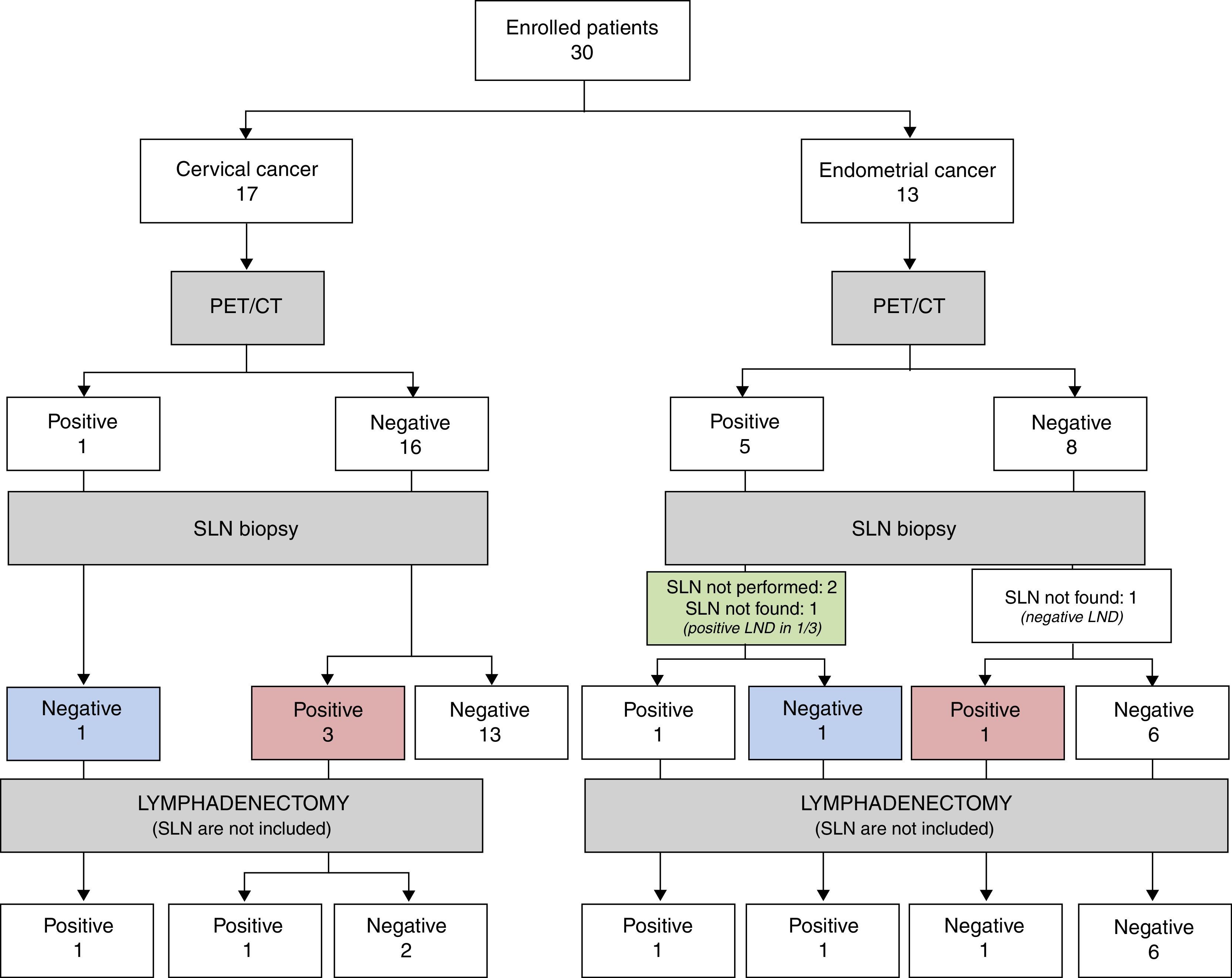
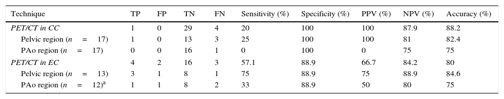
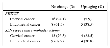
Results

In the pathology study, 4 patients with CC and 4 patients with EC had lymphatic metastasis. PET/CT showed hypermetabolic nodes in 1 patient with CC, and 5 with EC. Four of these had metastasis, one detected in the SLN biopsy. Four patients who had negative PET/CT had micrometastasis in the SLN biopsy, 1 patient with additional lymph nodes involvement. The overall patient-based sensitivity, specificity, positive and negative predictive values, and accuracy of PET/CT to detect lymphatic metastasis was 20.0%, 100.0%, 100.0%, 87.9%, and 88.2%, respectively, in CC, and 57.1%, 88.9%, 66.7%, 84.2% and 80.0%, respectively, in EC. DPI showed higher retention index in malignant than in inflammatory nodes, although no statistically significant differences were found.

Conclusions

PET/CT has low sensitivity in lymph node staging of CC and EC, owing to the lack of detection of micrometastasis. Thus, PET/CT cannot replace SLN biopsy. Although no statistically significant differences were found, DPI may help to differentiate between inflammatory and malignant nodes.

Keywords:
Cervical cancer
Endometrial cancer
PET/CT
Sentinel lymph node
Dual-time-point imaging
Resumen
Objetivo

La estadificación definitiva del cáncer de cérvix (CC) y de endometrio (CE) tiene lugar tras la cirugía. Nuestro objetivo fue evaluar la utilidad de la PET/TC para la detección de metástasis ganglionares en el CC y en el CE con imagen dual-time-point (DPI), considerando como gold standard la histopatología del ganglio centinela (GC) y la linfadenectomía.

Material y métodos

Diecisiete pacientes con CC inicial y 13 con CE de alto riesgo fueron incluidas prospectivamente. Preoperatoriamente se realizó una PET/TC, RM, detección del GC y linfadenectomía en los casos indicados. Se comparó la PET/TC con la histopatología.

Resultados

En el estudio anatomopatológico, 4 pacientes con CC y 4 con CE tuvieron metástasis ganglionares. La PET/TC mostró ganglios hipermetabólicos en una paciente con CC y en 5 con CE. Cuatro de ellas tenían metástasis, una detectada en el GC. Cuatro pacientes con PET/TC negativa presentaron micrometástasis en el GC, una paciente con ganglios adicionales infiltrados. La sensibilidad, especificidad, valor predictivo positivo y negativo y la exactitud diagnóstica de la PET/TC para detectar metástasis ganglionares fueron 20,0; 100,0; 100,0; 87,9 y 88,2% para el CC, y 57,1; 88,9; 66,7; 84,2 y 80,0% para el CE. La DPI mostró un índice de retención superior en ganglios infiltrados respecto a los inflamatorios, sin hallar diferencias estadísticamente significativas.

Conclusiones

La PET/TC tiene baja sensibilidad para estadificar el CC y CE por la incapacidad de detectar micrometástasis y, por tanto, no sustituye la detección del GC. Aunque no hubo diferencias estadísticamente significativas, la DPI podría ayudar a diferenciar ganglios inflamatorios de tumorales.

Palabras clave:
Cáncer de cérvix
Cáncer de endometrio
PET/TC
Ganglio centinela
Imagen dual-time-point

Artículo

Opciones para acceder a los textos completos de la publicación Revista Española de Medicina Nuclear e Imagen Molecular
Socio
Si es usted socio de la Sociedad Española de Medicina Nuclear e Imagen Molecular (SEMNIM) puede acceder al texto completo de los contenidos de la Revista Española de Medicina Nuclear e Imagen Molecular desde los enlaces a la revista publicados en la web de la SEMNIN (enlace a https://semnim.es/iniciar-sesion/), previo inicio de sesión como socio. Si tiene problemas de acceso puede contactar con la Secretaría Técnica de la SEMNIM en el correo electrónico secretaria.tecnica@semnim.es o en el teléfono: + 34 619 594 780
Suscriptor
Suscriptor de la revista

Si ya tiene sus datos de acceso, clique aquí.

Si olvidó su clave de acceso puede recuperarla clicando aquí y seleccionando la opción "He olvidado mi contraseña".
Suscribirse
Suscribirse a:

Revista Española de Medicina Nuclear e Imagen Molecular

Comprar
Comprar acceso al artículo

Comprando el artículo el PDF del mismo podrá ser descargado

Precio 19,34 €

Comprar ahora
Contactar
Teléfono para suscripciones e incidencias
De lunes a viernes de 9h a 18h (GMT+1) excepto los meses de julio y agosto que será de 9 a 15h
Llamadas desde España
932 415 960
Llamadas desde fuera de España
+34 932 415 960
E-mail
Opciones de artículo
es en pt

¿Es usted profesional sanitario apto para prescribir o dispensar medicamentos?

Are you a health professional able to prescribe or dispense drugs?

Você é um profissional de saúde habilitado a prescrever ou dispensar medicamentos