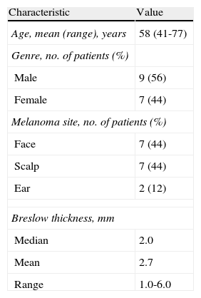
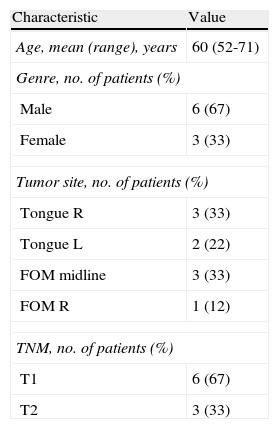
Recent innovations such as preoperative SPECT/CT, intraoperative imaging using portable devices and a hybrid tracer were evaluated in a multimodality approach for sentinel node (SN) mapping and biopsy in head and neck malignancies.
Material and methodsThe evaluation included 25 consecutive patients with head and neck malignancies (16 melanomas and 9 oral cavity squamous cell carcinomas). Patients were peritumorally injected with the hybrid tracer ICG-99mTc-nanocolloid. SNs were initially identified with lymphoscintigraphy followed by single photon emission computed tomography (SPECT/CT) 2hours after tracer administration. During surgery a portable gamma camera in combination with a near-infrared fluorescence camera was used in addition to a handheld gamma ray detection probe to locate the SNs.
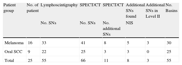
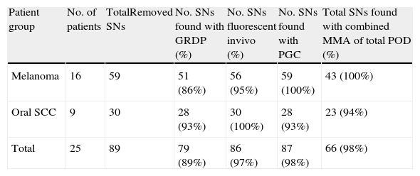
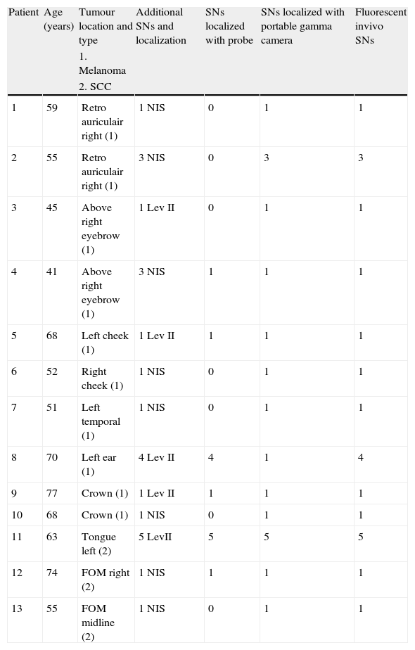
ResultsIn all patients the use of conventional lymphoscintigraphy, SPECT/CT and the additional help of the portable gamma camera in one case were able to depict a total of 67 SNs (55 of them visualized on planar images, 11 additional on SPECT/CT and 1 additional with the portable gamma camera). A total of 67 of the preoperatively defined SNs together with 22 additional SNs were removed intraoperatively; 12 out of the 22 additional SNs found during operation were located in the vicinity of the injection site in anatomical areas such as the periauricular or submental regions. The other 10 additional SNs were found by radioguided post-resection control of the excision SN site.
ConclusionIn the present series 26% additional SNs were found using the multimodal approach, that incorporates SPECT/CT and intraoperative imaging to the conventional procedure. This approach appears to be useful in malignancies located close to the area of lymphatic drainage such as the periauricular area and the oral cavity.
Se ha evaluado innovaciones recientes como la SPECT/TAC, dispositivos portátiles de imagen intraoperatorios y un trazador híbrido en el contexto de un abordaje multimodalidad para el mapeo y biopsia del ganglio centinela (GC) en tumores de cabeza y cuello.
Material y métodosSe evaluaron 25 pacientes consecutivos con tumores de cabeza y cuello (16 melanomas y 9 carcinomas de células escamosas). Se inyectaron peritumoralmente con el trazador híbrido ICG-99mTc-nanocoloide. Los GC se identificaron inicialmente mediante imágenes planares y a las 2 horas postinyección del trazador se realizó una SPECT/TAC. Intraoperatoriamente se utilizó una gammacámara portátil en combinación con una cámara infrarroja de fluorescencia y una sonda detectora de rayos gamma para localizar los GC.
ResultadosEn todos los casos las imágenes planares, la SPECT/TAC, y en un caso con la gammacámara portátil, se lograron identificar un total de 67 GC (55 con las imágenes planares, 11 adicionales con la SPECT/TAC y uno con la gammacámara portátil). Además de los 67 previamente definidos, se identificaron y extrajeron 22 GC adicionales intraoperatoriamente de los cuales 12 se encontraban cerca del punto de inyección en zonas como las regiones periauricular y submandibular. Los 10 restantes se encontraron mediante control radioguiado del lecho ganglionar postextracción.
ConclusiónEn nuestra serie se encontraron un 26% de GC adicionales. Este abordaje multimodalidad parece ser útil en tumores en los que el drenaje linfático es muy próximo al sitio de inyección como en la región periauricular y en cavidad oral.
Artículo
Comprando el artículo el PDF del mismo podrá ser descargado
Precio 19,34 €
Comprar ahora


