Clinical data are presented on patients with tumor thrombosis (TT) incidentally detected on FDG PET/CT imaging, as well as determining its prevalence and metabolic characteristics.
Materials and methodsOut of 12,500 consecutive PET/CT examinations of patients with malignancy, the PET/CT images of 15 patients with TT as an incidental finding were retrospectively investigated. A visual and semiquantitative analyses was performed on the PET/CT scans. An evaluation was made of the pattern of FDG uptake in the involved vessel as linear or focal via visual analyses. For the semiquantitative analyses, the metabolic activity was measured using SUVmax by drawing the region of interest at the site of the thrombosis and tumor (if any).
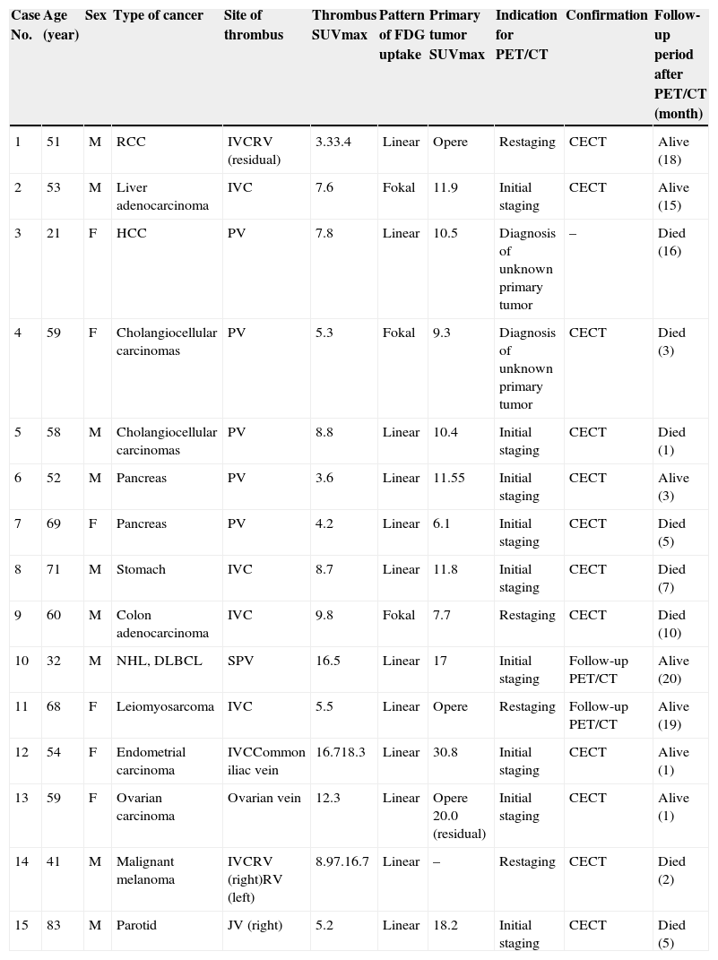
ResultsThe prevalence of occult TT was 0.12%. A total of 15 patients had various malignancies including renal (1 patient), liver (4), pancreas (2), stomach (1), colon (1), non-Hodgkin lymphoma (1), leiomyosarcoma (1), endometrial (1), ovarian (1), malign melanoma (1) and parotid (1). Nineteen vessels with TT were identified in 15 patients; three patients had more than one vessel. Various vessels were affected; the most common was the inferior vena cava (n=7) followed by the portal (n=5), renal (n=3), splenic (n=1), jugular (n=1), common iliac (n=1) and ovarian vein (n=1). The FDG uptake pattern was linear in 12 and focal in 3 patients. The mean SUVmax values in the TT and primary tumors were 8.40±4.56 and 13.77±6.80, respectively.
ConclusionOccult TT from various malignancies and locations was found incidentally in 0.12% of patients. Interesting cases with malign melanoma and parotid carcinoma and with TT in ovarian vein were first described by FDG PET/CT. Based on the linear FDG uptake pattern and high SUVmax value, PET/CT may accurately detect occult TT, help with the assessment of treatment response, contribute to correct tumor staging, and provide additional information on the survival rates of oncology patients.
Se presentan los datos clínicos de pacientes con trombosis tumoral (TT) detectada incidentalmente en estudios FDG PET/TC, y se determinan su prevalencia y sus características metabólicas.
Material y MétodosDe 12,500 exploraciones consecutivas PET/TC realizadas en pacientes con tumores malignos, se analizaron de forma retrospectiva las imágenes PET/TC de 15 pacientes con TT como un hallazgo incidental. Se realizaron un análisis visual y un análisis semicuantitativo de las exploraciones PET/TC. El patrón de captación de FDG en el vaso afecto, evaluado por análisis visual, fue lineal o focal. En el análisis semicuantitativo se midió la actividad metabólica usando SUVmax, dibujando regiones de interés en el sitio de la trombosis y en el tumor (si existía).
ResultadosLa prevalencia de TT fue 0.12%. Quince pacientes tenían diversos tumores malignos incluyendo riñón (1), hígado (4), páncreas (2), estómago (1), colon (1), linfoma no Hodgkin (1), leiomiosarcoma (1), endometrio (1), ovario (1), melanoma maligno (1) y parótida (1). Se identificaron 19 vasos con TT en 15 pacientes. Tres pacientes tenían más de un vaso afecto. El vaso más frecuentemente afectado fue la vena cava inferior (n=7), seguido de porta (n=5), renal (n=3), esplénica (n=1), yugular (n=1), ilíaca común (n=1) y venas ovárivas (n=1). El patrón de captación de FDG fue lineal en 12 y focal en 3 pacientes. El SUVmax medio en el TT y en los tumores primarios fue 8,40±4,56 y 13,77±6,80, respectivamente.
ConclusiónTrombosis tumoral oculta en diversos tumores malignos y en diferentes localizaciones se encontró incidentalmente en un 0,12%. Casos interesantes fueron el melanoma maligno y el carcinoma de parótida. La TT en la vena ovárica se describe por primera vez mediante FDG PET/TC. Basado en el patrón lineal captación de FDG y el elevado valor SUVmax, la PET/TC puede detectar con exactitud la TT oculta, ayudar en la evaluación de la respuesta al tratamiento, contribuir en la correcta estadificación del tumor y también puede proporcionar información adicional sobre la supervivencia en pacientes oncológicos.
Artículo
Comprando el artículo el PDF del mismo podrá ser descargado
Precio 19,34 €
Comprar ahora


