La combinación de la tomografía por emisión de positrones (PET) y la tomografía computarizada (TAC) en un único equipo (PET/TAC) supone una potente herramienta diagnóstica que abre nuevos horizontes en el diagnóstico por imagen.
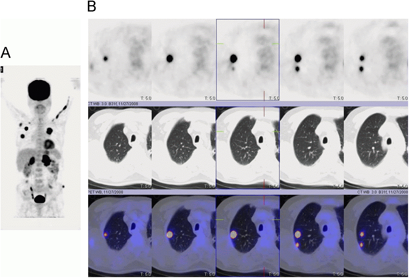
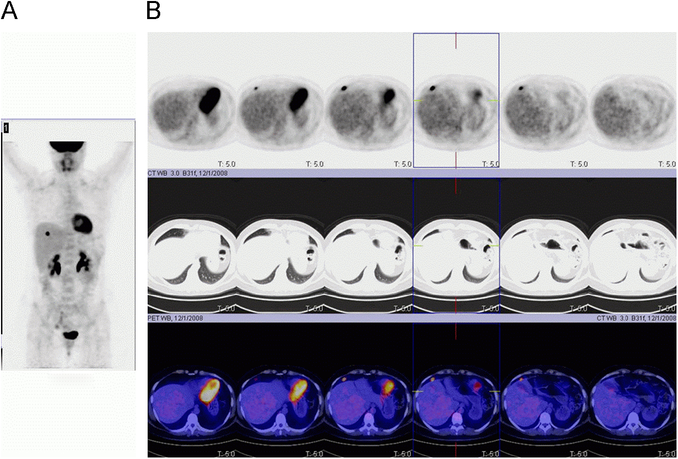
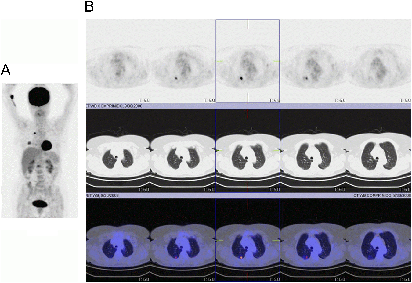
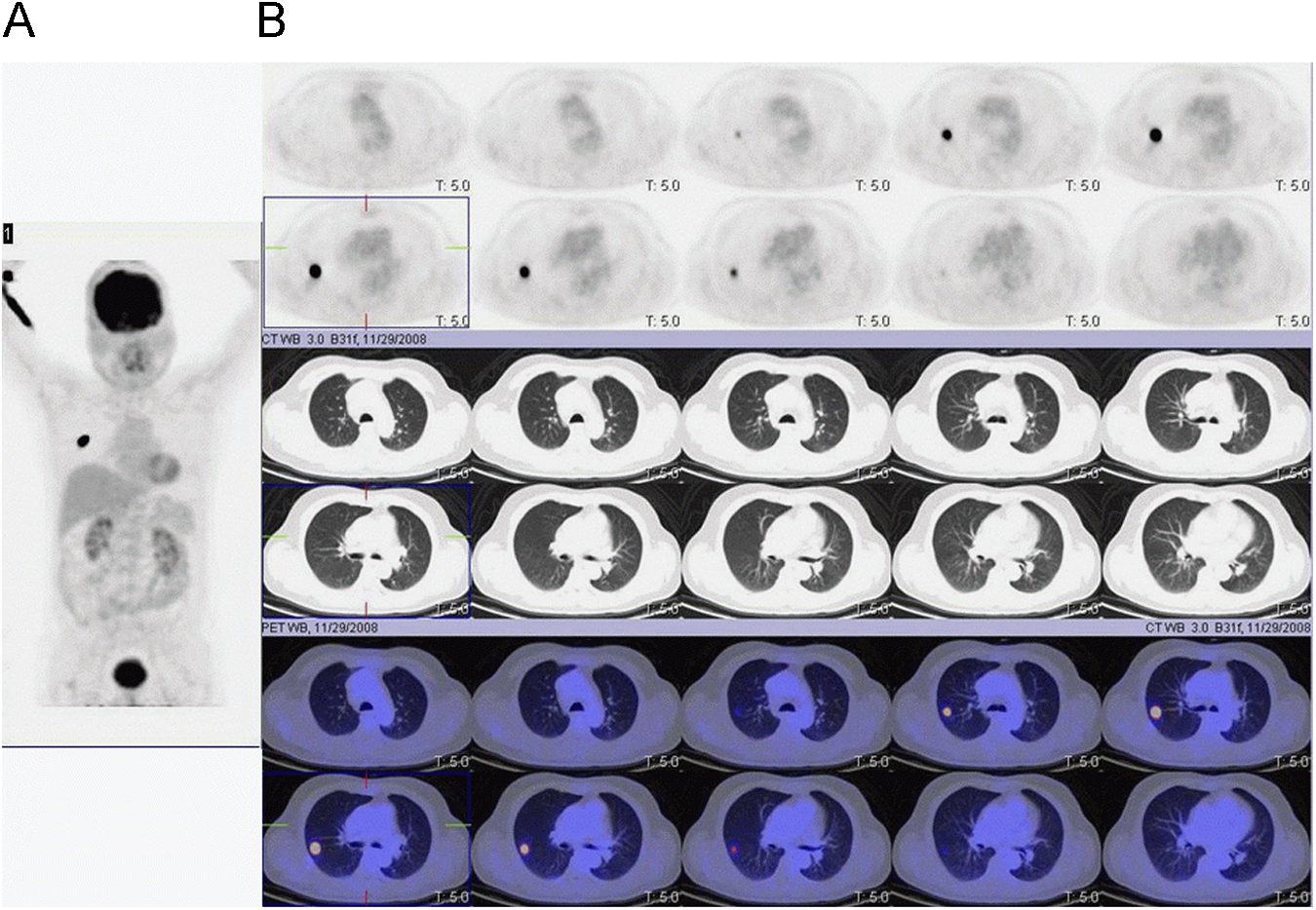
Para la correcta interpretación de los estudios PET/TAC, es necesario conocer la biodistribución de la 18F-fluorodeoxiglucosa, las variables fisiológicas así como los posibles artefactos ante los que nos podemos encontrar. Presentamos cuatro casos en los cuales en el contexto de seguimiento/diagnóstico de un proceso oncológico se realiza un estudio 18F-fluorodeoxiglucosa-PET/TAC. Dicho estudio mostró en todos los casos captación focal en el parénquima pulmonar en el estudio de la PET en ausencia de alteraciones estructurales en las imágenes de la TAC. La extravasación de radiotrazador en tres de ellos así como una modificación reciente de la técnica de inyección utilizada hicieron pensar en la existencia de un artefacto responsable de dichas discordancias.
The combination of positron emission tomography (PET) and computed tomography (CT) in a single device (PET/CT) offers a powerful diagnostic tool that opens up new horizons for imaging diagnosis.
In order to correctly interpret PET/CT studies, knowledge of the biodistribution of 18F-fluorodeoxyglucose (FDG), the physiological variants as well as the pitfalls, including artefacts, which may be found, is necessary. We report four cases performed during the follow-up diagnostic context of an oncology study performed with 18F-FDG-PET/CT. In every case, this study showed focal uptake in the lung parenchyma in the PET study with no structural lesions being found on the CT scan. Radiotracer extravasation in three of these patients and a recent change in the injection protocol used suggest that an artefact was responsible for these discrepancies.
Artículo
Comprando el artículo el PDF del mismo podrá ser descargado
Precio 19,34 €
Comprar ahora


