Evaluar el 18F-FDG-PET/TC en la sospecha de recidiva del cáncer de ovario con pruebas de imagen negativas/no concluyentes y en la reestadificación de recurrencia potencialmente resecable.
Material y métodosEstudiamos 36 casos y 140 localizaciones. Se realizaron PET/TC, TC con contraste i.v. y CA-125 en todos los casos: reestadificación (19), sospecha de recidiva (17).
Comparamos TC y PET/TC, valorados con histopatología y seguimiento radiológico, calculando sensibilidad y valor predictivo positivo (VPP) por casos y lesiones. Evaluamos la correlación entre tamaño, número y captación de las lesiones y el CA-125. Realizamos un análisis de supervivencia, utilizando curvas ROC para calcular el cut off óptimo de SUVmáx para predicción de supervivencia.
Comprobamos si la PET/TC modifica la actitud terapéutica vs. imagen convencional.
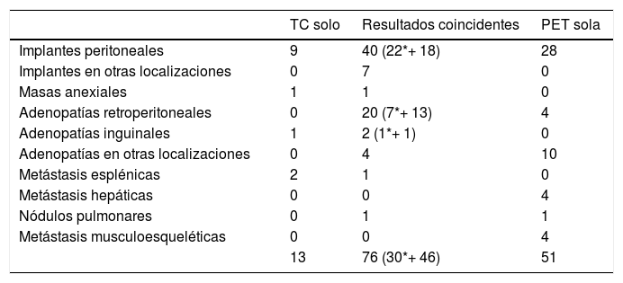
ResultadosPET/TC y TC fueron concordantes en 12 casos: 11 positivos (39 lesiones), todos confirmados. Hubo un FN. En los 24 no concordantes, PET/TC fue positivo en 19 (97 lesiones); TC en 21 (59 lesiones); 54% de las lesiones fueron concordantes.
Globalmente, PET/TC detectó 127 lesiones (sensibilidad = 97% y VPP = 100%) y TC 89 (sensibilidad = 61% y VPP = 90%).
No se encontró correlación significativa entre CA-125 y los otros parámetros. La PET/TC detectó 10 casos positivos, con CA-125 normal. Modificó el manejo terapéutico en 15 casos. Se encontraron diferencias significativas en supervivencia (SUVmáx = 11,8).
ConclusionesLa PET/TC juega un importante papel en la recurrencia del cáncer de ovario, con sensibilidad y VPP mayores que la TC, modificó el manejo terapéutico en un 42% de casos y podría ser una herramienta útil para predicción de supervivencia.
To evaluate 18F-FDG-PET/CT for suspected ovarian cancer relapse with negative/inconclusive conventional imaging, or restaging potentially resectable ovarian cancer relapse.
Material and methodsThirty-six cases and 140 locations were studied. PET/CT, ceCT and serum CA-125 was conducted in all cases. Nineteen cases were requested for restaging, 17 for suspected relapse.
We compared ceCT and PET/CT, assessed by histopathology or radiological follow-up, calculating sensitivity (S) and positive predictive value (PPV) by cases and lesions.
We evaluated the correlation between size, number, uptake of the lesions and CA-125. We conducted survival analysis, using ROC curves to calculate the optimal cut-off of SUVmax for survival prediction.
We checked whether PET/CT modify the therapeutic attitude vs. conventional imaging.
ResultsPET/CT and ceCT were concordant in 12 cases: 11 positives (30 lesions), all confirmed. There was 1 FN.
In the 24 non-concordant, PET/CT was positive in 19 (97 lesions); ceCT in 21 (59 lesions); 54% of the lesions were concordant.
Overall, PET/CT detected 127 lesions, with S = 97% and PPV = 100%. ceCT detected 89 lesions, with S = 61% and PPV = 90%.
No significant correlation was found between CA-125 and the other parameters. PET/CT detected 10 positive cases, with normal CA-125.
PET/CT modified therapeutic management in 15 cases.
Significant differences were found in survival with SUVmax = 11.8
ConclusionsPET/CT plays an important role in ovarian cancer relapse, with sensitivity and PPV higher than ceCT, modified therapeutic management in up to 42% of cases, and could be a valuable tool for predicting survival.
Artículo
Comprando el artículo el PDF del mismo podrá ser descargado
Precio 19,34 €
Comprar ahora


