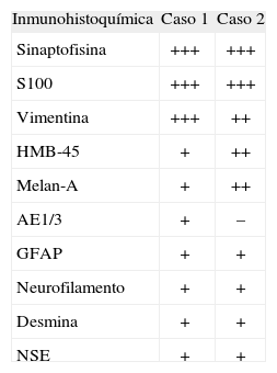
El meduloepitelioma intraocular es una neoplasia embrionaria poco frecuente desarrollada en el cuerpo ciliar y que ocasionalmente afecta el iris, la retina o el nervio óptico. Presentamos dos casos de meduloepitelioma intraocular maligno pigmentado, uno teratoide (con componente heterólogo: cartílago hialino) y otro no. Por histoquímica se reconoció la presencia de mucopolisacáridos ácidos y pigmento melánico. Los estudios de inmunohistoquímica mostraron positividad para marcadores de distintas líneas de diferenciación celular como neuroepitelial (proteína S100, sinaptofisina), glial (proteína ácida gliofibrilar), mesenquimatoso (vimentina, desmina), epitelial (citoqueratina AE1/3, EMA) y melanocítico (HMB-45, Melan-A). El meduloepitelioma intraocular está compuesto por células multipotenciales con expresión inmunofenotípica múltiple.
Intraocular medulloepithelioma is a rare embryonal tumour that occurs most often in the ciliary body, but may also arise from the iris, retina or optic nerve. We present two cases of pigmented malignant intraocular medulloepithelioma; one teratoid (with hyaline cartilage as a heterologous element), and one non-teratoid. Histochemistry showed the neoplastic cells synthesizing both an acidic substance that stained positive with alcian blue and melanin pigment positive for Fontana- Masson stain. Immunohistochemistry showed positive staining for markers of several lines of differentiation including neuroepithelial (S100 protein, synaptophysin), glial (GFAP), mesenchymal/muscle (vimentin, desmin), epithelial (cytokeratin AE1/3, EMA) and melanocytic (HMB-45, Melan-A). Intraocular medulloepithelioma is composed of multipotential cells capable of polyimmunophenotypic expression.
Artículo
Comprando el artículo el PDF del mismo podrá ser descargado
Precio 19,34 €
Comprar ahora


