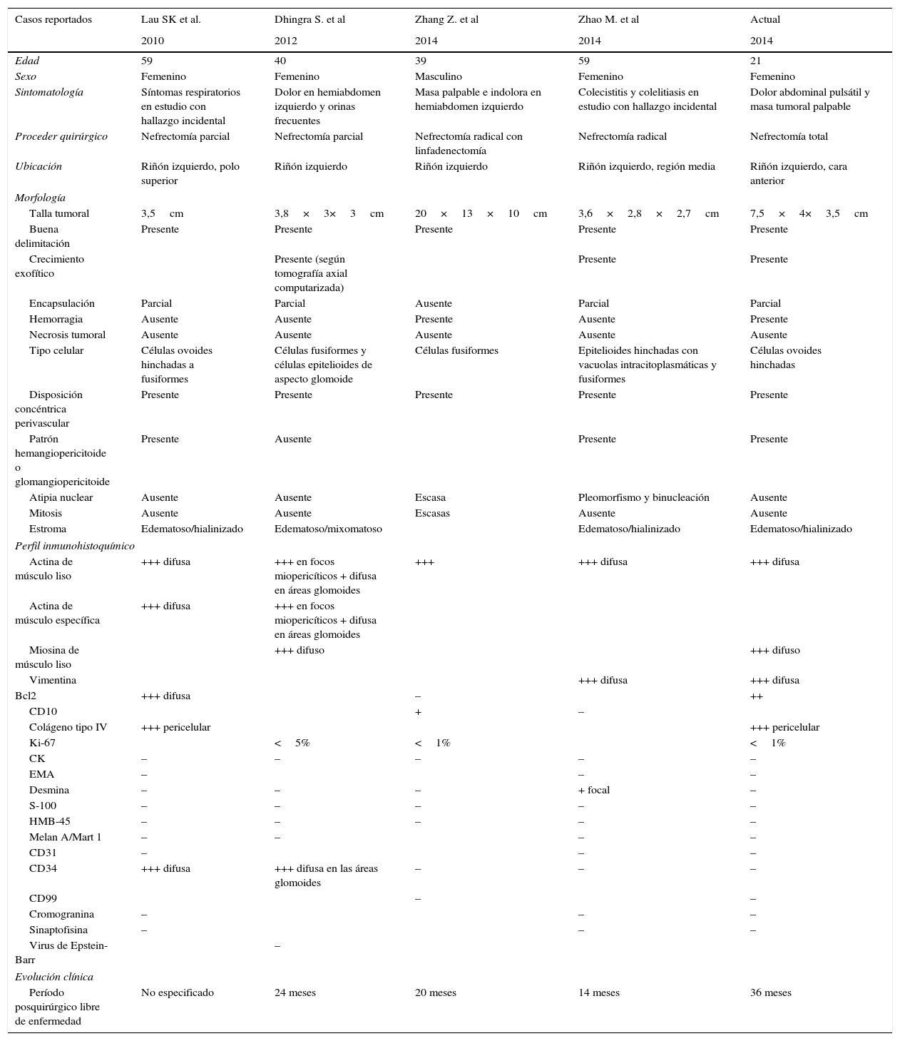
Presentamos el caso de una mujer de 21 años sin antecedentes de interés, que presentó dolor abdominal y masa perceptible en hipocondrio izquierdo. La tomografía axial computarizada informó de imagen tumoral de 7cm de diámetro, en la porción media y cara anterior del riñón izquierdo. Se le realizó nefrectomía total izquierda. El examen histológico reveló proliferación de células ovoides con distribución concéntrica perivascular e imagen glomangiopericitoide, inmunoreactividad para actina de músculo liso, y miosina de músculo liso y ausencia de reactividad para la desmina, con diagnóstico concluyente de miopericitoma renal. Este es un tumor infrecuente de células mioides perivasculares, considerado por la mayoría como una neoplasia de piel y partes blandas con escasos reportes de presentaciones en vísceras. Hasta el momento 4 casos de miopericitoma renal se han reportado en la literatura médica. Reportamos otro nuevo caso de esta inusual ubicación.
Myopericytoma is an infrequent neoplasm of perivascular myoid cells that usually originates in the skin and superficial soft tissues of distal extremities but is rarely found in the visceral organs. Only four cases of renal myopericytoma have been previously reported. We present a further case of myopericytoma of the kidney in a 21 year old woman with no clinical history of interest who presented with abdominal pain and a palpable mass in the left upper quadrant. Unenhanced computed tomography revealed a 7cm diameter mass in the middle portion and anterior aspect of the left kidney. The patient underwent left radical nephrectomy. Histological examination revealed a tumour composed of ovoid cells in a concentric arrangement and a «glomangiopericytoma» pattern. Immunohistochemistry showed positivity for smooth muscle actin, and smooth muscle myosin stains and negativity for desmin, confirming the diagnosis of renal myopericytoma.
Artículo
Comprando el artículo el PDF del mismo podrá ser descargado
Precio 19,34 €
Comprar ahora


