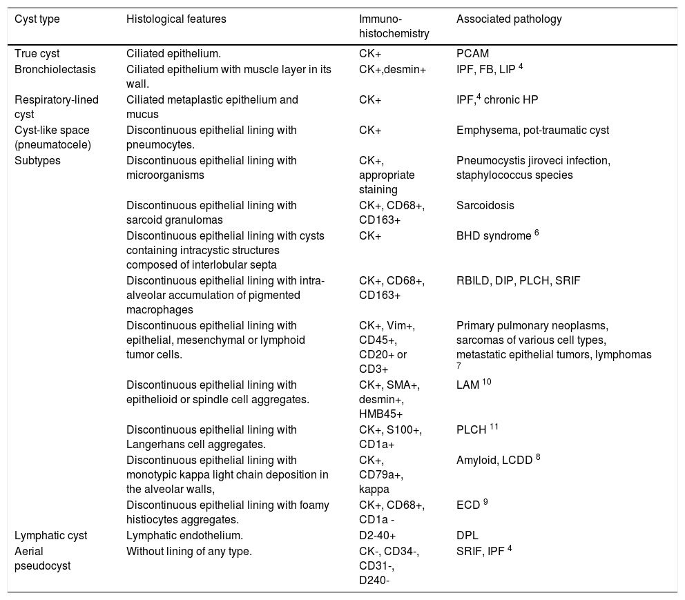
Smoking-related interstitial fibrosis is a distinct form of fibrosis, found in smokers, which has striking histopathological features. We present a case of pulmonary interstitial fibrosis with cysts in a 58-year-old woman who was a significant active smoker, presenting with a 7 month history of progressive dyspnea. TAC revealed thin-walled pulmonary cysts. An open lung biopsy was performed and the histopathological study showed hyaline fibrous thickening of the alveolar septa, respiratory bronchiolitis and cysts in the thickness of the interlobar septa. Immunohistochemically, the absence of an epithelial, vascular or lymphatic endothelial lining of the cysts would suggest that the cysts had been caused by pulmonary interstitial emphysema. Immunohistochemistry is essential in the differential diagnosis that includes, in this case, true cysts, pseudocysts and pulmonary lymphangiectasia.
La fibrosis intersticial relacionada con el tabaco es una forma especial de fibrosis con histología característica que ocurre en fumadores. Presentamos un caso de fibrosis intersticial pulmonar con quistes en una mujer de 58 años con historia de tabaquismo importante, que refería disnea progresiva en los últimos 7 meses. La TAC reveló quistes pulmonares de paredes delgadas. Se realizó una biopsia pulmonar abierta y el estudio histopatológico mostró engrosamiento fibroso hialino de los septos alveolares, bronquiolitis respiratoria y quistes en el espesor de los septos interlobares. Inmunohistoquímicamente, la ausencia de revestimiento epitelial, endotelial vascular y linfático de los quistes, apoya que estos son causados por enfisema intersticial pulmonar. La inmunohistoquímica es esencial en el diagnóstico diferencial que incluye en este caso, quistes verdaderos, seudoquistes y linfangiectasia pulmonar.
Artículo
Comprando el artículo el PDF del mismo podrá ser descargado
Precio 19,34 €
Comprar ahora


