


This case was designed to compensate for the absence of the lower first molar by means of traditional mesialization of the posterior segment thus restoring the lost function and aesthetics. Mesial movement of the lower second and third molars was performed, while controlling the incisor position, when losing anchorage is required. Mesialization of the posterior segment is important to solve problems involving the absence of the first molar since this procedure prevents tilting of adjacent teeth towards the edentulous space, extrusion of the antagonist, or periodontal problems, all of which induce a lack of support, stimulation and hygiene. The case reports a male patient of 31 years of age with a non-contributory medical history, who wished to «improve my crooked teeth». Clinical and radiographic analysis revealed an apparently symmetrical, dolichocephalic, hyperdivergent, skeletal class II due to maxillary protrusion. Intraorally, he presented a non-assessable molar class on the right side and a class I on the left; a non-assessable right and left canine class due to disocclusion and moderate crowding in upper and lower arch. Orthodontic treatment consisted in upper and lower first premolar extractions; anchorage and 0.022” Roth fixed appliances alignment and leveling was performed with the following archwire sequence: 0.014”-0.018” NiTi. For space closure, a 0.017” x 0.025” SS double keyhole loop archwire with Suzuki ligature was used. The segments were anchored for mesialization of teeth #47- 48, using bilateral class II elastics; second order bends were done for tooth #47 to parallelize the roots and short up and down elastics were placed for better intercuspation. Satisfactory facial, dental, aesthetic and functional results were obtained. In the retention phase, an upper and lower circumferential retainer were used.
Este caso fue diseñado para compensar la ausencia del primer molar inferior unilateral mediante mesialización tradicional del segmento posterior, devolviendo la función y estética perdida. Se realiza el movimiento mesial del segundo y tercer molar inferior, controlando la posición de los incisivos, cuando se requiere perder anclaje para cerrar el espacio del primer molar inferior. De esta manera la mesialización tradicional es importante en resolver los problemas que involucran la ausencia del primer molar, evitando la inclinación de las piezas adyacentes hacia el espacio, extrusión del molar antagonista o problemas periodontales que inducen una falta de apoyo, estimulación e higiene. Se presenta paciente masculino de 31 años de edad, quien a la consulta reporta «mejorar mis dientes chuecos». Al análisis clínico y radiográfico aparentemente simétrico, dolicocefálico, hiperdivergente, clase II esquelética por protrusión maxilar, intraoralmente presenta clase I molar izquierda y derecha no valorable; clase canina no valorable ambos lados por desoclusión; apiñamiento moderado en arcada superior e inferior. Se utilizó la técnica de slot 0.022”, el tratamiento ortodóntico consistió en extracciones de primeros premolares superiores e inferiores, se colocó anclaje, alineación-nivelación .014” a .018” NiTi, la mesialización tradicional se realizó con el arco 0.019” × 0.025” SS, a cada lado a la altura de los caninos, lleva dos ansas en forma de ojo de cerradura, su activación fue a través de una ligadura metálica que va desde el hook del molar hasta el ansa distal provocando su apertura 1 milímetro por mes. Una vez que se obtuvo la clase I canina, se colocaron elásticos cortos de arriba abajo para una mejor intercuspidación. Se lograron resultados faciales, dentales, estéticos y funcionales satisfactorios que se mantienen en la fase de retención con retenedor circunferencial superior e inferior.
Currently the specialist plays an important role when it comes to overcoming the loss of a first permanent molar by providing the patient solutions through orthodontic treatment. This approach prevents invasive therapies such as dental implants by means of a traditional mesialization of the posterior segment which is less aggressive and more durable for the patient, when compared to the prosthetic approach. Traditional mesialization of the posterior segment has the following benefits for the patient: a) it eliminates crowding in order to achieve a stable teeth alignment b) it retracts upper and lower incisors for improvement of the facial profile or occlusion in cases of bimaxillary protrusion. d) By mesializing the molars, the space for the eruption of third molars is increased.1
When performing traditional mesializacion, it must be carefully considered the quality of the remaining alveolar bone in the edentulous space. The space should be adequate in a bucco-lingual dimension of the alveolar ridge for placing the new molar and there should be no periodontal defects in the teeth adjacent to the gap.2
The diagnosis and treatment plan, determine if the molars are maintained in their position or moved mesially. Alternatives that should be considered to perform traditional mesialization are: age of the patient, position occupied by the tooth, bone quality and quantity, status of the posterior segment, stability, inclination, torsion and rotation.3
Little and Reidel (1990) stated that traditional mesialization as an alternative in orthodontic treatment will depend on an adequate biomechanical system to achieve correct posterior intercuspation and few facial changes.4
According to the authors the magnitude of the applied force in traditional mesialization of the posterior segment should be continuous and controlled. A closing loop supplies acontinuous and controlled force that induces dental movement of approximately 1 millimeter per month without allowing it to exceed 2 millimeters, so that the movement ceases if the patient does not attend his or her monthly appointment. This closing loop must provide an ideal force with a 1.5mm activation and must maintain a significant portion of it at least until 0.5mm are achieved.5,6
The aim of this case was to compensate for the absence of a first lower molar using traditional mesialization of the posterior segment thus restoring function and aesthetics.
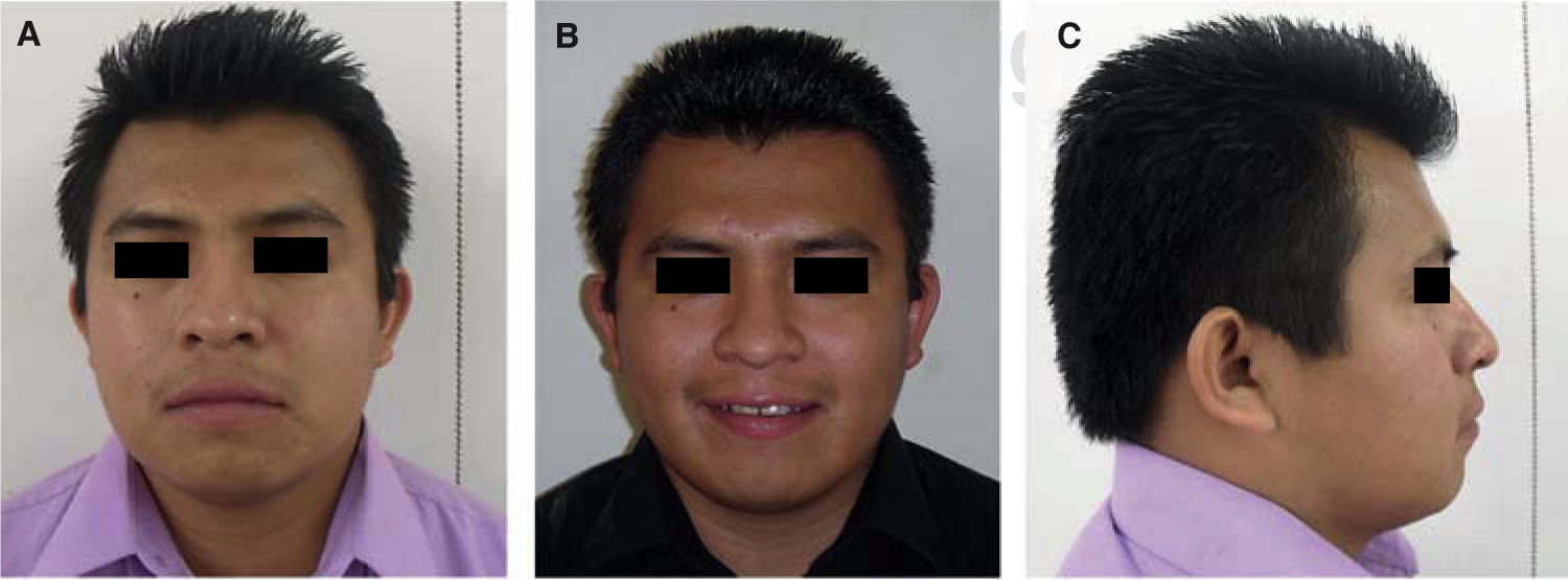
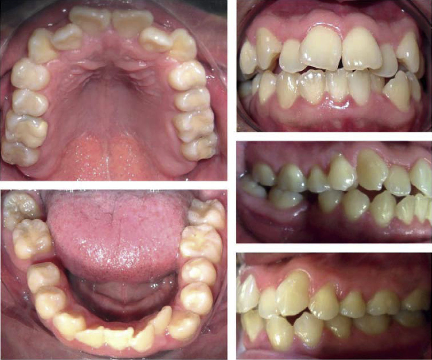
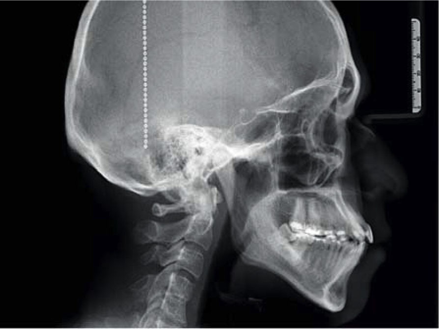
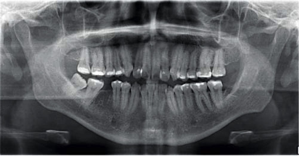
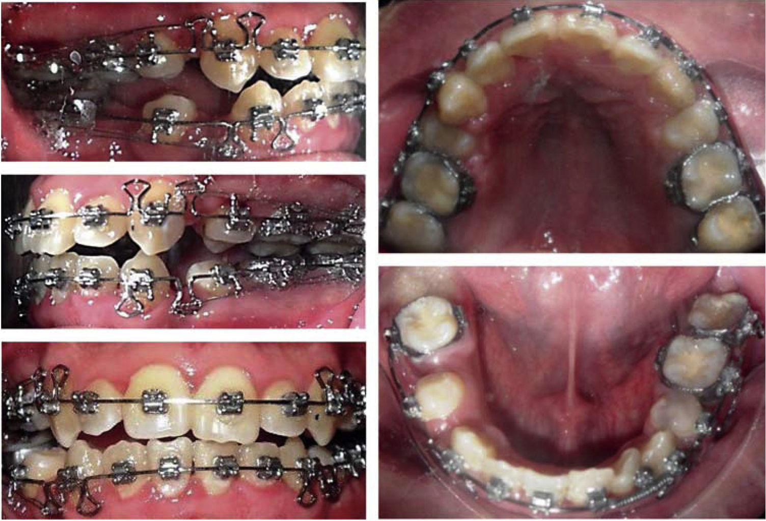
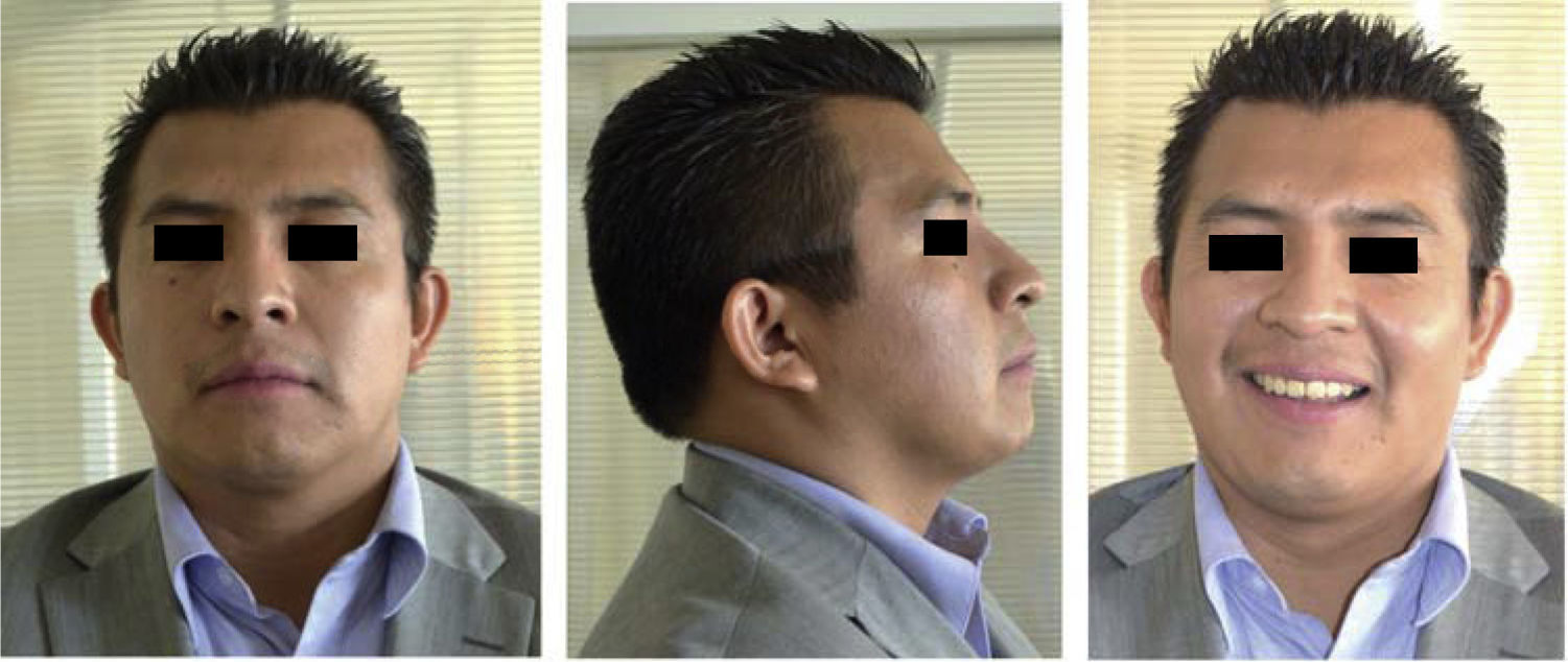
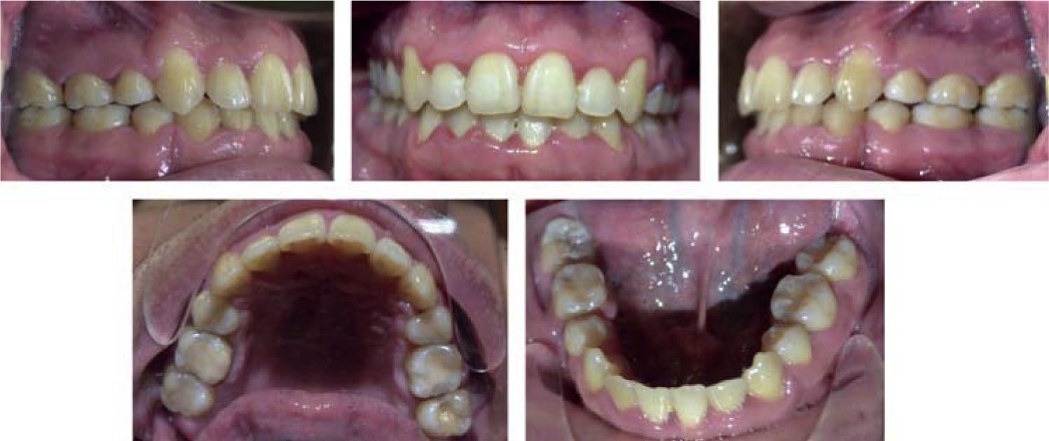
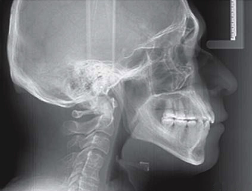
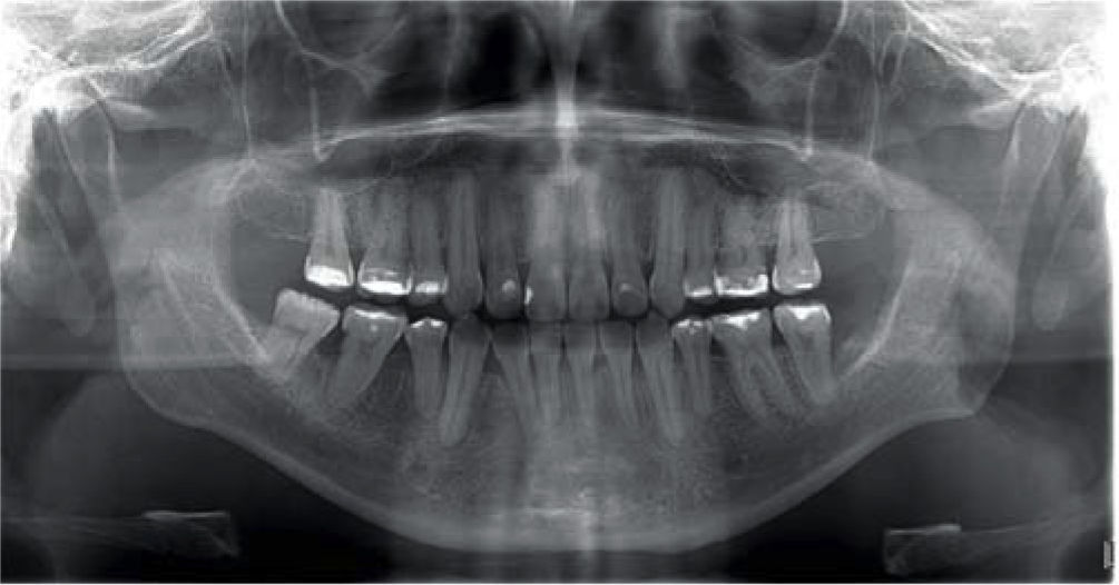
CASE PRESENTATIONMale patient, 31 years of age, refered as his chief complaint: «I want to improve my crooked teeth». In the extraoral assessment a dolichofacial biotype was observed as well as a lower dental midline deviated 2mm to the left in relation to the facial; vertical growth, convex profile and good lip contact. In the intraoral assessment an upper dentoalveolar discrepancy of -8mm and a lower of -7mm was noted; 1mm overbite and 3mm overjet, non-assessable left and right canine class due to disocclussion. The molar relationship was class I on the left side and nonassessable on the right due to first molar absence (Figures 1–4Figures 1 to 4).


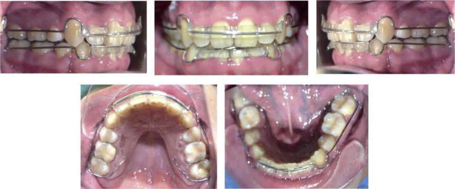
In this case, the continuous archwire with closing loops or double keyhole loops method was used. It consists of a stainless steel archwire that has two closing loops on each side for performing sagittal movements in the posterior segment with the purpose of closing spaces created by extractions. For this patient, a 0.022” slot technique was used/ with a 0.019” x 0.025” SS archwire. On each side of the canines two keyhole-shaped loops were made on the archwire. When this archwire is in place, the loops must be mesially and distally equidistant from the bracket of each canine. The archwire was placed once the upper and lower archeswere aligned, leveled and with good torque expression. Activation was through a wire ligature tied from the molar hook to the distal loop, causing it to open 1 millimeter. Once canine class I was obtained short up and down elastics were placed for better intercuspation (Figure 5).
RESULTS

In this work traditional mesialization proved to be beneficial. By providing a 15o positive torque in the posterior segment of the double keyhole loop archwire, the roots were moved away from the buccal cortical towards cancellous bone thus facilitating mesial movement of the right second and third molar. This provided the following benefits: it improved the resilience of the incisors torque, avoided extrusion of the anterior segment, moved the lower second molar mesially while minimizing the effect of crown tipping and reduced the intrusion effect on the lateral sector.
Traditional mesialization of the second and third molars was further favored by decreasing the archwire caliber in the posterior teeth where friction increased. Mesialization took 8 months of the whole orthodontic treatment time and the applied force was 150 grams.
Second order bends were made for root parallelization at the level of the lower right second molar, in which a slight root resorption occurred with no gingival recession.
At the end of orthodontic treatment good occlusal stability, bone status and molar inclination, torsion and rotation were obtained.
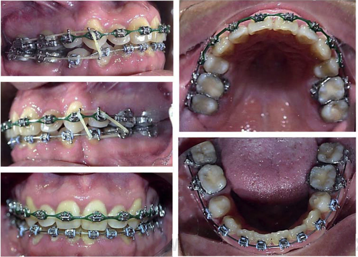
Extraoral examination revealed a proper facial balance and profile, a wide smile and the final orthopantomography, an acceptable root parallelism (Figures 6 to 10).
DISCUSSION


Lower molars move mesially less easily than upper molars. This phenomenon may be attributed to the narrowness of the alveolar process, however when analyzing root formation of the second and third molar, the possibility of an adequate occlusal stability increases. This case report illustrates a compensation for the absence of a lower first molar using traditional mesialization of the posterior segment thus restoring the lost function and aesthetics.
Brandt6 and Seddon7 recommend performing traditional mesialization of the posterior segment, in the three months following the loss of the first permanent molar; otherwise there is an increased risk of creating an undesirable inclination of the second molars and bone loss.
McLaughlin8 Carano9 and Williams10 studied traditional mesailization associating bone, inclination, torsion and rotation.
The results of this study agree with those of Luecke 11 who reported that biomechanics for traditional mesialization of the posterior segment are very complex and that treatment time was longer than when other kind of extractions were made, but in different case report articles and also in the current case report it was found to be a successful treatment whenever there is an adequate diagnosis and an effective biomechanical design.
Likewise we agree with Sandler12 and Moldez13 who concluded that traditional mesialization of the posterior segment should be performed with rectangular archwires that fill the slot for torque expression, rotation, anchorage and that compensation tipping or differential torque bends in the second and third molars are required to prevent undesirable movements.
CONCLUSIONSDeveloping a good diagnosis will result in an excellent treatment plan that guides the specialist into making the right decision for the patient. In this case it was determined to perform first upper and lower premolar extractions in order to obtain dental class I, achieving good results in the establishment of the desired molar relationship and null undesirable effects.
Compensation of the absence of the first lower molar using traditional mesailization of the posterior segment is an alternative method with an acceptable treatment time.
It is important to control occlusal stability through an effective retainer design.