


The lower incisor and its position in the arch are considered of vital importance when planning orthodontic treatment due to its effects on aesthetics and treatment stability. Facial biotype plays an important role in diagnosis and orthodontic treatment planning.
Objectiveof this study was to compare the lower incisor inclination of each facial biotype in patients whose maxillomandibular sagital relationship was class I as assessed by Ricketts lateral cephalometry.
Material and methods100 lateral headfilms of class I skeletal patients were selected and classified according to VERT's facial biotype and the lower incisor inclination to the A-Pog plane was measured.
Resultsshowed that incisor inclination between dolichofacial and mesofacial patients is not different, but among brachyfacial and dolichofacial there were statistically significant differences, as it happens between mesofacial and brachyfacial. In dolichofacial patients, there is more incisor inclination than in brachyfacial patients.
Conclusionthat dental inclinations vary according to facial biotype, so diagnosis is essential in order to make the correct choice of appliances. When treating brachyfacial patients a reduced lower incisor inclination should be considered compared to dolichofacial patients.
El incisivo inferior y su posición en el arco inferior se considera que es de vital importancia a la hora de planificar un tratamiento de ortodoncia, por sus efectos en la estética y la estabilidad de tratamiento. El biotipo facial juega un papel importante en el diagnóstico y la planificación del tratamiento ortodóncico.
ObjetivoEvaluar la inclinación del incisivo inferior en cada uno de los biotipos faciales en pacientes cuya relación maxilomandibular sagitalmente es clase I mediante la cefalometría lateral de Ricketts y determinar si existen diferencias estadísticamente significativas.
Material y métodosSe seleccionaron 100 radiografías laterales de cráneo clase I esqueletal, se clasificaron según el biotipo facial de acuerdo con el coeficiente de variación vertical de Ricketts y se midió la inclinación del incisivo inferior mediante el eje del incisivo inferior y el plano A-Pog.
ResultadosLa inclinación del incisivo inferior entre dolicofaciales y mesofaciales no es diferente, pero entre dolicofaciales y braquifaciales si presenta diferencia estadísticamente significativa, tal como sucede también entre mesofaciales y braquifaciales. En dolicofaciales es mayor la inclinación que en braquifaciales.
ConclusiónLas inclinaciones dentales varían de acuerdo al biotipo facial, por lo que el diagnóstico es fundamental, ya que de este depende la correcta elección de la aparatología. Al atender pacientes con biotipos faciales braquifaciales se debe considerar una inclinación menor del incisivo inferior respecto a los dolicofaciales.
The relationship between function and form, as described in evolutionist principles, can be applied to orthodontic patients through skeletal compensation and more evidently, through dentoalveolar compensations where nature needs to have, in order to compensate, a genetic basis in each person.1,2
Dr Ricketts analysis was described since in 1960, classifying clinical problems by analyzing 1000 lateral head films thus providing standards of dental inclinations and leaving as a legacy for the clinician an aid for orthodontic treatments.1 Years later, in 1976, Corelius and Linder-Aronson reported that incisor inclination varies depending on the skeletal class.2 and subsequently, Hernández linked inclination to different malocclusions and different facial patterns in Europeans patients.3
When planning an orthodontic treatment several parameters are considered among which the lower incisor and its position in the lower arch are key for diagnosis. This is of crucial importance in orthodontics due to its effects on aesthetics and treatment stability.4
Another parameter that must be considered is facial biotype, which plays an important role in diagnosis and orthodontic treatment planning, since the correct choice of the appliances rely on it, even more when the patient is in a growth period and the use of orthopedic means is necessary.2
A practical method for obtaining the facial biotype is through the calculation of the VERT, which is carried out using cephalometric measurements thus obtaining an average by which the facial biotype is determined.2
The role of dentoalveolar compensation in the development of a normal occlusion has been described at length.5–8 Similarly, there is adaptation in the changes that occur in the maxillo-mandibular relationship during the growth.9–11 This is known as a dentoalveolar compensation mechanism.12,13
The aim of this study was to compare the lower incisor inclination in each one of the facial biotypes in patients whose sagittal maxillo-mandibular relationship is class I as assessed through Ricketts analysis
MATERIAL AND METHODSFor the present study, cephalograms were selected using the following criteria:
Inclusion: lateral head films of patients of 14 years of age or more for women and 16 years or more for men, who were about to begin Orthodontic treatment. The cephalograms were obtained with the Orthoceph OC200 D® apparatus in the area of Radiology of the Department of Post-Graduate Studies and Research of the Faculty of Odontology of the National Autonomous University of Mexico.
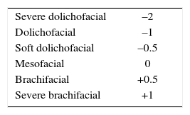
Exclusion: lateral head films which were not clearly visible to the researcher or those with poor mechanical handling; radiographs that did not have a good anatomical image quality or those that showed restorations of more than three quarters of the lower incisor. The x-rays were taken by standard methods and the linear and angular cephalometric measurements were performed by the same operator manually as described above. Afterwards, the Ricketts VERT was analyzed and the subjects were classified according to the resulting facial biotype (Table I).
The lower incisor inclination was analyzed with the method used in the Ricketts cephalometry.
FilesPrior authorization by the head of the Orthodontics Department, we proceeded to analyze 313 files of treatments initiated between August 2011 and June 2013. One hundred lateral head films were selected, which turned out to be skeletal class I according to Ricketts’ cephalometric tracing.
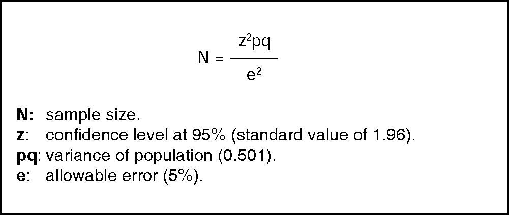
To determine sample size the following formula was used (Figure 1):3

According to this formula, with a population of 1,768 patients, it was determined that a standard error of 5 per cent requires a sample size of 96 patients.
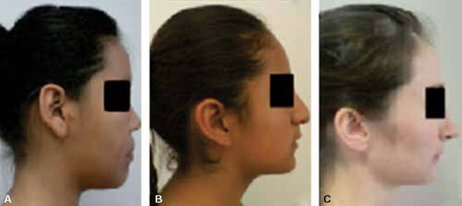
Previous studies on the topic3,13,14 used similar total samples of patients. On this basis and according to the sample size analysis, it was determined that in order to be statistically significant, 100 skeletal class I lateral head films as determined by Ricketts lateral cephalometry were used. Based on these analyzes, they were divided into groups according to facial biotype (Figure 2), and then analyze lower incisor inclination according to the plane-Pg.

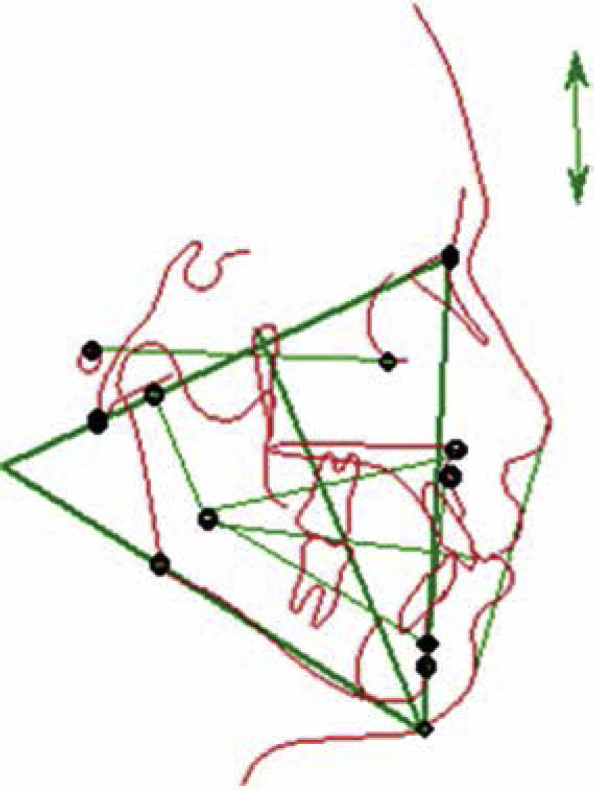
Ricketts cephalometric tracing was performed using a DENTAURUM®, 0.003mm acetate paper, mechanical pencil and mines of 0.5mm and protractor. Plotted points, reference lines and measurements of the cephalometric analysis were performed (Figure 3):

Reference points: Nasion (Na), Basion (Ba), Gnation (GN), Gonion (Go), Center of the condyle (DC), Pogonion (Pg), Point A, Orbits (OR), Porion (for), Center of the mandibular ramus (Xi) Anterior nasal spine (ENA), Menthon (PM).
Angular measurements: Facial depth, maxillary depth, lower facial height, facial axis, mandibular plane angle, mandibular arch, lower incisor inclination with the A-Po plane.
Linear measures: Facial convexity.
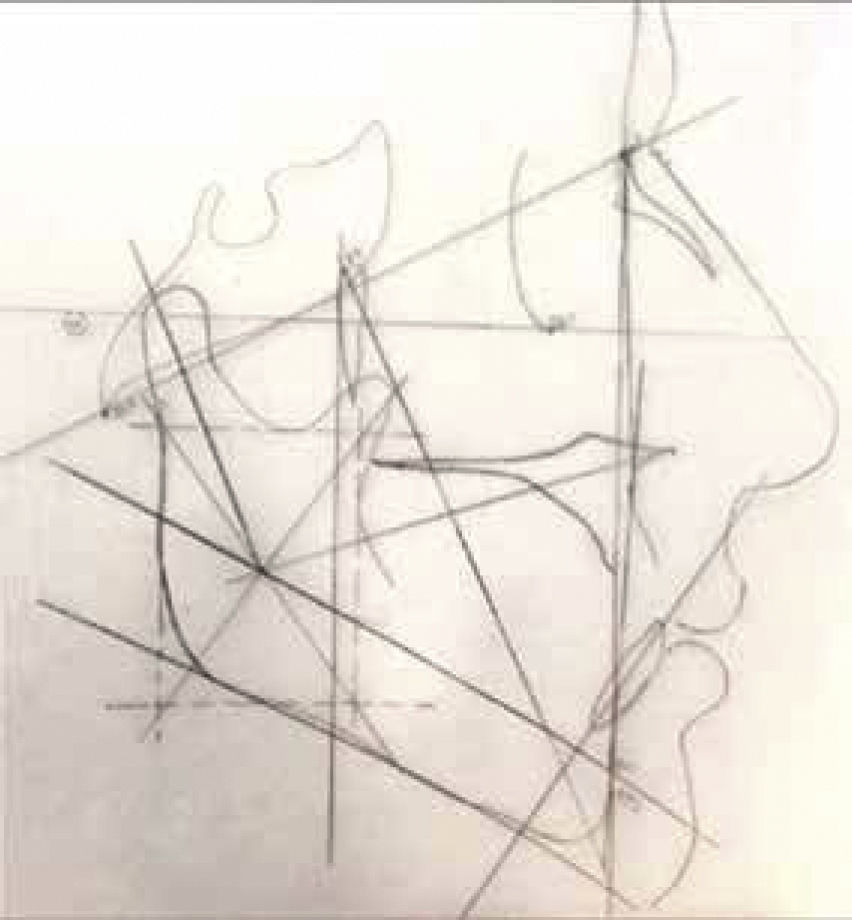
A linear measurement and three angula r measurements were used to assess the patient's skeletal class, five angular measurements to determine facial biotype through the analysis of VERT and an angular measurement to assess lower incisor inclination (Figure 4).

For calibration, a pilot test was conducted selecting 20 lateral head films in which the cephalometric tracing was performed. The same radiographs were measured once again by the operator after two weeks to verify coincidence, and that there were no errors thus achieving intra-operator reliability. Two weeks later, the same radiographs were traced by the tutor of the investigation to verify that there were no errors (inter-operator reliability).
Registration methods and procedureAll measurements were recorded on a capture sheet and later captured by a single individual in an Excel spreadsheet. Subsequently, they exported to the KaleidaGraph® version 3.6.2 for Mac program (Synergy Software; Reading, PA, USA) for statistical analysis. Descriptive and analytical statistical analysis of the different variables was conducted and data distribution verified in order to determine if there were statistically significant differences or not.
Statistical testAn analysis of variance with Bonferroni post hoc test was used. The values that were considered statistically different were the ones that presented a value of p < 0.05.
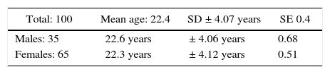
RESULTSA population of 100 analyzed radiographs was obtained. Sixty-five percent of them corresponded to female sex and 35% to males. The mean age was 22.4 years, with a. standard deviation (SD) of ± 4.07° and a standard error (SE) of 0.40. The mean age for females was 22.3 years with a.SD of ± 4.12 years and a SE of 0.51 (Table II).
The average age for the male sex was 22.6 years with a.SD of ± 4.06 years and a SE of 0.68.
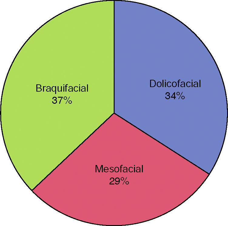
Facial biotypes were represented in the following manner: Dolichofacial 34%, Mesofacial 29% and brachifacial 37% (Figure 5).

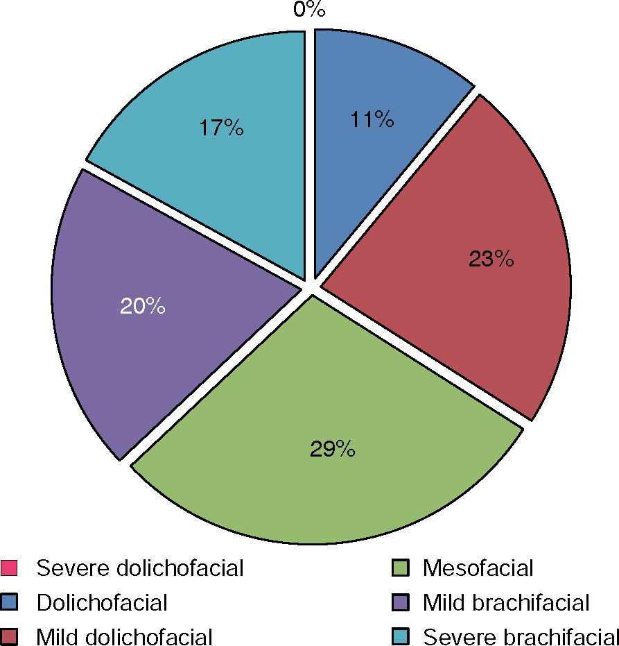
In turn, the biotypes were divided in the following way, as described in the literature, represented by the population: severe dolichofacial, 0%; dolichofacial, 11%; mild dolichofacial 23%; brachifacial, 29%; mild mesofacial, 20%; severe brachifacial, 17% (Figure 6).

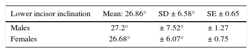
The mean inclination of the lower incisor was 26.86°, with a ± 6.58° SD and a SE of ± 0.65. The mean inclination in men was 27.2° with a SD of ± 7.52°, and a SE of 1.27. The mean inclination in women was 26.68o with a SD of ± 6.07°, and a standard error of ± 0.75 (Table III).
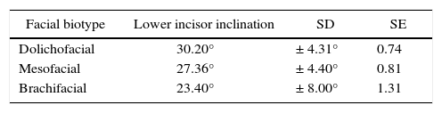
Incisor inclination for each facial biotype was as follows: dolichofacial subjects presented a mean inclination of 30.20°, with a ± 4.31° SD and a standard error of 0.74. Mesofacial patterns presented a mean inclination of 27.36°, with a SD of ± 4.40° and a SE of 0.81. Brachifacials presented a mean inclination of 23.40°, with a SD of ± 8.00° and a standard error of 1.31 (Table IV).
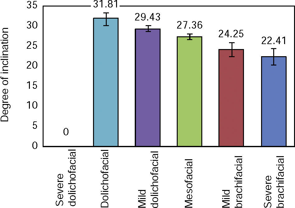
For each subgroup the lower incisor inclination was as follows: dolichofacial, 31.81° with a ± 4.99° SD and a SE of 1.50; mild Dolichofacial, 29.43° with a SD ± 3.83° and a SE of 0.79; Mesofacial, 27.36° with a ± 4.40° SD and a standard error of 0.81; mild brachifacial, 24.25° with a SD of ± 7.81° and a SE of 1.74; severe brachifacial, 22.41° with a ± 8.34° SD and a 2.02 SE. We did not find any patient with a severe dolichofacial biotype (Figure 7).

Inclination between dolichofacials and mesofacials presented no statistically significant differences (p = 0.12). In contrast, the values between dolichofacials and brachifacials was found to be statistically significant (p = 0.004). Similarly, between brachifacials and mesofacials there was also a statistically significant difference (p = 0.02). Lower incisor inclination does not vary in terms of gender (p = 0.71). The inclination of the lower incisor differs between dolichofacial and brachifacial patients (p = 0.0004); between dolichofacials and mild brachifacials there is a statistically significant difference (p = 0.004). Likewise, there is a difference between mild dolichofacial and mild brachifacial biotypes (p = 0.01), and between mild dolichofacials and brachifacials (p = 0001). There is no statistically significant difference in the inclination of the lower incisor among patients with dolichofacial and mild dolichofacial biotypes (p = 0.64), nor between a dolichofacial and a mild mesofacial (p = 0.54). The inclination of the lower incisor among brachifacial patients is not significantly different (p = 0.75).
DISCUSSIONThe position of the lower incisor at the beginning of treatment depends on many factors of which a proper diagnosis must be made in order to make a good treatment plan.
The lower incisor is located ahead to A-Po line both in position and inclination as established by Raleigh Williams.1 According to the results of this study the facial biotype of the patient must also be considered to increase the possibility of success after treatment.
As Raleigh Williams15 mentioned, in order to prevent relapse, one must avoid solving cases with large dental discrepancies through dental proclination as would happen in patients with dolichofacial biotypes. In the present study, dolichofacial patterns showed an increased dental inclination in relation to other facial biotypes.
Hernandez3 mentions that there is a statistically significant difference of reduced lower incisor inclination when the mandibular plane is less inclined as in patients with brachifacial biotype. The same is true for cases with less inclined occlusal planes (p = 0.04). In our study, similar results were present: dolichofacial patients showed increased lower incisor inclinations when compared to those of other facial biotypes (p = 0.004).
Schulhof16 reports in a study of 60 patients a significant correlation between lower incisor inclination and the skeletal class of the patient, and that the inclination of this tooth is different according to the gender of the patient. However in this study we found no significant differences in the inclination of the lower incisor regarding gender, so it is necessary to conduct more studies with a larger sample.
Tweed17–20 established the importance of the relationship between the lower incisor inclination and the mandibular plane thus establishing a determined angular relationship among them. In our results we found similar results: it was observed a statistically significant relation between the lower incisor inclination in brachifacial and dolichofacial biotypes.
CONCLUSIONSLower incisor inclination in the different facial biotypes was found to have a mean of 26.86° (SD ± 6.58°) and a standard error of 0.65. It was also found that the inclination of the lower incisor does not vary in terms of gender. Lower incisor inclination presented statistically significant differences between dolichofacial and brachifacial patients. The inclination of the lower incisor among dolichofacial patients showed no statistically significant difference.
Facial biotype and dental inclinations play an important role in orthodontic diagnosis and treatment planning, since it has an impact on the correct choice of appliances and when the patient is in growth stages and requires the use of orthopedic means.
Dental inclinations vary according to facial biotype: dolichofacial and brachifacial patients showed different dental inclinations.
To treat patients with a brachifacial biotype a reduced incisor inclination should be considered when compared to a dolichofacial biotype due to the reduced inclination of their mandibular plane. Based on the abovementioned statements dental proinclinación may be considerd as an orthodontic treatment strategy.
Dolichofacial patients have increased dental inclinations hence they might be candidates for orthodontic therapies that consider extractions if the treatment plan so requires it. For example, in dolichofacial patients with dental crowding, orthodontic therapies without extractions will probably lead the clinician into obtaining increased dental inclinations than the initial.
Graduate, Orthodontics Department.
This article can be read in its full version in the following page: http://www.medigraphic.com/ortodoncia