


Regardless of the different orthodontic treatment options for patients with subdivision class II malocclusions, the involvement of skeletal structures is significant. It is desirable to combine orthodontic and surgical treatment to achieve a stable and more esthetic result, as illustrated in this case report, which describes the treatment of a 41-year-old woman with a skeletal class II malocclusion and a history of temporomandibular joint pain. She also presented an anteroposterior discrepancy and a convex profile. Intraorally, she had an overjet of 7mm. Mandibular surgical rotation coupled with a non-extraction orthodontic approach was performed to reduce the overjet and minimize the temporomandibular joint disorder. The purpose of this report is to show the importance of developing an individualized treatment plan, tailored to the patient's specific dental and skeletal problems, as well as to his or her needs and desires.
A pesar de las diferentes opciones de tratamiento de ortodoncia para pacientes con maloclusiones de clase II subdivisión, la participación de las estructuras esqueléticas es significativo. Es conveniente combinar el tratamiento ortodóncico y quirúrgico para lograr un resultado estable y mejor estética, como ilustra este caso clínico, el cual describe el tratamiento de una mujer de 41 años de edad con maloclusión clase II esquelética, y una historia de dolor en la articulación temporomandibular. Con una discrepancia anteroposterior y un perfil convexo. Intraoralmente, tenía una sobremordida horizontal de 7mm. Rotación quirúrgica mandibular, asociada a la no extracción, se llevó a cabo para reducir el resalte, y minimizar el trastorno de la articulación temporomandibular. El propósito de este reporte es demostrar la importancia del desarrollo y planeación del tratamiento individualizado, adaptado a los problemas dentales y esqueléticos específicos del paciente, así como a sus necesidades y deseos.
Class II malocclusions, according to Angle, are named class II or distoclusions. The term refers to those malocclusions characterized by a distal relationship of the lower dental arch with regard to the upper, taking as reference the mesiolabial cusp of the first maxillary molars and the mandibular first molar groove which is located distally. Among class II malocclusions, Angle distinguishes two subdivisions: division 1 and division 2 depending on the incisor relationship.1
Class II malocclusions constitute a high percentage of orthodontically treated cases. Approximately 70% of these patients have been associated with a skeletal discrepancy that is commonly the result of a retrognathic mandible. Some patients have severe skeletal discrepancies which require orthodontic-surgical treatment. The objective of orthodontic-surgical treatment is to correct the facial profile, occlusion, and function. Patients typically undergo an initial decompensation phase (alignment and arch coordination). Mandibular advancement surgery is commonly used for the correction of class IImalocclusions.2 Patients with a class II malocclusion or mandibular retrognathia and an increased occlusal plane angle have a high incidence of temporomandibular joint problems.3 There is controversy about the appropriate management of patients with temporomandibular disorders (TMD) who require orthognathic surgery. Some researchers suggest that orthognathic surgery helps reduce the symptoms.4,5 However, other authors contend that orthognathic surgery in these patients causes deterioration of the temporomandibular joint (TMJ) thus worsening symptoms and causing post-surgical dysfunction.3,6
In a patient who has completed his or her growth, there are two treatment options for class II malocclusions with skeletal discrepancy: dental compensation in order to camouflage the problem and surgical correction of the existing skeletal discrepancy.7 Clinical examination and cephalometric measurements are required to determine if a patient will be treated with orthodontic camouflage or with orthognathic surgery. With regard to the clinical factors affecting this decision, Proffit described a series of parameters to help in treatment planning.8
METHODSCase presentationA female patient of 41 years of age attended the orthodontics clinic of the Center of Medical Specialties of the State of Veracruz (CEMEV). The reason for consultation was «My jaw muscles hurt and my bite is not correct».
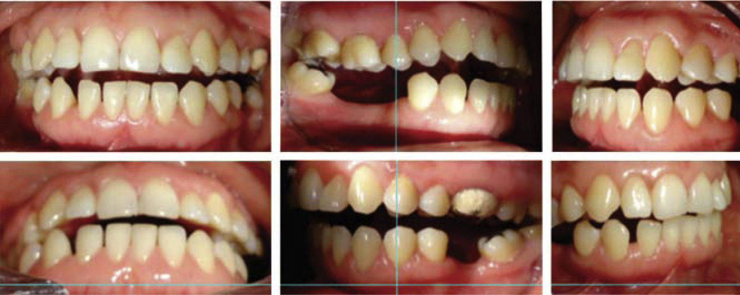
At the extraoral analysis, during palpation the patient felt pain in the masseter and external pterygoid muscles bilaterally. Facial form was oval; the biotype, dolichofacial; asymmetric facial fifths, increased lower facial third and convex profile (Figure 1). Intraorally she had loss of multiple dental organs, presence of restorations and fixed prosthesis, an oval arch form, spacing, extrusion of teeth #15 and 16, a 7mm overjet, bilateral canine class II and a non-assessable molar class (Figure 2). The orthopantomography revealed the presence of root canal treatments in teeth #24 and 25, multiple restorations, edentulous areas, presence of third molars (18, 28 and 38) and bony structures without pathological findings (Figure 3).


Table I shows the results of the cephalometric measurements performed in the lateral headfilm (Figure 4). The patient was diagnosed with myofascial pain syndrome, skeletal class II due to mandibular retrognathism, bilateral canine class II and non-assessable molar class.
Initial cephalometric analysis.
| Skeletal analysis | Patient |
|---|---|
| (S-N-Ar) | 123o |
| (S-Ar-Go) | 144o |
| (Ar-Go-Me) | 135o |
| SUM | 402o |
| (S-N) | 68mm |
| (S-Ar) | 33mm |
| (N-Go-Ar) | 53o |
| (N-Go-Me) | 82o |
| (Ar-Go) | 44mm |
| (Go-Gn) | 72mm |
| Relationship between mandibular body with | 1:0.9 |
| regard to anterior craneal base | 72:68 mm |
| SNA | 80° |
| SNB | 74° |
| ANB | 6° |
| Go-Gn-SN | 42° |
| Facial depth (N-Go) | 108 mm |
| Facial length/Y axis (s-Gn) | 127 mm |
| Y axis/SN | 68° |
| Anterior facial height (N-Me) | 120 mm |
| Posterior facial height (S-Go) | 73 mm |
| Facial plane (SN-Pg) | 79° |
| Facial convexity (NA-Pg) | 6° |
| Dental analysis | |
| Occlusal plane/Go-Gn | 23° |
| Interincisal angle | 130° |
| L1/Go-Gn | 85° |
| U1/SN | 102° |
| U1/facial plane | 7 mm |
| L1/facial plane | -4 mm |
| E line/upper lip | -3 mm |
| E line/lower lip | -4 mm |

To provide function, stability, esthetics and periodontal health.
Treatment planA deprogramming splint was placed and third molar extractions were performed. A combined method of 0.022” slot Roth prescription fixed appliances combined with mandibular advancement and rotation orthognathic surgery is described.
- a)
Presurgical phase: treatment initiated with a rigid deprogrammer type splint made of thermocurable acrylic which was placed for six months as palliative for the painful symptoms of the masticatory muscles (Figure 5).
During the use of the splint extractions of teeth #18, 28 and 38 were performed because they created premature contact points which aggravated the anterior open bite (Figure 6). 0.022” x 0.028” slot Roth fixed appliances were placed in order to produce a decompensation by aligning and leveling, space closure, tipping and torque. The archwire sequence was as follows: 0.014” NiTi, 0.016” NiTi, 0.016” x 0.022” NiTi and SS; 0.017” x 0.025” NiTi and SS; and 0.019” x 0.025” NiTi and SS. Additionally, cantilevers made with 0.017” x 0.025” SS wire were used for uprighting the molars (Figure 7); prior to surgery 0.019” x 0.025” SS surgical archwires were placed (Figure 8). At this point, a prediction surgery was made in the cephalogram and study models where a mandibular advancement of 6mm was suggested (Figure 9).
- b)
Surgical phase: based on the analysis and treatment plan, a bilateral sagittal osteotomy of the mandibular ramus (BSOMR)was performed for advancing the mandible 6mm using osteosynthesis screws for fixing (Figure 10).
- c)
Postsurgical phase: after surgery we started the use of 4.5 oz. 3/8” elasticsin «N» form for a month and began settling the occlusion. A new mounting on the articulator in centric relation was made for occlusal adjustment (Figure 11).


Facially, a better harmony due to the sagittal correction of the mandible was obtained as well as a straight profile and a regulation of the neuromuscular system and stomatognathic function in occlusion and centric relation. Anterior and canine guidance, which were absent prior to orthodontic and surgical treatment, were obtained as well as a decrease in the overjet and a bilateral canine class I.
Cephalometric post-surgical values were normal as shown in table II. Currently the patient refers that her myalgia has been eradicated entirely.
Post-surgical cephalometric values.
| Skeletal analysis | Patient |
|---|---|
| (S-N-Ar) | 123° |
| (S-Ar-Go) | 144° |
| (Ar-Go-Me) | 133° |
| SUM | 400° |
| (S-N) | 68mm |
| (S-Ar) | 33mm |
| (N-Go-Ar) | 53° |
| (N-Go-Me) | 80° |
| (Ar-Go) | 44mm |
| (Go-Gn) | 72mm |
| Relationship between mandibular body with regard to anterior craneal base | 1:1mm |
| SNA | 80° |
| SNB | 78° |
| ANB | 2° |
| Go-Gn-SN | 40° |
| Facial depth (N-Go) | 108mm |
| Facial length/Y axis (s-Gn) | 127mm |
| Y axis/SN | 70o |
| Anterior facial height (N-Me) | 117mm |
| Posterior facial height (S-Go) | 73mm |
| Facial plane (SN-Pg) | 79° |
| Facial convexity (NA-Pg) | 1° |
| Dental analysis | |
| Occlusal plane/Go-Gn | 21° |
| Interincisal angle | 130° |
| L1/Go-Gn | 89° |
| U1/SN | 104° |
| U1/facial plane | 7mm |
| L1/facial plane | -2mm |
| E line/upper lip | -3mm |
| E line/lower lip | -1mm |
Removable circumferential retainers were placed after the appliance removal (Figure 12).
DISCUSSION
Authors of previous studies have concluded that the majority of patients have some craniofacial asymmetry, including those who are perceived as normal.9,10 Numerous investigations have shown the remodeling that takes place in the head of the condyle in response to occlusal alterations.11–13
When orthognathic surgery is required in combination with orthodontics, a therapy without extractions shortens the orthodontic phase substantially and prevents incisor retraction which is often associated with depression of the lip profile. However, in some patients, extractions are necessary to reduce the maxillary dental protrusion as well as to decrease mandibular incisor proinclination that results from leveling the mandibular arch.14
It has been suggested that, in the treatment of class II malocclusions, premolar extractions must be performed asymmetrically. In the case hereby presented, the arch length discrepancy was not significant therefore asymmetric extractions were not planned. In addition, the patient's profile did not allow incisor retraction. Therefore, asymmetric extractions would not have been beneficial for this problem. Patients who were treated surgically and orthodontically have reported a high range of benefits of treatment, including psychological stability, self-esteem, and an improvement in function and dental aesthetics.15–19
The goal of our orthodontic preparation was to allow the surgeon to perform sufficient mandibular advancement in order to compensate for the sagittal discrepancy thus positioning the arches in a normal transverse occlusion and canine class I.
In 1993 the World Health Organization (WHO), defined quality of life as the people's perception of their position in life, in the context of culture and value system in which they live and in relation to their goals, expectations, standards, and concerns.9 The importance of interdisciplinary work between dental specialties in the benefit of improving the quality of life of patients has been suggested since remote times.
CONCLUSIONSClass II malocclusions treatment, after careful analysis, may be carried out orthodontically through different protocols. However, if a discrepancy is associated with a skeletal malocclusion it may be resolved in a surgical and orthodontic manner as it has been hereby shown thus providing a better aesthetic result for the patient. In spite of the fact that there are different protocols of care for patients with class II malocclusion it is of vital importance to take into consideration the patient's treatment expectations from the first day of consultation.
Student of the Orthodontics Specialty.
This article can be read in its full version in the following page: http://www.medigraphic.com/ortodoncia