


Background: Liver is the most common site of infection and several methods of surgery have been described to treat this common disease. In this study we aim to compare the results of two common methods of surgery; simple drainage versus omentoplasty. Methods: In this prospective study 65 patients with hepatic hydatid cyst underwent surgery from 10 May 1995 to 1 July 2002. 35 patients were treated with omentoplasty (group I) and 30 of them were treated with drainage (group II). The results of surgery including mortality, complications and recurrences were recorded. Results: There was no case of mortality in each group of patients. Postoperative complications were seen in 5.7% of group I, 16.6% of group II patients. The mean duration of hospital stay was 6.5 and 15.6 days in group I and group II patients. During a mean period of 18.6 month follow up there was no recurrence in each group of patients. Conclusion: According to the results of this study we suppose that omentoplasty of cyst cavity –if feasible– is preferred to tube drainage.
Hydatidosis is an endemic parasitic disease in countries where sheep are prolific. This is particularly the case in Mediterranean countries including North Africa, Spain and Portugal.17 The parasite, Echinococcus granulosus, is a cestode that grows in the small intestine of its definite host, usually a dog. The host puts off eggs of the parasite within stool and when an intermediate host (sheep or human) ingests vegetables contaminated with definite host feces, larvae of the parasite exit the eggs in the duodenum.
The larvae pass through the intestinal wall and reach the liver via the portal system where they form cysts. The liver is the most common site of infection in hydatid disease (50-93%).3,15 Without treatment, cysts grow and eventually form fistulas into adjacent organs or rupture into the peritoneal cavity. Older cysts have an increased risk of daughter cyst formation, which is an important factor for recurrence of disease after surgery.11,18 Several types of treatment have been described to treat hydatid cyst of the liver. Medical therapy alone is insufficient to cure the disease, although stabilization of disease has been reported with albendazole alone or in combination with praziquantel.8,13 Surgical approaches vary from complete resection (e.g. total pericystectomy or hepatectomy) to minimal invasive procedures (e.g. percutaneous aspiration of cysts).4,10,19 More recently, reports have been published on laparoscopic surgery for hepatic hydatid cysts.9,21 The choice of therapy depends on several factors: general condition of the patient, number and localization of the cysts, the surgeon’s experience and the presence of special services such as intensive care unite. This study presents the long-term results of two surgical techniques in treating hepatic hydatid cyst and compares their postoperative complications, morbidity and recurrence of the disease.
Patients and methodsSixty-five patients, who underwent surgery for hepatic hydatid cysts between 10 May 1993 and 1 July 2002, were included in this study. Patients were divided into two groups. The first group consisted of 35 patients treated with omentoplasty and the second group were 30 patients treated surgically with tube drainage of the cyst cavity. In 62 (95.3%) patients sonography was the imaging study of choice done to make the diagnosis (Figure 1) and in remaining 3 (4.6%) patients CT scan was done as the first imaging study. Hepatic infestation with Echinococcus granulosus was confirmed histologically in all patients. Surgical techniques were compared with respect to post-operative complications, hospital stay and recurrence of the disease. Patients were invited to be visited every 3 months, at which clinical examination, ultrasonography and serological tests were performed.

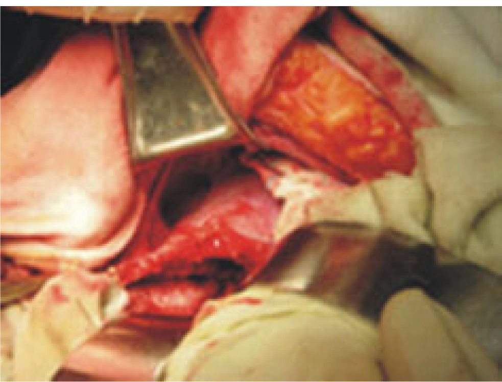
Surgical techniques: In both techniques we used an extended right subcostal incision. After entering the abdominal cavity, routine exploration of abdominal viscera was done as well as the liver and the cyst. The surgical field was covered with packs immersed in scolicidal agent (silver nitrate 0.5%) to prevent the spread of the parasite and reduce the risk of intraperitoneal soiling and contamination. Then the cyst is evacuated by aspiration with a closed system suction device. After complete aspiration of cyst contents, the cyst was unroofed and the germinal layer and the remaining daughter cysts were removed. Then we put clean sponges over the inner layer of the cyst to define bile leakage from biliary openings. If there was any biliary opening, each one was ligated to prevent postoperative bile leakage. Then the cyst cavity was filled with silver nitrate immersed sponges for a few minutes to sterilize the cavity of the cyst and then sponges were removed. At this point, the first group of patients were considered to fill the cavity of their cysts with omentum and the second group were treated with simple tube drainage (closed suction) of the cyst cavity (Figure 2).
Results
65 patients (16 male and 49 female) were included in this study with a mean age of 37.8 years (rang 14-81 years). 55 (88.6%) cysts were located in the right lobe and 10(15.6%) in the left lobe of the liver. The mean diameter of the cysts was 12 cm (rang 8-35 cm). Postoperative complications including atelectasia, wound infection and abscess formation were compared in two groups of patients. Atelectasia as a common complication of abdominal surgery was common in both groups of patients with no significant difference. Wound infection was seen in 4 (13.3%) patients of group II and 2 (5.7%) patients of group I. Intraabdominal abscess formation occurred in 1 (3.3%) patient of group II. Overall complication rate in group I patients was 5.7% and in group II patients it was 16.6% (p < 0.05) (Table I).
The mean postoperative hospital stay was significantly longer after drainage procedures (15.6 days) than those treated with omentoplasty (6.5 days) (p < 0.05).
In a mean period of 18.6 (range 13-29) month of follow up there was no case of recurrence based on physical examination, sonography and serologic tests in each group of patients.
DiscussionIn general, hepatic hydatid cysts are single, uncomplicated, and located in the right lobe of the liver. These findings are consistent with published literature. Asymptotic cysts may persist for years without complaint from the patient.
Surgical treatment still remains the treatment of choice in the management of hydatid disease. Treatment of the cystic cavity of the hepatic hydatid cyst is the principal issue with this disease that could not be completely resolved by surgical techniques. Two main operative approaches have been described: (i) drainage procedures, and (ii) the obliteration of the cyst cavity after evacuation of the cystic content without drainage. Many debatable results have been reported in relation to the surgical treatment of the cystic cavity. Cysts located peripherally, and pedunculated cysts can be excised entirely. Such operations have high morbidity and mortality rates and can be considered radical procedures for such a benign disease. Hepatic resection should only be considered for Echinococcus alveolaris cases that are located on one hepatic lobe.
Drainage procedures can be followed by various postoperative complications, such as hepatic abscess, biliary fistulas, and a longer hospital stay. Therefore omentoplasty for a single uncomplicated hydatid cyst caused significantly fewer complications than external drainage, and patients left hospital in a shorter period of time.5,7,12,14
However, in our experience in the group treated by drainage, tube drainage of the cystic cavity increased the post-operative morbidity in cases of hepatic hydatid cyst in comparison with other group of patients. The difference was statistically significant (P < 0.05). The restriction of activity in patients due to the drainage tube was also an important predisposing factor for pulmonary infections and thromboembolism. Furthermore, the drain was an important factor in the entrance of microorganisms into the peritoneum thus increasing peritoneal infections. Although in the majority of cases, however, external tube drainage retains its value as a simple and safe procedure.16,23
Omentoplasty seems to be the best possible surgical alternative for the radical treatment of hepatic hydatid cysts. The management of hydatid cysts should be flexible, taking into consideration a number of factors and variables.20
Therefore no single method can be recommended for the treatment of hepatic hydatid cysts but the choice of the surgical method must be made according to the complications of the cyst. Omentoplasty is the procedure of choice for uncomplicated cysts with a low complication rate and relatively short hospital stay. External tube drainage is recommended for infected cysts and a biliary drainage procedure must be added to external tube drainage for cysts with intrabiliary rupture.2
Omentoplasty should be the standard surgical procedure because it is safe, simple, and effective and meets all criteria of surgical treatment for hydatid disease: entire elimination of the parasite, no intraoperative spillage especially by using a cone, and saving healthy tissue.6
Infected cases were drained, and choledochotomy and internal or external drainage were performed for Intrabiliary ruptured cases.22
Recently, Laparoscopy is quite feasible to perform in hydatid disease of the liver, and the use of helical fasteners allows effective omental flap fixation.1
As a result of this study we conclude, as have many authors, that excision of the cyst and omentoplasty is the preferred method: having the lowest morbidity rate and an acceptable rate of recurrence. Thus, surgical procedures do not employ drainage should be the treatment of choice in the management of patients with hydatid cysts of the liver. External drainage could occasionally be reserved for more complicated cases, where there is sepsis of the biliary tree.


