metricas
covid
Buscar en
Enfermedades Infecciosas y Microbiología Clínica (English Edition)
Toda la web
Inicio Enfermedades Infecciosas y Microbiología Clínica (English Edition) Helminthosis and eosinophilia in Spain (1990–2015)
Información de la revista
Vol. 36. Núm. 2.
Páginas 120-136 (febrero 2018)
Visitas
4078
Vol. 36. Núm. 2.
Páginas 120-136 (febrero 2018)
Review article
Acceso a texto completo
Helminthosis and eosinophilia in Spain (1990–2015)
Helmintosis y eosinofilia en España (1990–2015)
Visitas
4078
Cristina Carranza-Rodrígueza,b,1, Miriam Escamilla-Gonzálezc,1, Isabel Fuentes-Corripioc, María-Jesús Perteguer-Prietoc, Teresa Gárate-Ormaecheac,2, José-Luis Pérez-Arellanoa,b,2,
Autor para correspondencia
jlperez@dcmq.ulpgc.es

Corresponding author.
a Departamento de Ciencias Médicas y Quirúrgicas, Universidad de Las Palmas de Gran Canaria, Las Palmas de Gran Canaria, Spain
b Unidad de Enfermedades Infecciosas, Complejo Hospitalario Universitario Insular Materno Infantil, Las Palmas de Gran Canaria, Spain
c Servicio de Parasitología, Centro Nacional de Microbiología, Instituto de Salud Carlos III, Majadahonda, Madrid, Spain
Este artículo ha recibido
Información del artículo
Resumen
Texto completo
Bibliografía
Descargar PDF
Estadísticas
Figuras (3)
Mostrar másMostrar menos
Tablas (12)
Table 1. Main cestodiasis.
Table 2. Neurocysticercosis in Spain.
Table 3. Schistosomiasis imported by travellers (cases and series).
Table 4. Schistosomiasis imported by immigrants (isolated cases).
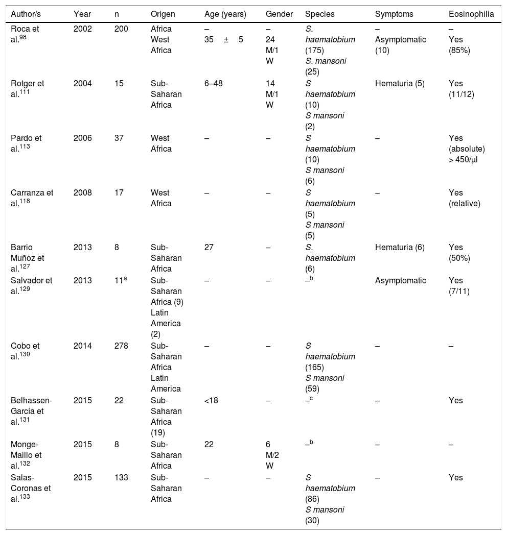
Table 5. Schistosomiasis imported by immigrants (series).
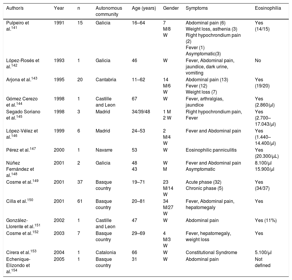
Table 6. Fasciolosis in Spain.
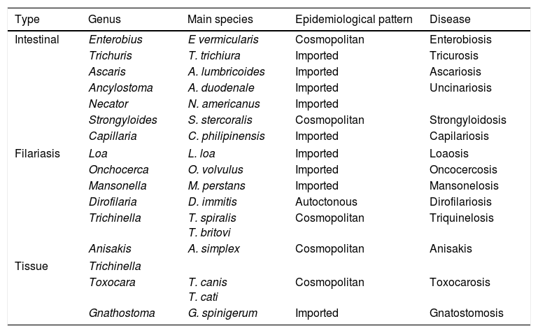
Table 7. Principal nematodosis in Spain.
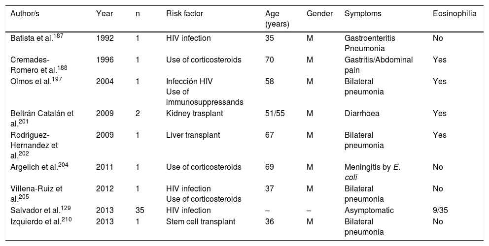
Table 8. Strongyloidosis and risk factors.
Table 9. Autochthonous strongyloidosis in Spain.
Table 10. Imported strongyloidosis in Spain.
Table 11. Imported Filariasis in Spain (cases).
Table 12. Imported filariasis in Spain (series).
Mostrar másMostrar menos
Abstract

The finding of blood eosinophilia in a patient is a relatively frequent reason to refer him/her to a Clinical Department of Infectious Diseases. The doctor usually intends to rule out a parasitic disease in the autochthonous population, travellers or immigrants. It is uncommon for an eosinophilia to be produced by protozoa infection, whereas helminth parasites are more frequently associated with an increase of eosinophil counts in the infected patient. Eosinophilia can be the only abnormal finding, or it could be part of more complex clinical manifestations suffered by the patient. Furthermore, many, but not all, helminth infections are associated with eosinophilia, and the eosinophil level (low, high) differs according to parasite stages, helminth species, and worm co-infections. The purpose of the present article is to carry out a systematic review of cases and case series on helminth infections and eosinophilia reported in Spain from 1990 to 2015, making a distinction between autochthonous and imported (immigrants and travellers) cases, and studying their relationship with immunodepression situations.

Keywords:
Helminths
Eosinophilia
Spain
Imported diseases
Resumen

La detección de eosinofilia periférica es un motivo relativamente frecuente para la remisión de un paciente a una Unidad/Servicio de Enfermedades Infecciosas. En general, se pretende descartar una enfermedad parasitaria, tanto en personas autóctonas como en viajeros o inmigrantes. Excepcionalmente la eosinofilia relacionada con parásitos corresponde a una protozoosis, siendo los helmintos los principales agentes causales de este hallazgo hematológico. La eosinofilia puede ser el único hallazgo anormal o formar parte del cuadro clínico-biológico del paciente. Por otro lado, no todas las helmintosis se asocian de forma sistemática a eosinofilia, y el grado de la misma difiere entre las fases de la infección y el tipo de helminto. El propósito de esta revisión es un estudio sistemático de la relación entre helmintosis y eosinofilia en la literatura española, distinguiendo los casos autóctonos e importados, así como la relación con situaciones de inmunodepresión.

Palabras clave:
Helmintos
Eosinofilia
España
Enfermedades importadas
Texto completo
Introduction

The term “eosinophilia” indicates the raising in the number or percentage of polymorphonuclear-eosinophil leukocytes in any solid or liquid tissue.1 Although no limit has been established, it is considered that eosinophilia exists when blood values surpass 450cells/μl.1 Their detection in the blood requires an investigation of the cause responsible, as it may arise from highly diverse causes, from mild conditions (e.g. allergic rhinitis) to severe processes (e.g. tumours of the hermatopoietic and lymphoid tissues).1 One of the main conditions leading to detection of eosinophilia is the presence of a parasitic disease. Furthermore, and with few exceptions (Isospora belli, Dientamoeba fragilis, Sarcocystis spp.), the protozoa are not the agents connected to the appearance of eosinophilia, and their presence suggests helminthosis.1

In Spain extensive literature exists regarding the association between infection by helminthes and the presence of eosinophilia. The global map of this association in Spain is complex, since in several cases parasitism is detected in isolated cases, and in others, in form of outbreaks. Several parasitic diseases also only appear as imported diseases (travellers or immigrants) whilst others have a more cosmopolitan distribution.2 Thirdly, the presence of eosinophilia depends on the life cycle stage of the parasite and even of response to treatment. Finally, several factors such as age, the patient's geographical origin, their immunological status and the presence of polyparasitism are determining factors in the detection of eosinophilia.

The aim of this study was to review the helminths associated with eosinophilia in Spain during the last 25 years. This study is based on a systematic search in PubMed, which included originals, brief originals, clinical notes and scientific letters (Fig. 1). The electronic search strategy was as follows: country (Spain) AND disease OR agent. The following MESH terms were considered in the inclusion of diseases: Helminthiasis, Taeniasis, Hymenolepiasis, Dipylidiasis, Cysticercosis, Echinococcosis, Sparganosis, Diphyllobothriasis, Schistosomiasis, Fascioliasis, Paragonimiasis Opisthorchiasis, Clonorchiasis, Dicrocoeliasis, Enterobiasis, Ancylostomiasis, Necatoriasis, Ascariasis, Trichuriasis, Strongyloidiasis, Dirofilariasis, Filariasis, Loiasis, Onchocerciasis, Mansonelliasis, Dracunculiasis, Trichinellosis, Anisakiasis, Toxocariasis, Gnathostomiasis. With regard to the agents, the following MESH terms were included: Helminth, Taenia, Hymenolepis, Dipylidium, Cysticercus, Echinococcus, Spirometra, Sparganum, Schistosoma, Fasciola, Paragonimus, Opisthorchis, Clonorchis, Heterophyes, Metagonimus, Dicrocoelium, Ancylostoma, Necator, Hookworm, Ascaris, Trichuris, Strongyloides, Capillaria, Dirofilaria, Wuchereria, Brugia, Loa, Onchocerca, Mansonella, Dracunculus, Trichinella, Anisakis, Toxocara, Gnathostoma. Results were restricted to studies carried out in humans. The search period lasted from January 1st 1990 to 31st August 2015.

Fig. 1.

Strategy for the selection of articles.

(0.18MB).
Cestodiasis

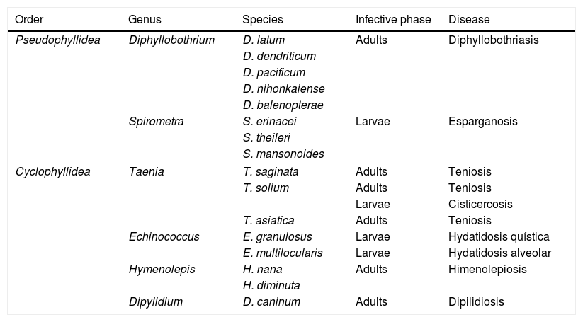
Cestodiasis are diseases caused by flat worms (phylum Plathelminthes) with a segmented body (Cestoda classification). The main agents of the disease in humans include 2 types, Pseudophyllidea and Cyclophyllidea, and may cause the disease either through the adult form of the parasite or through the larva stage or both. Table 1 indicates the main types of cestodiasis. In general, we may state that the eosinophilia associated with the cestodiasis is mild or moderate, and often does not appear during the course of the disease. Eosinophilia is also more common in larval cestodiasis (with tissue compromise) than in those produced by adult worms (with isolated intestinal compromise). Finally, the rupture or surgical manipulation of larval forms (especially in cystic echinococcosis and to a lesser degree in alveolar echinococcosis) is associated with a notable raising in the number eosinophils in the bloodstream.

Table 1.

Main cestodiasis.

Order  Genus  Species  Infective phase  Disease 
PseudophyllideaDiphyllobothriumD. latum  AdultsDiphyllobothriasis
D. dendriticum 
D. pacificum 
D. nihonkaiense 
D. balenopterae 
SpirometraS. erinacei  LarvaeEsparganosis
S. theileri 
S. mansonoides 
CyclophyllideaTaeniaT. saginata  Adults  Teniosis 
T. soliumAdults  Teniosis 
Larvae  Cisticercosis 
T. asiatica  Adults  Teniosis 
EchinococcusE. granulosus  Larvae  Hydatidosis quística 
E. multilocularis  Larvae  Hydatidosis alveolar 
HymenolepisH. nana  AdultsHimenolepiosis
H. diminuta 
Dipylidium  D. caninum  Adults  Dipilidiosis 

The cestodiasis diagnosed most frequently in Spain is, without a doubt, that of infections caused by the parasites of the order Cyclophyllidea. Among them, the lowest number of references corresponds to the intestinal types. Although the recording of intestinal infection caused by Taenia sp., is relatively frequent, as shown by indirect data (personal cases in the Hospital Universitario Insular de Gran Canaria) and samples sent to the Instituto de Salud Carlos III, few cases have been published in the literature.3 Infection by Hymenolepis nana has been reported in Spain, mainly in cases of Sahrawi children from Tindouf who are spending their summer holidays in Spain. Prevalence is 6.5–7.5% and there is frequent co-parasitism with intestinal protozoa (Giardia intestinalis).4,5 A case of infection by this helminth has also been reported anecdotally in a child from Ecuador.6 We only found one case published on Hymenolepis diminuta in a child aged 5 in the province of Guadalajara.7 In the review carried out we did not locate cases of infection in humans by Dipylidium caninum. However, the detection of eggs from the parasite in dog faeces in several regions of Spain,8–11 suggests that this parasitism could be under-diagnosed.

In contrast, the larval forms of cestodiasis are common, both as autochthonous parasitism or imported. The 2 major ones are cystic hydatidosis and cysticercosis, and particularly in its neurological form (neurocysticercosis).

Cystic hydatidosis produced by Echinococcus granulosus is an autochthonous zoonotic disease, endemic in the Iberian peninsula, which had major socio-economic repercussions up to the end of the 20th century.12–15 Historically the most affected regions were the Northern communities (Basque Country, Navarre, Aragon, La Rioja, Cantabria) and the central regions (Castille and Leon, Extremadura, Castilla-La Mancha).15–22 Moreover, in recent years a large number of cases were reported in the Community of Valencia.23 However, we found no published cases of cystic hydatidosis in the Canary Island Community or the Balearic Island communities. Hydatidosis was significantly reduced thanks to the control programmes introduced in the eighties and nineties, although direct and indirect data exist, such as the presence of new cases of infants or the maintenance of high levels of infection in young people in the last few years, which suggest a re-emergence of the disease.12,13 In Spain, just as in other parts of the world, the most common clinical manifestations of this disease are liver24–27 and bile duct28,29 compromise and secondly derive from respiratory system lesions.18,30–33 Furthermore, several Spanish groups have reported cases or series of “atypical” forms of cystic hydatidosis which were intra-abdominal (splenic,34–36 pancreatic,20 renal,37 ovarian38 and other less common forms17,39–41) such as in other organs (heart,42–45 muscles and skeleton,21,46,47 bone48,49 or skin and soft tissues50). Several cases of imported cystic hydatidosis have also been reported, although they are scarce and not well characterised.51–53 The presence of eosinophilia is highly uncommon in the classic cystic hydatidosis, but a high rate of incidence has been published in several extra pulmonary forms, and particularly renal,37 or after rupture of the cysts (spontaneous or during surgery).54–56

Alveolar hydatidosis, cause by Echinococcus multilocularis, is exceptional in Spain, both in its autochthonous form and its imported form. There have only been 2 references in the literature in the last 25 years. These cases presented in the usual manner with similar symptoms to a primary liver tumour.57,58

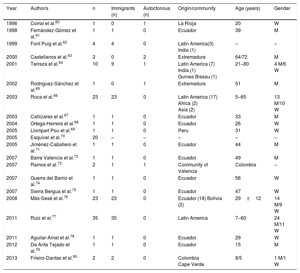
Cysticercosis is a disease caused by the larvae of Taenia solium (Cysticercus cellulosae). The 3 main locations of the larval forms are cutaneous, ocular and neurological. The most severe cases are logically those which affect the central nervous system and a few cases have been published in Spain, with mixed forms.59Table 2 displays the epidemiological data of the cases and the series of neurocysticercosis published in Spain.60–80 As maybe observed, and has already been reported in other references,60–82 there are 2 different patterns of the disease: (a) imported, which includes the greater part of cases detected in the last few years, and particularly those observed in immigrants with an age range from infancy to middle age, and (b) autochthonous, with rare cases, and reported in Spaniards over the age of 18. The main origin of the imported cases is Latin American (mostly Ecuador, Peru, Colombia and Bolivia), although a few cases of patients also come from Africa (Guinea Bissau, Cape Verde, the Ivory Coast) and Asia (India and China.)64,66–69,74–80 Most autochthonous cases were described in Extremadura, La Rioja, Madrid and the Community of Valencia. In the imported cases there is a similar number of males and females, whilst in the autochthonous cases there is a clear predominance of males. In general, neurocysticercosis presents in immune-competent people although in Spain several cases have been reported in people infected with HIV73 and those with transplants.72 From the clinical viewpoint, the most frequent manifestations of neurocysticercosis are epileptic crises (of different types)and headaches. However, in Spain other unusual presentations have been reported, such as blepharospasm,83 Bruns syndrome (sudden headache associated with acute vestibular syndrome related to sudden movements of the head),71,78 medullar lesions,60 psychiatric changes79 and sudden death.69 The presence of eosinophilia in patients with neurocysticercosis is exceptional and for the most part, poorly documented.84–86

Table 2.

Neurocysticercosis in Spain.

Year  Author/s  Immigrants (n)  Autoctonous (n)  Origin/community  Age (years)  Gender 
1996  Corral et al.60  La Rioja  20 
1998  Fernández-Gómez et al.61  Ecuador  39 
1999  Font Puig et al.62  Latin America(3) India (1)  –  – 
2000  Castellanos et al.63  Extremadura  64/72 
2001  Terraza et al.64  10  Latin America (7)
India (1)
Guinea Bissau (1) 
21–80  4 M/6 W 
2002  Rodríguez-Sánchez et al.65  Extremadura  51 
2003  Roca et al.66  23  23  Latin America (17)
Africa (2)
Asia (2) 
5–65  13 M/10 W 
2003  Cañizares et al.67  Ecuador  33 
2004  Ortega-Herrera et al.68  Ecuador  26 
2005  Llompart Pou et al.69  Peru  31 
2005  Esquivel et al.70  20  –  –  –  –  – 
2005  Jiménez-Caballero et al.71  Ecuador  44 
2007  Barra Valencia et al.72  Ecuador  49 
2007  Ramos et al.73  Community of Valencia  Colombia  – 
2007  Guerra del Barrio et al.74  Ecuador  56 
2007  Sierra Bergua et al.75  Ecuador  47 
2008  Más-Sesé et al.76  23  23  Ecuador (18) Bolivia (2)  29±12  14 M/9 W 
2011  Ruiz et al.77  35  35  Latin America  7–60  24 M/11 W 
2011  Aguilar-Amat et al.78  Ecuador  29 
2012  De Anta Tejado et al.79  Ecuador  15 
2013  Frieiro-Dantas et al.80  Colombia
Cape Verde 
8/5  1 M/1 W 

W: woman; M: man; –: no data.

Infections by parasites of the Pseudophyllidea order are rare in Spain. Among these, most references correspond to cases of the diphyllobothriasis.87–91 Only one case of sparganosis (infection by difference species of Spirometran) was recently reported. This was an imported sparganosis by a male aged 29 from Bolivia, with symptoms of convulsions and multi-cystic cerebral lesion and who was diagnosed with suspected dysembryoplastic neuroepithelial tumour (DNET).92 With regard to diphyllobothriasis, the symptoms of this disease may not be present or may present non-specific abdominal upset which may or may not be associated with megaloblastic anaemia. Of the published cases in Spain, most are autochthonous, and diagnosed in a broad range of ages (3–71 years). The most frequently detected species is Diphyllobothrium latum, although cases caused by Diphyllobothrium pacificum and Diplogonoporus balaenopterae90 have also been reported. These are possibly related to the consumption of imported fish or travel abroad. Eosinophilia is exceptional in the cases published in Spain.

Trematodosis

Trematodosis are diseases produced by flatworm (phylum Plathelminthes) with unsegmented foliaceus body (Trematoda class). Most are hermaphrodites, with the exception of the genus Schistosoma, which present sexual dimorphism and characteristic morphology.

Schistosomiasis

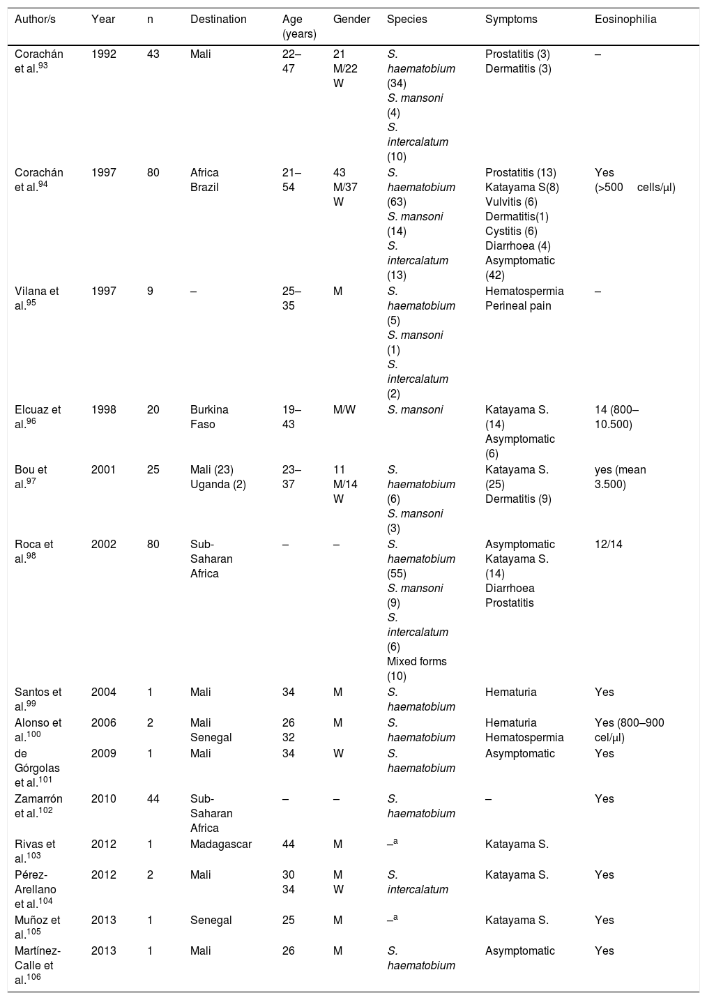
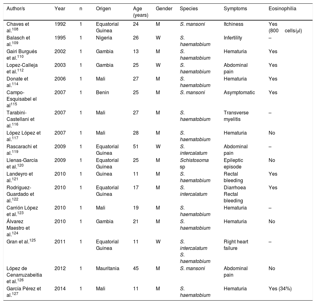
All cases published in Spain on this disease correspond to imported forms. However, symptoms and biology differ notably between infections detected in travellers and those diagnosed in immigrants (Tables 3–5).

Table 3.

Schistosomiasis imported by travellers (cases and series).

Author/s  Year  Destination  Age (years)  Gender  Species  Symptoms  Eosinophilia 
Corachán et al.93  1992  43  Mali  22–47  21 M/22 W  S. haematobium (34)
S. mansoni (4)
S. intercalatum (10) 
Prostatitis (3)
Dermatitis (3) 
– 
Corachán et al.94  1997  80  Africa
Brazil 
21–54  43 M/37 W  S. haematobium (63)
S. mansoni (14)
S. intercalatum (13) 
Prostatitis (13)
Katayama S(8)
Vulvitis (6)
Dermatitis(1)
Cystitis (6)
Diarrhoea (4)
Asymptomatic (42) 
Yes (>500cells/μl) 
Vilana et al.95  1997  –  25–35  S. haematobium (5)
S. mansoni (1)
S. intercalatum (2) 
Hematospermia
Perineal pain 
– 
Elcuaz et al.96  1998  20  Burkina Faso  19–43  M/W  S. mansoni  Katayama S. (14)
Asymptomatic (6) 
14 (800–10.500) 
Bou et al.97  2001  25  Mali (23)
Uganda (2) 
23–37  11 M/14 W  S. haematobium (6)
S. mansoni (3) 
Katayama S. (25)
Dermatitis (9) 
yes (mean 3.500) 
Roca et al.98  2002  80  Sub-Saharan Africa  –  –  S. haematobium (55)
S. mansoni (9)
S. intercalatum (6)
Mixed forms (10) 
Asymptomatic
Katayama S. (14)
Diarrhoea
Prostatitis 
12/14 
Santos et al.99  2004  Mali  34  S. haematobium  Hematuria  Yes 
Alonso et al.100  2006  Mali
Senegal 
26
32 
S. haematobium  Hematuria
Hematospermia 
Yes (800–900 cel/μl) 
de Górgolas et al.101  2009  Mali  34  S. haematobium  Asymptomatic  Yes 
Zamarrón et al.102  2010  44  Sub-Saharan Africa  –  –  S. haematobium  –  Yes 
Rivas et al.103  2012  Madagascar  44  a  Katayama S.   
Pérez-Arellano et al.104  2012  Mali  30
34 
M
S. intercalatum  Katayama S.  Yes 
Muñoz et al.105  2013  Senegal  25  a  Katayama S.  Yes 
Martínez-Calle et al.106  2013  Mali  26  S. haematobium  Asymptomatic  Yes 

W: woman; M: man; –: no data.

a

Serological diagnosis.

Table 4.

Schistosomiasis imported by immigrants (isolated cases).

Author/s  Year  Origen  Age (years)  Gender  Species  Symptoms  Eosinophilia 
Chaves et al.108  1992  Equatorial Guinea  24  S. mansoni  Itchiness  Yes (800cells/μl) 
Balasch et al.109  1995  Nigeria  26  S. haematobium  Infertility  – 
Gairi Burgués et al.110  2002  Gambia  13  S. haematobium  Hematuria  Yes 
Lopez-Calleja et al.112  2003  Gambia  25  S. haematobium  Abdominal pain  Yes 
Donate et al.114  2006  Mali  27  S. haematobium  Hematuria  Yes 
Campo-Esquisabel el al115  2007  Benin  25  S. mansoni  Asymptomatic  Yes 
Tarabini-Castellani et al.116  2007  Mali  27  S. haematobium  Transverse myelitis  – 
López López et al.117  2007  Mali  28  S. haematobium  Hematuria  No 
Rascarachi et al.119  2009  Equatorial Guinea  51  S. intercalatum  Abdominal pain  – 
Llenas-García et al.120  2009  Equatorial Guinea  25  Schistosoma sp  Epileptic episode  No 
Landeyro et al.121  2010  Guinea  11  S. haematobium  Rectal bleeding  Yes 
Rodríguez-Guardado et al.122  2010  Equatorial Guinea  17  S. intercalatum  Diarrhoea
Rectal bleeding 
Yes 
Carrión López et al.123  2010  Mali  19  S. haematobium  Hematuria  – 
Álvarez Maestro et al.124  2010  Gambia  21  S. haematobium  Hematuria  No 
Gran et al.125  2011  Equatorial Guinea  11  S. intercalatum
S. haematobium 
Right heart failure  – 
López de Cenarruzabeitia et al.126  2012  Mauritania  45  S. mansoni  Abdominal pain  No 
García Pérez et al.127  2014  Mali  11  S. haematobium  Hematuria  Yes (34%) 

W: woman; M: man; –: no data.

Table 5.

Schistosomiasis imported by immigrants (series).

Author/s  Year  Origen  Age (years)  Gender  Species  Symptoms  Eosinophilia 
Roca et al.98  2002  200  Africa
West Africa 

35±

24 M/1 W 
S. haematobium (175)
S. mansoni (25) 

Asymptomatic (10) 

Yes (85%) 
Rotger et al.111  2004  15  Sub-Saharan Africa  6–48  14 M/1 W  S haematobium (10)
S mansoni (2) 
Hematuria (5)  Yes (11/12) 
Pardo et al.113  2006  37  West Africa  –  –  S haematobium (10)
S mansoni (6) 
–  Yes (absolute)
> 450/μl 
Carranza et al.118  2008  17  West Africa  –  –  S haematobium (5)
S mansoni (5) 
–  Yes (relative) 
Barrio Muñoz et al.127  2013  Sub-Saharan Africa  27  –  S. haematobium (6)  Hematuria (6)  Yes (50%) 
Salvador et al.129  2013  11a  Sub-Saharan Africa (9)
Latin America (2) 
–  –  b  Asymptomatic  Yes (7/11) 
Cobo et al.130  2014  278  Sub-Saharan Africa
Latin America 
–  –  S haematobium (165)
S mansoni (59) 
–  – 
Belhassen-García et al.131  2015  22  Sub-Saharan Africa (19)  <18  –  c  –  Yes 
Monge-Maillo et al.132  2015  Sub-Saharan Africa  22  6 M/2 W  b  –  – 
Salas-Coronas et al.133  2015  133  Sub-Saharan Africa  –  –  S haematobium (86)
S mansoni (30) 
–  Yes 

W: woman; M: man; –: no data.

a

Patients infected with HIV.

b

Only serology.

c

Serological diagnosis in most cases.

With regard to travellers, isolated cases and series have been reported in several Spanish regions.93–106 Practically all patients had travelled to Africa, with the most visited countries being Mali (particularly Dogon country), Burkina Fasso, Uganda (Sesé island), Malawi, Senegal and Madagascar. In general, the disease is more common in males and affects middle aged people(which are congruent with the common international traveller profile). The main species responsible is Schistosoma haematobium, followed by Schistosoma mansoni and Schistosoma intercalatum. Mixed forms of parasitism are also common. Symptoms are similar to acute schistosomiasis, with Katayama syndrome98,103–105 and bather's dermatitis97 as the most common symptoms. Other major changes of the disease are genital-urinary (hematuria, prostatitis, hematospermia).93–95,98,99 We should note that the first description of hematospermia as a sign of schistosomiasis was reported by Spanish authors.107 One interesting factors is the detection of asymptomatic patients, which together with the different interval between exposure and appearance of symptoms in people with the same outbreak, suggests the presence of host factors modulate the expression of the disease. With regard to eosinophilia, this is present in acute forms, generally high but variable, among people of the same outbreak.

Schistosomiasis in Immigrants presents similarities but also major differences with regard to the forms described in travellers.101,108–133 Thus most cases originate from sub-Saharan Africa, particularly West Africa and specifically the countries of Mali, Equatorial Guinea and Mauritania. In immigrants, schistosomiasis also predominates in males, although the age interval is higher, with cases in children and older adults. S. haematobium is the most common agent, followed by S. mansoni and S. intercalatum. Symptoms are highly varied, with hematuria (micro or macroscopic) being the most common factor, and related to the infection by S. haematobium and secondly abdominal pain in infections by S. mansoni and S. intercalatum. Furthermore, in Spain there have been several cases with atypical symptoms and complications of the disease. Specifically, some of the most serious forms of the parasitosis (neuroschistosomiasis)134 have been reported, and specifically transverse myelitis 115,116 and a hemispheric focal lesion, with hemiparesis and convulsions.125 Other complications described in immigrants in Spain are female infertility,109 pulmonary hyptertension125 and acute appendicitis.126 Also, in the population under study a large number of cases of schistosomiasis are asymptomatic. Eosinophilia in imported schuistosomosis in immigrants is highly variable. Absolute eosinophilia (mild-moderate) has been detected in several cases, relative eosinophilia in others and no identification of this change in the remainder. A screening for these disease in immigrants coming from areas under risk would be reasonable, bearing in mind the relevance of the long-term complications (e.g. portal hypertension, squamous cancer of the bladder).

A major problem of schistosomiasis is complex diagnosis. In acute cases it is very common for no eggs to be present in urine or faeces and in chronic cases the elimination of eggs is intermittent. Radiologic studies135 and cytoscope123 may help when there is suspected diagnosis, particularly in cases of infection by S. haematobium. Serology has been a standard test to detect several acute cases and other parasitological negative cases, although there are many limitations.136,137 Application of standard molecular biological techniques (PCR) has not been useful in clinical samples nor proven their usefulness in clinical practice.138,139 New diagnostic procedures (LAMP) are current under development, with promising results.140

Trematodosis by hermaphrodite species

The most common autochthonous trematodosis in Spain is fasciolosis, a disease caused by Fasciola hepatica in Spain and related to the ingestion of metacercariae which are present in vegetables (mainly cress).141–154 With few exceptions, the cases reported are autochthonous and their analysis in the last 25 years (Table 6) has enabled us to made several generalisations: (1) Most published cases correspond to patients from the northern half of the Iberian peninsula (Basque country, Galicia, Cantabria, Navarre and Castille Leon). (2) During the reviewed period a notable reduction in cases was observed, thoroughly documented in Guipúzcoa.150 In fact, we are unaware of cases published in Spain since 2005, which does not exclude the presence of isolated cases diagnosed in hospitals and benchmark centres. (3) Most cases are reported in adults, with similar prevalence in men and women. (4) The most common clinical symptoms correspond to the acute phase of the disease with constitutional syndrome associated with right hypochondriac pain, and chronic changes (compromise of bile duct) are less habitual. (5) Atypical symptoms are infrequent (subcutaneous nodules, eosinophilic panniculitis, pulmonary infiltrates, pleuropericarditis, meningitis or swollen lymph nodes)141,143,147 and local complications (e.g., pancreatitis) or hepatic subcapsular abscess.151,154 (6) Eosinophilia is constant in the disease, usually with very high rates and is the main reason for suspicion of this disease.

Table 6.

Fasciolosis in Spain.

Author/s  Year  Autonomous community  Age (years)  Gender  Symptoms  Eosinophilia 
Pulpeiro et al.141  1991  15  Galicia  16–64  7 M/8 W  Abdominal pain (6)
Weight loss, asthenia (3)
Right hypochrondium pain (2)
Fever (1)
Asymptomatic(3) 
Yes (14/15) 
López-Rosés et al.142  1993  Galicia  46  Fever, Abdominal pain, jaundice, dark urine, vomiting  No 
Arjona et al.143  1995  20  Cantabria  11–62  14 M/6 W  Abdominal pain (13)
Fever (12)
Weight loss (7) 
Yes (19/20) 
Gómez Cerezo et al.144  1998  Castille and Leon  67  Fever, arthralgias, jaundice  Yes (2.860/μl) 
Segado Soriano et al.145  1998  Madrid  34/39/48  1 M
2 W 
Right hypochrondium pain, Fever  Yes (2.700–17.043/μl) 
López-Vélez et al.146  1999  Madrid  24–53  2 M/4 W  Fever and Abdominal pain  Yes (1.440–14.400/μl) 
Pérez et al.147  2000  Navarre  53  Eosinophilic panniculitis  Yes (20.300/μL) 
Núñez Fernández et al.148  2001  Galicia  48
43 
W
Fever and Abdominal pain
Asymptomatic 
8.100/μl
15.900/μl 
Cosme et al.149  2001  37  Basque country  19–71  23 M/14 W  Acute phase (32)
Chronic phase (5) 
Yes (34/37) 
Cilla et al.150  2001  61  Basque country  20–81  34 M/27 W  Fever, Abdominal pain, hepatomegaly  Yes 
González-Llorente et al.151  2002  Castille and Leon  47  Abdominal pain  Yes (11%) 
Cosme et al.152  2003  Basque country  29–69  4 M/3 W  Fever, hepatomegaly, weight loss  Yes 
Cirera et al.153  2004  Catalonia  66  Constitutional Syndrome  5.100/μl 
Echenique-Elizondo et al.154  2005  Basque country  31  Abdominal pain  Not defined 

Paragonimosis, caused by different species of Paragonimus, is an uncommon trematodosis in Spain and always diagnosed as an imported disease (2 patients in Equatorial Guinea and one in Ecuador).155–157 Clinical symptoms are most commonly respiratory, imitating tuberculosis, which is also frequently associated with it. It is very common to find eosinophilia in patients with paragonimosis.

Infections by oriental varieties (Opisthorchis spp., Clonorchis spp., Metagonimus spp. or Heterophyes spp.) are exceptional in Spain, with the description of a single case in one south eastern Asiatic immigrant.158

To conclude this section we should point out the presence of false parasitisms by Dicrocoelium dendriticum in immigrants (elimination of eggs in faeces without causing the disease). This trematode which parasitises the bile ducts of herbivores may cause disease in humans, although the most frequent occurrence is the expulsion of eggs in faeces after the ingestion of raw infected animal liver. The cases described in Spain principally correspond to people of sub-Saharan origin and more infrequently from North Africa.159–161

Nematodosis

Nematodosis are helminthosis produced by parasites of the phylum Nematoda, characterised by the cylindrical shape of the worms and the presence of sexual dimorphism. From a clinical viewpoint, they may be classified into 3 large groups: intestinal, haematic/dermal/ocular (filariosis) and tissue (Table 7).

Table 7.

Principal nematodosis in Spain.

Type  Genus  Main species  Epidemiological pattern  Disease 
IntestinalEnterobius  E vermicularis  Cosmopolitan  Enterobiosis 
Trichuris  T. trichiura  Imported  Tricurosis 
Ascaris  A. lumbricoides  Imported  Ascariosis 
Ancylostoma  A. duodenale  Imported  Uncinariosis 
Necator  N. americanus  Imported   
Strongyloides  S. stercoralis  Cosmopolitan  Strongyloidosis 
Capillaria  C. philipinensis  Imported  Capilariosis 
FilariasisLoa  L. loa  Imported  Loaosis 
Onchocerca  O. volvulus  Imported  Oncocercosis 
Mansonella  M. perstans  Imported  Mansonelosis 
Dirofilaria  D. immitis  Autoctonous  Dirofilariosis 
Trichinella  T. spiralis
T. britovi 
Cosmopolitan  Triquinelosis 
Anisakis  A. simplex  Cosmopolitan  Anisakis 
TissueTrichinella       
Toxocara  T. canis
T. cati 
Cosmopolitan  Toxocarosis 
Gnathostoma  G. spinigerum  Imported  Gnatostomosis 
Intestinal

Parasitism by Enterobius vermicularis is one of the most common helminths. However, references in the Spanish literature, and especially recent references, are scarce and often discrepant.3,4,131,132,162–167 The first aspects to consider in the differences of prevalence reported are the study design and scope of study. In Gran Canaria, in a work based on the parasitological data of all health centres during one year, enterobiosis accounted for approximately a third of cases (31.5%; 301/957).3 Furthermore the prevalence of this parasitism depends on whether the study was conducted on asymptomatic peoples162–164 or in the presence of symptoms.165 A further aspect of interest is the clear predominance of those infected at paediatric age, data which could be variable depending on the geographical region second (31.5% in Gran Canaria, 20.4% in the Guadalquivir valley, 10.8% in Valencia and 1.34% in Cuenca).3,162,163 In the wholes series described, co-parasitism is normal with other intestinal nematodes and/or protozoa. Data on this infection in immigrants is scarce, with it being more common in Maghreb immigrant children3,4 and exceptionally in sub-Saharan children130,131,166 and adults.167 In addition to pathogenic reasons, the low detection of cases could be related to the absence of a systematic use of the “Graham test”.165 In general, enterobiosis is not a serious disease. It is characterised by anal and/or genital itchiness. However, several complications have been described such as eosinophilic colitis (related to the parasite larvae) or the reduction of serum concentration of metals (copper, zinc and magnesium).168 Finally, eosinophilia is mild or does not exist in most cases, with the exception of invasive forms.

Standard intestinal nematodosis are caused by uncinarias (Ancylostoma duodenale, Necator americanus), Ascaris lumbricoides and Trichuris spp. (mainly Trichuris trichiura and exceptionally Trichuris vulpis).169 These diseases were well represented throughout Spain in other times but incidence has since decreased considerately thanks to improvements in hygiene and health.170 For this reason, with the exception of isolated cases and usually in people of advanced age, these diseases only appear in immigrants. Analysis of published cases in the last 25 years in Spain171–183 have led to several generalisations: (1) In the series where this type of parasitism has been studied in adults, most cases have corresponded to uncinarias, followed by Trichuris spp. and A. lumbricoides,3,113,118,133,167 with this pattern being inverted in series involving children.166 (2) Most cases correspond to immigrants from Sub-Saharan Africa, although cases in patients from Latin America130,172,176,181,182 and Asia179 have also been reported. (3) Co-parasitism is very common between the classical intestinal nematodes173 and other helminths and protozoa.108,115,121 (4) Symptoms are very varied and include a large number of asymptomatic caseas,2,178 either associated or not with absolute eosinophilia113,133 or relative eosinophilia,118 non-specific abdominal pains178 or the “standard” disease symptoms. The latter are the least common form of the infection by intestinal nematodes, although they are over represented in the literature. One case of infection by Trichuris spp. with a rectal polyp was reported,121 several cases of iron-deficiency anaemia in infections by uncinarias172,175,178–180,184 and local or systemic complications in the infection by A. lumbricoides (e.g. intestinal obstruction,182 bile duct/pancreatic mass obstruction,174,181,183,185 and Löffler176 syndrome or elimination of adult worm177) were reported. (5) Detection of eosinophilia and its degree of severity is highly varied, although as a general rule, it is detected in approximately half of cases and is mild or moderate.

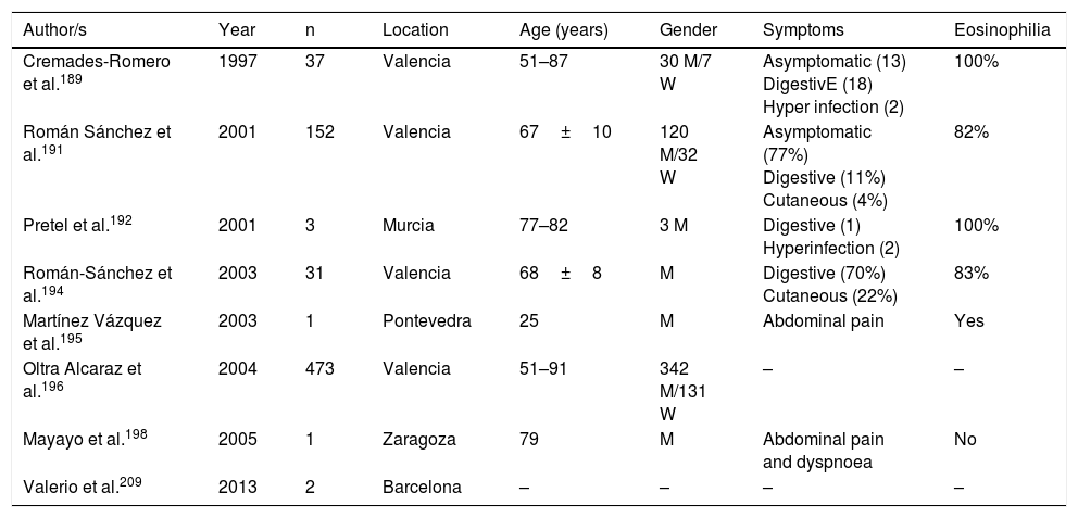
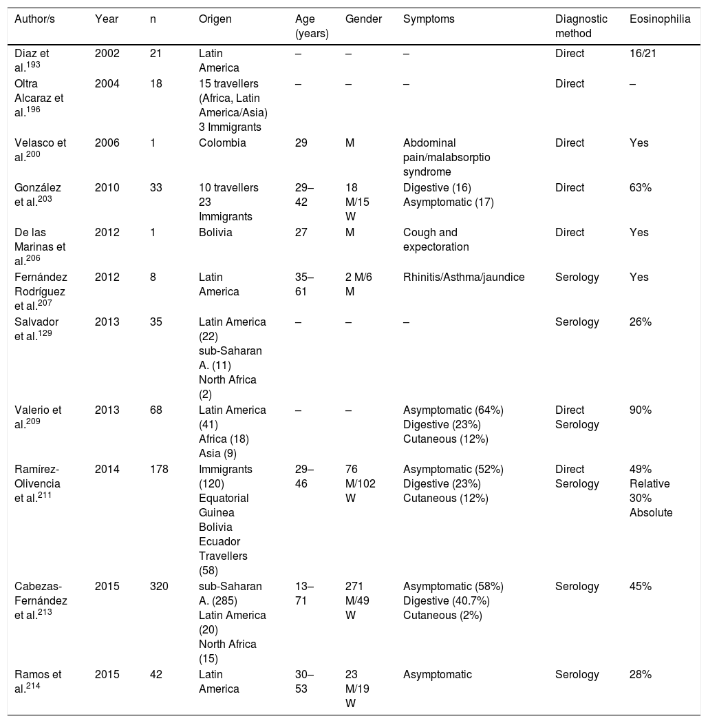
Strongyloidosis is a major parasitic disease in Spain. Analysis of isolated cases and patient series reveals several interesting characteristics.129,186–205,207–213 Firstly, there is the difficulty of completing precise diagnosis of the nematode. The standard co-proparasitological study detects a minimum proportion of cases,3,132,167 a figure which increases on using more specific techniques (e.g. the Baermann concentration test, the Harada-Mori technique and the Koga agar plate culture). However, several Spanish strongyloidosis series are based on serological diagnosis, with inherent limitations to this technique.133,211 Strongyloidosis in Spain is a disease which mainly affects adults, although there have been isolated cases in children.199 One essential aspect in strongyloidosis is the differentiation of clinical symptoms between immunocompetent and immunodepressed patients. In the immunocompetent person, this nematode is usually asymptomatic or the course of the disease is with one or several of the data of the eosinophilia-diarrhoea-skin lesion triad. However, in the immunodepressed patient the eosinophilia disappears and serious systemic symptoms may present, such as the systemic infection by intestinal microorganisms led by Strongyloids. We are interested in pointing out that the main forms of immunodepression associated with the hyperinfection syndrome correspond to the use of corticosteroids and other immunosuppressants, whilst the association with the HIV infection is infrequent and on many occasions linked to other risk factors (e.g. corticoid steroids or HTLV-I infection)208 (Table 8). From an epidemiological viewpoint in Spain there are 2 strongyloidosis patterns: the autochthonous and the imported forms. At present, strongyloidosis continues to be an autochthonous disease, which is why it is included in the differential diagnosis of any patient with digestive or “allergic” symptoms. Symptoms (Table 9). Notwithstanding, most cases are sporadic,195,198,209 with the exception of a specific area in Valencia (Gandía and Oliva), where an accumulation of patients with a well defined profile arose: male adults with a compatible professional history.189,191,194,215 Imported strongyloidosis however is a disease mainly described in immigrants, and exceptionally in travellers203,212 (Table 10). Unlike other imported helminths, where sub-Saharan origin predominates, a high number of cases of strongyloidosis come from Latin America.129,193,196,200,206,208,212–214 Most cases are asymptomatic, and it therefore seems reasonable to include this disease in the screening of immigrants from the before-mentioned geographical areas. In the remainder of cases, the normal symptoms are digestive and to a lesser extent, cutaneous. We should point out the detection of several cases in patients with allergic symptoms,206,207 especially of Latin American origin since the use of corticoid steroids in this context leads to hyperinfection syndrome. Eosinophilia in immunocompetent patients (autochthonous or immigrants) is highly variable (Tables 9 and 10).

Table 8.

Strongyloidosis and risk factors.

Author/s  Year  Risk factor  Age (years)  Gender  Symptoms  Eosinophilia 
Batista et al.187  1992  HIV infection  35  Gastroenteritis
Pneumonia 
No 
Cremades-Romero et al.188  1996  Use of corticosteroids  70  Gastritis/Abdominal pain  Yes 
Olmos et al.197  2004  Infección HIV
Use of immunosuppressands 
58  Bilateral pneumonia  Yes 
Beltrán Catalán et al.201  2009  Kidney trasplant  51/55  Diarrhoea  Yes 
Rodriguez-Hernandez et al.202  2009  Liver transplant  67  Bilateral pneumonia  Yes 
Argelich et al.204  2011  Use of corticosteroids  69  Meningitis by E. coli  No 
Villena-Ruiz et al.205  2012  HIV infection
Use of corticosteroids 
37  Bilateral pneumonia  No 
Salvador et al.129  2013  35  HIV infection  –  –  Asymptomatic  9/35 
Izquierdo et al.210  2013  Stem cell transplant  36  Bilateral pneumonia  No 

W: woman; M: man; –: no data.

Table 9.

Autochthonous strongyloidosis in Spain.

Author/s  Year  Location  Age (years)  Gender  Symptoms  Eosinophilia 
Cremades-Romero et al.189  1997  37  Valencia  51–87  30 M/7 W  Asymptomatic (13)
DigestivE (18)
Hyper infection (2) 
100% 
Román Sánchez et al.191  2001  152  Valencia  67±10  120 M/32 W  Asymptomatic (77%)
Digestive (11%)
Cutaneous (4%) 
82% 
Pretel et al.192  2001  Murcia  77–82  3 M  Digestive (1)
Hyperinfection (2) 
100% 
Román-Sánchez et al.194  2003  31  Valencia  68±Digestive (70%)
Cutaneous (22%) 
83% 
Martínez Vázquez et al.195  2003  Pontevedra  25  Abdominal pain  Yes 
Oltra Alcaraz et al.196  2004  473  Valencia  51–91  342 M/131 W  –  – 
Mayayo et al.198  2005  Zaragoza  79  Abdominal pain and dyspnoea  No 
Valerio et al.209  2013  Barcelona  –  –  –  – 

W: woman; M: man; –: no data.

Table 10.

Imported strongyloidosis in Spain.

Author/s  Year  Origen  Age (years)  Gender  Symptoms  Diagnostic method  Eosinophilia 
Diaz et al.193  2002  21  Latin America  –  –  –  Direct  16/21 
Oltra Alcaraz et al.196  2004  18  15 travellers (Africa, Latin America/Asia) 3 Immigrants  –  –  –  Direct  – 
Velasco et al.200  2006  Colombia  29  Abdominal pain/malabsorptio syndrome  Direct  Yes 
González et al.203  2010  33  10 travellers
23 Immigrants 
29–42  18 M/15 W  Digestive (16) Asymptomatic (17)  Direct  63% 
De las Marinas et al.206  2012  Bolivia  27  Cough and expectoration  Direct  Yes 
Fernández Rodríguez et al.207  2012  Latin America  35–61  2 M/6 M  Rhinitis/Asthma/jaundice  Serology  Yes 
Salvador et al.129  2013  35  Latin America (22)
sub-Saharan A. (11)
North Africa (2) 
–  –  –  Serology  26% 
Valerio et al.209  2013  68  Latin America (41)
Africa (18)
Asia (9) 
–  –  Asymptomatic (64%)
Digestive (23%)
Cutaneous (12%) 
Direct
Serology 
90% 
Ramírez-Olivencia et al.211  2014  178  Immigrants (120)
Equatorial Guinea
Bolivia
Ecuador
Travellers (58) 
29–46  76 M/102 W  Asymptomatic (52%)
Digestive (23%)
Cutaneous (12%) 
Direct
Serology 
49% Relative
30% Absolute 
Cabezas-Fernández et al.213  2015  320  sub-Saharan A. (285)
Latin America (20)
North Africa (15) 
13–71  271 M/49 W  Asymptomatic (58%)
Digestive (40.7%)
Cutaneous (2%) 
Serology  45% 
Ramos et al.214  2015  42  Latin America  30–53  23 M/19 W  Asymptomatic  Serology  28% 

W: woman; M: man; –: no data.

We only found one reference in the Spanish literature to the infection by Capillaria philipinensis.216 This disease is not limited to the Philippines, but is also present in Far and Middle Eastern countries, or South American, and its detection is therefore possible in imported cases. Generally capilarosis is a diarrhoea process with vomiting, although patients without treatment for several months may die due to the loss of electrolytes, or the sepsis associated with secondary bacterial infection (auto-infection processes).

Filariasis

Filariasis in Spain presents 2 different patterns: autochthonous (of cosmopolitan distribution) where there are no microfilariae, and imported forms, characterised by the presence of microfilariae in blood, skin or eyeball.

The cosmopolitan forms are caused mainly by 2 species of Dirofilaria (Dirofilaria immitis and Dirofilaria repens).217–220 In Spain, the 2 regions where this parasitism has been reported are Salamanca and Gran Canaria. The most common form of infection is vectoral transmission by different species of mosquitoes (Aedes spp., Anopheles spp. and Culex spp.) from infected mammals (mainly dogs). The infection takes places through immature worms, is generally asymptomatic and occasionally presents with subcutaneous, pulmonary (persitent or transitory) and ocular nodules. Eosinophilia is exceptional in these cases.

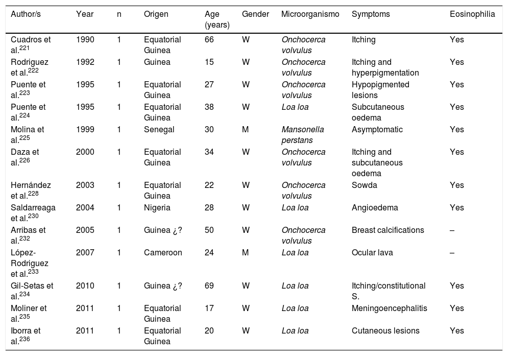
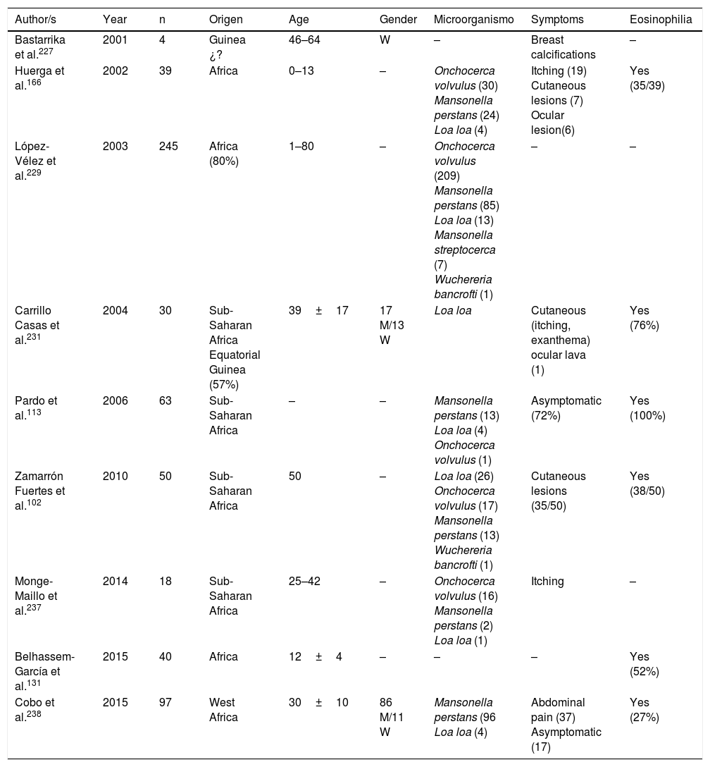
Filariasis associated with the presence of microfilariae in blood or tissues are in all cases imported diseases in Spain.102,113,131,166,221–238 Practically all of them are described in African patients, particularly sub-Saharan and with a clear predominance in West Africa (mainly Equatorial Guinea). The imported cases correspond mainly to immigrants, although they have also been described in travellers.102,237 Analysis of published cases (Tables 11 and 12) does not show any significant differences in the age of detection (1–80 years) or gender of patients. The 3 main imported filariasis are mansonelosis by Mansonella perstans, loaosis by Loa loa and oncocercosis by Onchocerca volvulus. Detection of Wuchereria bancrofti and Mansonella streptocerca is anecdotal,229 and there are no published case of infections by Brugia malayi, Brugia timori or Mansonella ozzardi. These data are subject to complexity in the diagnosis of these nematodes. Direct parasitological studies (blood smears and/or Knott test for the detection of microfilaremia, or “cutaneous pinching” in infection by O. volvulus and M. streptocerca) are very specific, but present limited sensitivity. Moreover, serological technique are very sensitive but have inherent limitations (e.g. cross over reactions with other helminths, no differentiation between active and past infections, etc.). In fact, the use of molecular biological techniques enables the detection of a large number of cases of undiagnosed loaosis from standard techniques.239,240 Furthermore, temporary evolution of imported filariasis presents a clear pattern, with a progressively lower number of cases of oncocercosis (very possibly related to control measures ing endemic countries, such as Equatorial Guinea) and a progressively lower number of case of mansonelosis (possibly linked to the screening of these entities in immigrants). Clinical symptoms are highly variables, with a large number of cases being asymptomatic. In symptomatic cases, common signs of these nematodes are: cutaneous (e.g. itching, exanthema, nodules) and ocular. It is therefore of interest to point out the presence of atypical manifestations, such as breast calcifications.227,232 The present of eosinophilia is very common, although its absence does not exclude diagnosis.

Table 11.

Imported Filariasis in Spain (cases).

Author/s  Year  Origen  Age (years)  Gender  Microorganismo  Symptoms  Eosinophilia 
Cuadros et al.221  1990  Equatorial Guinea  66  Onchocerca volvulus  Itching  Yes 
Rodriguez et al.222  1992  Guinea  15  Onchocerca volvulus  Itching and hyperpigmentation  Yes 
Puente et al.223  1995  Equatorial Guinea  27  Onchocerca volvulus  Hypopigmented lesions  Yes 
Puente et al.224  1995  Equatorial Guinea  38  Loa loa  Subcutaneous oedema  Yes 
Molina et al.225  1999  Senegal  30  Mansonella perstans  Asymptomatic  Yes 
Daza et al.226  2000  Equatorial Guinea  34  Onchocerca volvulus  Itching and subcutaneous oedema  Yes 
Hernández et al.228  2003  Equatorial Guinea  22  Onchocerca volvulus  Sowda  Yes 
Saldarreaga et al.230  2004  Nigeria  28  Loa loa  Angioedema  Yes 
Arribas et al.232  2005  Guinea ¿?  50  Onchocerca volvulus  Breast calcifications  – 
López-Rodriguez et al.233  2007  Cameroon  24  Loa loa  Ocular lava  – 
Gil-Setas et al.234  2010  Guinea ¿?  69  Loa loa  Itching/constitutional S.  Yes 
Moliner et al.235  2011  Equatorial Guinea  17  Loa loa  Meningoencephalitis  Yes 
Iborra et al.236  2011  Equatorial Guinea  20  Loa loa  Cutaneous lesions  Yes 

W: woman; M: man; –: no data.

Table 12.

Imported filariasis in Spain (series).

Author/s  Year  Origen  Age  Gender  Microorganismo  Symptoms  Eosinophilia 
Bastarrika et al.227  2001  Guinea ¿?  46–64  –  Breast calcifications  – 
Huerga et al.166  2002  39  Africa  0–13  –  Onchocerca volvulus (30)
Mansonella perstans (24)
Loa loa (4) 
Itching (19)
Cutaneous lesions (7)
Ocular lesion(6) 
Yes (35/39) 
López-Vélez et al.229  2003  245  Africa (80%)  1–80  –  Onchocerca volvulus (209)
Mansonella perstans (85)
Loa loa (13)
Mansonella streptocerca (7)
Wuchereria bancrofti (1) 
–  – 
Carrillo Casas et al.231  2004  30  Sub-Saharan Africa
Equatorial Guinea (57%) 
39±17  17 M/13 W  Loa loa  Cutaneous (itching, exanthema) ocular lava (1)  Yes (76%) 
Pardo et al.113  2006  63  Sub-Saharan Africa  –  –  Mansonella perstans (13)
Loa loa (4)
Onchocerca volvulus (1) 
Asymptomatic (72%)  Yes (100%) 
Zamarrón Fuertes et al.102  2010  50  Sub-Saharan Africa  50  –  Loa loa (26)
Onchocerca volvulus (17) Mansonella perstans (13) Wuchereria bancrofti (1) 
Cutaneous lesions (35/50)  Yes (38/50) 
Monge-Maillo et al.237  2014  18  Sub-Saharan Africa  25–42  –  Onchocerca volvulus (16)
Mansonella perstans (2)
Loa loa (1) 
Itching  – 
Belhassem-García et al.131  2015  40  Africa  12±–  –  –  Yes (52%) 
Cobo et al.238  2015  97  West Africa  30±10  86 M/11 W  Mansonella perstans (96
Loa loa (4) 
Abdominal pain (37)
Asymptomatic (17) 
Yes (27%) 

W: woman; M: man; –: no data.

Tissue

The 4 main tissue nematodosis reported in Spain are: triquinelosis, anisakidosis, toxocarosis and gnatosthomosis.

Infections produced by tissue nematodes of the genus Trichinella are autochthonous parasites, well referenced in the Spanish literature and in exceptional cases as an imported pathology.241–256 The 2 main species involved in the clinical cases described in Spain are Trichinella spiralis and Trichinella britovi. The common form of contagion is ingestion of raw meat or not well cooked meant of infected pigs and wild boar, which implies that the cases published are grouped around outbreaks. After the control of the domestic cycle in Spain (pigs), wild animals, like wild boars, have been the origin for most of the recent outbreaks. In any case, the rate of cases of this parasitism is increasingly lower, probably due to veterinary surveillance prior to consumption of game. The main outbreaks (indicated in Fig. 2) are concentrated on mountainous areas: (1) Cantabrian and Pyrenean mountain ranges; (2) Iberian range; (3) Central range; (4) Toledo mountain range and (5) Baetic rangea. During the last 25 years no autochthonous cases have been reported in Galicia, Murcia or the Canary island community. Clinical symptoms of the disease come from the tissue invasion of the parasite and the immunological response this triggers. The acute forms include, in variable proportions in each outbreak, the following data: myalgias, fever, exanthema, diarrhoea and palpebral oedema. Atypical forms have also been described, such as thoracic muscle calcification.254 Eosinophilia and the raising of quinase creatinine are common lab data in the described cases.

Fig. 2.

Number of outbreaks of triquinelosis in Spain by autonomous community (1990–2015).

(0.24MB).

Since 1991, the description of isolated cases and clinical series of anisakis has been constant in Spain.257–287 Humans are infected by ingestion of fresh, raw or little cooked fish (e.g. by microwaves268), with the most common epidemiological background being the consumption of anchovies and in some cases, cebiche.284 Practically all cases correspond to acquired autochthonous infections and are mainly reported by autonomous communities, in the central area of the peninsula (Fig. 3). The most common agents are nematodes of the genus Anisakis (particularly Anisakis simplex) and to a lesser extent Pseudoterranova decipiens.258 The pathological consequences of the infection by these nematodes is apparent in3 different patterns: digestive, allergic or mixed. The digestive forms may affect several parts of the same, leading to compromise of the gastroduodenal region,ileal region, caecum and colon. Clinical symptoms depend on 2 complementary mechanisms: direct aggression by the nematode and the response of local hypsensitivity to the same. In the upper digestive tract the most common signs are acute epigastric pain after the intake of fish, frequently associated with allergic symptoms. However, when the lower digestive tracts involved abdominal pain presents with characteristics which are indistinguishable from an acute appendicitis or intestinal obstruction. Other atypical manifestations have also been described, such as splenic rupture286 or the appearance of an abdominal mass.271 Allergic signs are highly variables, both in their association with intestinal symptoms, and in their severity (from simple cutaneous forms to anaphylaxis).260,261,284 Other uncommon signs of hypersensitivity to these nematodes, described in Spain, are the appearance of a nephrotic syndrome281 and gingivoestomatitis.277 Eosinophilia is an inconstant finding in this parasitism (4–41%), and it depends on the before-mentioned clinical forms.

Fig. 3.

Provinces with few published cases of anisakis (1990–2015).

(0.27MB).

Data on toxocarosis, produced by Toxocara spp. are scarce in Spain and complex to interpret.287–295 The studies on seroprevalence of this infection, conducted in the nineties in several areas of the country, show a high rate of positivity, with figures of up to 66% in children from low socio-economic classes in Guipúzcoa, 3.4% in the general population of the Canary Island Community and between 17% and 32% in Galicia.288,292 These data should be further studied, since the etiological diagnosis of toxocarosis is based on serology, which displays cross-over reactivity with other nematodes such as Anisakis spp.294 The few reported clinical cases of toxocarosis correspond to both imported and autochthonous forms295 and within the latter, to ocular toxocarosis290 and visceral larva migrans.289 In the cases with visceral manifestations the presence of eosinophilia is common.291

Finally, gnathostomosis, mainly produced by Gnathostoma spinigerum, is a rare tissue nematodosis, but occasionally reported in Spain.296–300 This helminthosis appears as a consequence of food consumption (e.g. raw fish, frogs, snakes) and usually the sign are cutaneous lesions (similar to the cutaneous lava migrans) and in severe cases, myeloradiculitis or a radiculomyelo-meningoencephalitis.298,299 In general, it is a disease which is imported after travelling to Latin America297,298 and Asia (South East Asia and China),297,298,300 although 2 autochthonous cases have also been diagnosed in women from Granada who had not travelled to the tropics.296

Conclusions

To conclude, helminthosis (autochthonous or imported, in travellers or immigrants, with or without immunosuppression) is a major problem in the Spanish population, both with regards to its prevalence and medical consequences. Association with eosinophilia (absolute or relative) presents great variability and is dependent upon a number of factors. For this reason, knowledge of the current situation may help etiological diagnosis of helminthosis. An appropriate therapeutic approach is required, with the avoidance of “empirical” attitudes that may be unsatisfactory, inappropriate or even harmful.301

Financing

The authors declare they have not received any financing for carrying out this study.

Conflict of interests

The authors have no conflict of interests to declare.

References
[1]
J.L. Pérez Arellano, J. Pardo, M. Hernández Cabrera, C. Carranza, A. Angel-Moreno, A. Muro.
Manejo práctico de una eosinofilia.
An Med Intern (Madrid), 21 (2004), pp. 244-252
[2]
A. Vilajeliu Balagué, P. de Las Heras Prat, G. Ortiz-Barreda, M.J. Pinazo Delgado, J. Gascón Brustenga, A. Bardají Alonso.
Parasitosis importadas en población inmigrante en España.
Rev Esp Salud Publica, 88 (2014), pp. 783-802
[3]
I. Novo-Veleiro, A.M. Martín Sánchez, R. Elcuaz-Romano, A. Muro, O. Afonso-Rodriguez, D. García Bardeci, et al.
Parasitosis en Gran Canaria (España): estudio prospectivo multicéntrico durante un año.
Rev Ibero-Latinoam Parasitol, 71 (2012), pp. 34-41
[4]
J.M. Soriano, G. Domènech, M.C. Martínez, J. Mañes, F. Soriano.
Intestinal parasitic infections in hosted Saharawi children.
Trop Biomed, 28 (2011), pp. 557-562
[5]
J.M. Paricio Talayero, L. Santos Serrano, A. Fernández Feijoo, M. Ferriol Camacho, F. Rodríguez Serrano, P. Brañas Fernández.
Examen de salud de niños de la República Árabe Saharaui Democrática de vacaciones en España.
An Esp Pediatr, 49 (1998), pp. 33-38
[6]
C. Martínez-Peinado, M.M. López-Perezagua, F.J. Arjona-Zaragozí, M.M. Campillo-Gallego.
Parasitosis en niña ecuatoriana.
Enferm Infecc Microbiol Clin, 24 (2006), pp. 207-208
[7]
D. Tena, M. Pérez Simón, C. Gimeno, M.T. Pérez Pomata, S. Illescas, I. Amondarain, et al.
Human infection with Hymenolepis diminuta: case report from Spain.
J Clin Microbiol, 36 (1998), pp. 2375-2376
[8]
C. Martínez-Carrasco, E. Berriatua, M. Garijo, J. Martínez, F.D. Alonso, R.R. de Ybáñez.
Epidemiological study of non-systemic parasitism in dogs in southeast Mediterranean Spain assessed by coprological and post-mortem examination.
Zoonoses Public Health, 54 (2007), pp. 195-203
[9]
F.J. Martínez-Moreno, S. Hernández, E. López-Cobos, C. Becerra, I. Acosta, A. Martínez-Moreno.
Estimation of canine intestinal parasites in Córdoba (Spain) and their risk to public health.
Vet Parasitol, 143 (2007), pp. 7-13
[10]
G. Miró, M. Mateo, A. Montoya, E. Vela, R. Calonge.
Survey of intestinal parasites in stray dogs in the Madrid area and comparison of the efficacy of three anthelmintics in naturally infected dogs.
Parasitol Res, 100 (2007), pp. 317-320
[11]
G. Miró, A. Montoya, S. Jiménez, C. Frisuelos, M. Mateo, I. Fuentes.
Prevalence of antibodies to Toxoplasma gondii and intestinal parasites in stray, farm and household cats in Spain.
Vet Parasitol, 126 (2004), pp. 249-255
[12]
J. Pardo, A. Muro, I. Galindo, M. Cordero, A. Carpio, M. Siles-Lucas.
Hidatidosis en la provincia de Salamanca: ¿Debemos bajar la guardia?.
Enferm Infecc Microbiol Clin, 23 (2005), pp. 266-269
[13]
F.A. Rojo-Vazquez, J. Pardo-Lledias, M. Francos-von Hunefeld, M. Cordero-Sanchez, R. Alamo-Sanz, A. Hernandez-Gonzalez, et al.
Cystic echinococcosis in Spain: current situation and relevance for other endemic areas in Europe.
PLoS Negl Trop Dis, 5 (2011), pp. e893
[14]
A. Lopez-Bernus, M. Belhassen-García, A. Carpio-Perez, L. Perez del Villar, A. Romero-Alegria, V. Velasco-Tirado, et al.
Is cystic echinoccocosis re-emerging in western Spain?.
Epidemiol Infect, 8 (2015), pp. 1-7
[15]
C. Armiñanzas, M. Gutiérrez-Cuadra, M.C. Fariñas.
Hidatidosis: aspectos epidemiológicos, clínicos, diagnósticos y terapéuticos.
Rev Esp Quimioter, 28 (2015), pp. 116-124
[16]
M.A. Asencio, O. Herraez, J.M. Tenias, E. Garduño, M. Huertas, R. Carranza, et al.
Seroprevalence survey of zoonoses in Extremadura, southwestern Spain, 2002–2003.
Jpn J Infect Dis, 68 (2015), pp. 106-112
[17]
A.M. Calvo, J.M. Cires, S. Montón, H. Sarasibar, P. Lasanta, F.J. Artázcoz.
Tumoración inguinal: una forma de presentación de la hidatidosis ósea. A propósito de un caso.
An Sist Sanit Navar, 30 (2007), pp. 475-479
[18]
J. Hueto Pérez de Heredia, M. Pérez de las Casas, J. Domínguez del Valle, E. Vila Mayo, M. Urquía Braña, M. Gómez Dorronsoro.
Hidatidosis torácica. Nuestra experiencia en los últimos quince años.
Rev Clin Esp, 199 (1999), pp. 13-17
[19]
H. Carabin, F.J. Balsera-Rodríguez, J. Rebollar-Sáenz, C.T. Benner, A. Benito, J.C. Fernández-Crespo, et al.
Cystic echinococcosis in the Province of Álava, North Spain: the monetary burden of a disease no longer under surveillance.
PLoS Negl Trop Dis, 8 (2014), pp. e3069
[20]
M. Echenique-Elizondo, J.A. Amondarain Arratíbel.
Hydatid disease of the pancreas.
JOP, 5 (2004), pp. 51-52
[21]
M.M. Echenique Elizondo, J.A. Amondarain Arratibel.
Muscular hydatid disease.
J Am Coll Surg, 197 (2003), pp. 162
[22]
C.A. Amado-Diago, M. Gutiérrez-Cuadra, C. Armiñanzas, F. Arnaiz de las Revillas, M. Gómez-Fleitas, M.C. Fariñas.
Equinococosis: epidemiología, clínica y resultados en una pano-rámica de 15 años.
Rev Clin Esp, 215 (2015), pp. 380-384
[23]
J. Moreno, C.J. Téllez, F.J. Pardo.
Casos de hidatidosis en el Departamento de Salud 2 de la Comunidad Valenciana.
Rev Esp Quimioter, 22 (2009), pp. 62-67
[24]
J.M. Ramia, R. de-la-Plaza, J. Quiñones, F. Adel, C. Ramiro, J. García-Parreño.
Frank intrabiliary rupture in liver hydatidosis located in the hilar plate: a surgical challenge.
Dig Surg, 30 (2013), pp. 439-443
[25]
J.M. Suárez Grau, M.A. Gómez Bravo, J.M. Alamo Martínez, C. Rubio Cháves, L.M. Marín Gómez, G. Suárez Artacho, et al.
Giant hydatid cyst involving the right hepatic lobe.
Rev Esp Enferm Dig, 101 (2009), pp. 133-135
[26]
E. Hernando, J.L. García Calleja, E. Córdoba, L. Lahuerta, F. del Río, V. Ferreira.
Hidatidosis hepática. Revisión de una serie de 677 pacientes tratados quirúrgicamente.
Gastroenterol Hepatol, 19 (1996), pp. 140-145
[27]
J.M. Ramia, I. Poves, C. Castellón, L. Diez-Valladares, C. Loinaz, A. Serrablo, et al.
Radical laparoscopic treatment for liver hydatidosis.
World J Surg, 37 (2013), pp. 2387-2392
[28]
C. Guarner-Argente, C. Gomez-Oliva, M. Poca, S. Sainz, F.G. Marinello, C. Villanueva, et al.
Cholangitis caused by biliary hydatidosis.
Gastrointest Endosc, 72 (2010), pp. 1264-1265
[29]
J. Molina Infante, M. Fernández Bermejo, E. Martín Noguerol, B. Pérez Gallardo.
Biliary hydatidosis.
Rev Esp Enferm Dig, 101 (2009), pp. 136-138
[30]
A. García Ruiz de Gordejuela, L. Lladó, J. Torras, E. Ramos, A. Rafecas.
Hidatidosis hepática gigante con fístula quistopleural e hidatosis pleural.
Cir Esp, 82 (2007), pp. 177-179
[31]
G. Ramos, A. Orduña, M. García-Yuste.
Hydatid cyst of the lung: diagnosis and treatment.
World J Surg, 25 (2001), pp. 46-57
[32]
R. Burgos, A. Varela, E. Castedo, J. Roda, C.G. Montero, S. Serrano, et al.
Pulmonary hydatidosis: surgical treatment and follow-up of 240 cases.
Eur J Cardiothorac Surg, 16 (1999), pp. 628-634
[33]
R. Arroyo Pérez, J.L. Echarte Pazos, A. Aguirre Tejedo, L. Pijuan Andújar, E. Lerma Chippirraz, E. Terrer Galera.
Derrame pleural derecho, compatible con empiema, en mujer de 82 años.
Rev Clin Esp, 208 (2008), pp. 46-48
[34]
J.M. Ramia-Angel, A. Gasz, R. de la Plaza-Llamas, J. Quinones-Sampedro, E. Sancho, J. Garcia Parreno.
Hidatidosis of the spleen.
Pol Przegl Chir, 83 (2011), pp. 271-275
[35]
M. Prieto, T. Marquina, P. Mifsut, T. Moreno.
Hidatidosis esplénica: 5 casos de esta rara localización.
Enferm Infecc Microbiol Clin, 29 (2011), pp. 634-635
[36]
M. Fernández-Ruiz, J.M. Guerra-Vales, A.B. Enguita-Valls, J. Vila-Santos, F.J. García-Borda, C. Morales-Gutiérrez.
Splenic hydatid cyst, a rare location of extrahepatic echino-coccosis: report of six cases.
Eur J Intern Med, 19 (2008), pp. e51-e53
[37]
J.C. Angulo, M. Sanchez-Chapado, A. Diego, J. Escribano, J.C. Tamayo, L. Martin.
Renal echinococcosis: clinical study of 34 cases.
J Urol, 157 (1997), pp. 787-794
[38]
M.E. Alonso García, P. Suárez Mansilla, P. Mora Cepeda, E. Bayón Álvarez, C. Alvarez Colomo, J.I. González Martín.
Ovarian hydatid disease.
Arch Gynecol Obstet, 289 (2014), pp. 1047-1051
[39]
J.F. Ruiz-Rabelo, M. Gomez-Alvarez, J. Sanchez-Rodriguez, S. Rufian Peña.
Complications of extrahepatic echinococcosis: fistulization of an adrenal hydatid cyst into the intestine.
World J Gastroenterol, 14 (2008), pp. 1467-1469
[40]
N. Senarriaga Ruiz de la Illa, A. Loizaga Iriarte, I. Iriarte Soldevilla, I. Lacasa Viscasillas, M. Unda Urzaiz.
Hidatidosis pelviana como ejemplo de masas pelvianas de etiología dudosa.
Actas Urol Esp, 33 (2009), pp. 1129-1132
[41]
M. Colomina Pascual, V. Giner Galvan, A. Domenech Iglesias, M. Planelles Asensio.
Hidatidosis abdominal diseminada.
Med Clin (Barc), 136 (2011), pp. 182
[42]
A. Cecconi, L. Maroto, I. Vilacosta, M. Luaces, L. Ortega, N. Escribano, et al.
Acute pericarditis secondary to hydatid cyst rupture: diagnosis by multimodality imaging.
Circulation, 128 (2013), pp. 2073-2074
[43]
M. Díaz-Menéndez, J.A. Pérez-Molina, F.F. Norman, A. Pérez-Ayala, B. Monge-Maillo, P.Z. Fuertes, et al.
Management and outcome of cardiac and endovascular cystic echinococcosis.
PLoS Negl Trop Dis, 6 (2012), pp. e1437
[44]
E. Perez-David, M.A. Fernandez, F. Fernández-Avilés.
Isolated cardiac hydatidosis.
Eur Heart J, 28 (2007), pp. 2829
[45]
J.G. Tejada, J. Saavedra, L. Molina, A. Forteza, C. Gomez.
Hydatid disease of the interventricular septum causing pericardial effusion.
Ann Thorac Surg, 71 (2001), pp. 2034-2035
[46]
R. Seijas, J.M. Catalán-Larracoechea, O. Ares-Rodríguez, N. Joshi, J.P. de la Fuente, M. Pérez-Domínguez.
Primary hydatid cyst of skeletal muscle affecting the knee: a case report.
Arch Orthop Trauma Surg, 129 (2009), pp. 39-41
[47]
F. García-Alvarez, J. Torcal, J.C. Salinas, A. Navarro, I. García-Alvarez, M. Navarro-Zorraquino, et al.
Musculoskeletal hydatid disease: a report of 13 cases.
Acta Orthop Scand, 73 (2002), pp. 227-231
[48]
M. Belhassen-Garcia, A. Carpio-Perez, J.F. Blanco, V. Velasco-Tirado, J. Pardo-Lledías.
Recurrent spinal echinococcosis.
Int J Infect Dis, 15 (2011), pp. e435-e436
[49]
A. Herrera, A.A. Martínez, J. Rodríguez.
Spinal hydatidosis.
Spine (Phila Pa 1976), 30 (2005), pp. 2439-2444
[50]
S. Gómez-Senent, N. Manceñido-Marcos, J.C. Erdozain-Sosa, J. Segura-Cabral.
Hidatidosis cutánea.
Med Clin (Barc), 127 (2006), pp. 480
[51]
G. Gironé, C. Mateo, V. Gaya, J. Usó, C. Mínguez, B. Roca, et al.
Admissions for imported and non-imported parasitic diseases at a General Hospital in Spain: a retrospective analysis.
Travel Med Infect Dis, 13 (2015), pp. 322-328
[52]
J.V. Arcos Machancoses, A. Parra Llorca, J. Martín Benlloch, A. Ortí Martín.
Waterlily sign.
[53]
G. Herráiz Gastesi, C. Bonnet Carrón, B. Madariaga Ruiz, E. Freile García, M. Gracia Casanova.
Hidatidosis pulmonar bilateral en paciente inmigrante.
An Pediatr (Barc), 76 (2012), pp. 361-362
[54]
M.E. Jiménez-Mejías, J.C. Alarcón-Cruz, F.J. Márquez-Rivas, J. Palomino-Nicás, J.M. Montero, J. Pachón.
Orbital hydatid cyst: treatment and prevention of recurrences with albendazole plus praziquantel.
J Infect, 41 (2000), pp. 105-107
[55]
S. Sanjuán Rodríguez, J.M. Morán Penco, P. Rincón Rodera, G. González Díez.
Abdomen agudo secundario a quiste hidatídico pancreático.
Cir Pediatr, 18 (2005), pp. 36-38
[56]
P.L. Minciullo, A. Cascio, A. David, L.M. Pernice, G. Calapai, S. Gangemi.
Anaphylaxis caused by helminths: review of the literature.
Eur Rev Med Pharmacol Sci, 16 (2012), pp. 1513-1518
[57]
C. Novo Alonso, J. Garín Ferreira, A. Rodríguez Fernández, E. Hernández Antequera, J.A. Zapata Blanco, G. Vaquero Gajate.
Hidatidosis alveolar humana.
Rev Esp Enferm Dig, 84 (1993), pp. 127-129
[58]
M.A. Arrechea Irigoyen, A. Córdoba Iturriagagoitia, M.A. Tuñón Álvarez, M.L. Gómez Dorronsoro, J.M. Martínez-Peñuela Virseda.
Equinococosis alveolar humana. Presentación de un caso.
Rev Esp Patol, 41 (2008), pp. 203-206
[59]
L. Iglesias Oliva, G. Pérez Roji, E. Pérez-Llantada Amunarriz, A. González Mandly, L. García-Castrillo Riesco.
Múltiples calcificaciones musculares en un varón de 68 años de edad.
Rev Clin Esp, 202 (2002), pp. 669-671
[60]
I. Corral, C. Quereda, A. Moreno, R. López-Vélez, J. Martínez-San-Millán, A. Guerrero, et al.
Intramedullary cysticercosis cured with drug treatment. A case report.
Spine (Phila Pa 1976), 21 (1996), pp. 2284-2287
[61]
J.M. Fernández-Gómez, J.L. García-Garmendia, J.M. López-Domínguez, J.L. Casado-Chocán.
Neurocisticercosis y crisis convulsivas.
Rev Neurol, 26 (1998), pp. 1072-1073
[62]
C. Font Puig, J.A. Ruiz Postigo, C. Muñoz Batet, F. Pardós Arnal, M. Corachan Cuyás.
Neurocisticercosis en España. A propósito de 4 casos observados en pacientes inmigrados de países endémicos.
An Med Intern, 16 (1999), pp. 89-91
[63]
F. Castellanos, I. Montes, L.F. Porras, E. Peragallo, J. Ampuero, R. Rueda.
Quistes subaracnoideos gigantes por neurocisticercosis: a propósito de dos casos observados en un área rural de Extremadura.
Rev Neurol, 30 (2000), pp. 433-435
[64]
S. Terraza, T. Pujol, J. Gascón, M. Corachán.
Neurocisticercosis: ¿una enfermedad importada.
Med Clin (Barc), 116 (2001), pp. 261-263
[65]
G. Rodríguez-Sánchez, F. Castellanos-Pinedo, J. Giménez-Pando, M.T. Adeva-Bartolomé, F. Zancada-Díaz.
Hidrocefalia y quiste subaracnoideo por neurocisticercosis. Un nuevo caso en una zona rural de Extremadura.
Rev Neurol, 34 (2002), pp. 348-351
[66]
C. Roca, J. Gascón, B. Font, T. Pujol, M.E. Valls, M. Corachán.
Neurocysticercosis and population movements: analysis of 23 imported cases in Spain.
Eur J Clin Microbiol Infect Dis, 22 (2003), pp. 382-384
[67]
R. Cañizares, P. Roig, A. Esparcia, A. Zorraquino, V. Ortiz de la Tabla, J. Merino.
Crisis convulsiva en varón joven.
Rev Clin Esp, 203 (2003), pp. 601-603
[68]
R. Ortega-Herrera, M.E. Fernández-Segura, Y. Gómez de Travecedo, I. Calvo.
Inmigrante ecuatoriana con cefalea.
Enferm Infecc Microbiol Clin, 22 (2004), pp. 248-249
[69]
J.A. Llompart Pou, A. Gené, J.I. Ayestarán, C. Saus.
Neurocysticercosis presenting as sudden death.
Acta Neurochir (Wien), 147 (2005), pp. 785-786
[70]
A. Esquivel, F. Diaz-Otero, S. Gimenez-Roldan.
Growing frequency of neurocysticercosis in Madrid (Spain).
Neurologia, 20 (2005), pp. 116-120
[71]
P.E. Jiménez-Caballero, M. Mollejo Villanueva, C. Marsal Alonso, A. Álvarez Tejerina.
Síndrome de Bruns: descripción de un caso de neurocisticercosis con estudio anatomopatológico.
Neurología, 2 (2005), pp. 86-89
[72]
V. Barra Valencia, A. Moreno Elola-Olaso, Y. Fundora Suárez, J.C. Meneu Díaz, S.F. Jiménez de los Galanes, B. Pérez Saborido, et al.
Second case of neurocysticercosis in a patient with liver transplantation (first case in Spain): a case report.
Transplant Proc, 39 (2007), pp. 2454-2457
[73]
J.M. Ramos, M. Masia, S. Padilla, E. Bernal, A. Martin-Hidalgo, F. Gutiérrez.
Fatal infection due to larval cysts of cestodes (neurocysticercosis and hydatid disease) in human immunodeficiency virus (HIV) infected patients in Spain: report of two cases.
Scand J Infect Dis, 39 (2007), pp. 719-723
[74]
E. Guerra del Barrio, R. López Roger.
Lesión quística intracerebral en un paciente inmigrante.
Rev Clin Esp, 207 (2007), pp. 301-302
[75]
B. Sierra Bergua, J. Navarro Calzada, I. Sanjoaquín Conde, S. Santos Lasaosa.
Convulsión generalizada tónico-clónica en inmigrante.
Neurologia, 22 (2007), pp. 253-255
[76]
G. Más-Sesé, I. Vives-Piñera, A. Fernández-Barreiro, J.F. Martínez-Lage, E. Martínez-Salcedo, H. Alarcón-Martínez, et al.
Estudio descriptivo de neurocisticercosis en un hospital terciario.
Rev Neurol, 46 (2008), pp. 194-196
[77]
S. Ruiz, E. García-Vázquez, R. Picazo, A. Hernández, J.A. Herrero, J. Gómez.
La neurocisticercosis en Murcia.
Rev Clin Esp, 211 (2011), pp. 133-138
[78]
M.J. Aguilar-Amat, P. Martínez-Sánchez, J. Medina-Baez, E. Díez-Tejedor.
Síndrome de Bruns causado por neurocisticercosis intraventricular.
Med Clin (Barc), 137 (2011), pp. 43-46
[79]
De Anta Tejado, K.T. Pozo, C.B. Palomino, J.L. de Dios de Vega.
Psychiatric manifestations of neurocysticercosis in paediatric patients.
[80]
C. Frieiro-Dantas, R. Serramito-García, R.M. Reyes-Santías, M. Rico-Cotelo, A.G. Allut, M. Gelabert-González.
Neurocisticercosis pediátrica: a propósito de dos casos.
Rev Neurol, 56 (2013), pp. 86-90
[81]
S. Giménez-Roldán, F. Diaz, A. Esquivel.
Neurocisticercosis e inmigración.
Neurologia, 18 (2003), pp. 385-388
[82]
E. Alvarez-Rodríguez, R. Torres-Gárate, J. Cabello, C. Lozano Tonkin.
Neurocisticercosis en España.
Rev Clin Esp, 205 (2005), pp. 518
[83]
F.J. Jiménez-Jiménez, J.A. Molina-Arjona, A. Roldán-Montaud, A. Agullá, J. Santos, A. Fernández-Ballesteros.
Blepharospasm associated with neurocysticercosis.
Acta Neurol (Napoli), 14 (1992), pp. 56-59
[84]
S. Accomando, M. Caserta, A. Trizzino, G.M. Amato.
Two strange cases of hyper-eosinophilia and child's relapsing angio-oedema.
Pediatr Med Chir, 25 (2003), pp. 367-369
[85]
R.B. Singh, N.M. Pavithran, N. Bakshi.
Intraperitoneal rupture of cysticercosal cyst mimicking appendicular perforation.
Trop Doct, 36 (2006), pp. 180-181
[86]
P.L. Minciullo, E.V. Spagnolo, A. Cascio, G. Cardia, S. Gangemi.
Fatal anaphylactic shock and Taenia solium infestation: a possible link?.
Ann Allergy Asthma Immunol, 103 (2009), pp. 449-450
[87]
J. Colomina, J. Villar, G. Esteban.
Parasitación asintomática por Diphyllobothrium latum en un niño español de 3 años.
Med Clin (Barc), 118 (2002), pp. 279
[88]
A. Gil-Setas, A. Mazón, P. Pascual, H. Sagua.
Helmintiasis poco frecuente en nuestro medio en un varón de 71 años.
Enferm Infecc Microbiol Clin, 22 (2004), pp. 553-554
[89]
F. Marcos Sánchez, I. Albo Castaño, A. Viana Alonso, L. Caballero Sánchez-Robles.
Dolor abdominal en una mujer guineana de 21 años.
Rev Clin Esp, 205 (2005), pp. 459-460
[90]
J. Pastor-Valle, L.M. González, J.P. Martín-Clemente, F.J. Merino, B. Gottstein, T. Gárate.
Molecular diagnosis of diphyllobothriasis in Spain, most presumably acquired via imported fish, or sojourn abroad.
New Microbes New Infect, 2 (2014), pp. 1-6
[91]
J.G. Esteban, C. Muñoz-Antoli, M. Borras, J. Colomina, R. Toledo.
Human infection by a ‘fish tapeworm’, Diphyllobothrium latum, in a non-endemic country.
Infection, 42 (2014), pp. 191-194
[92]
A. Lo Presti, D.T. Aguirre, P. de Andrés, L. Daoud, J. Fortes, J. Muñiz.
Cerebral sparganosis: case report and review of the European cases.
Acta Neurochir (Wien), 157 (2015), pp. 1339-1343
[93]
M. Corachan, L. Ruiz, M.E. Valls, J. Gascon.
Schistosomiasis and the Dogon country (Mali).
Am J Trop Med Hyg, 47 (1992), pp. 6-9
[94]
M. Corachán, J. Almeda, T. Vinuesa, M.E. Valls, T. Mejías, P. Jou, et al.
Esquistosomiasis importada por viajeros españoles: estudio clínico-epidemiológico de 80 casos.
Med Clin (Barc), 108 (1997), pp. 721-725
[95]
R. Vilana, M. Corachán, J. Gascón, E. Valls, C. Bru.
Schistosomiasis of the male genital tract: transrectal sonographic findings.
J Urol, 158 (1997), pp. 1491-1493
[96]
R. Elcuaz, M. Armas, M. Ramírez, F.J. Noguera, M. Bolaños, I. Quiñones, et al.
Brote de esquistosomiasis en un grupo de viajeros procedentes de Burkina Fasso.
Enferm Infecc Microbiol Clin, 16 (1998), pp. 367-369
[97]
A. Bou, J. Gascón, M.E. Valls, M. Corachán.
Fiebre de Katayama en turistas españoles: análisis de 25 casos.
Med Clin (Barc), 116 (2001), pp. 220-222
[98]
C. Roca, X. Balanzó, J. Gascón, J.L. Fernández-Roure, T. Vinuesa, M.E. Valls, et al.
Comparative, clinico-epidemiologic study of Schistosoma mansoni infections in travellers and immigrants in Spain.
Eur J Clin Microbiol Infect Dis, 21 (2002), pp. 219-223
[99]
Y. Santos, E. Balliu, D. Villán.
Hematuria en un varón de 34 años.
Med Clin (Barc), 123 (2004), pp. 312-316
[100]
D. Alonso, J. Muñoz, J. Gascón, M.E. Valls, M. Corachan.
Failure of standard treatment with praziquantel in two returned travelers with Schistosoma haematobium infection.
Am J Trop Med Hyg, 74 (2006), pp. 342-344
[101]
M. de Górgolas, V. Casado, G. Renedo, J.F. Alen, M.L. Fernández Guerrero.
Nodular lung schistosomiais lesions after chemotherapy for dysgerminoma.
Am J Trop Med Hyg, 81 (2009), pp. 424-427
[102]
P. Zamarrón Fuertes, A. Pérez-Ayala, J.A. Pérez Molina, F.F. Norman, B. Monge-Maíllo, M. Navarro, et al.
Clinical and epidemiological characteristics of imported infectious diseases in Spanish travelers.
J Travel Med, 17 (2010), pp. 303-309
[103]
P. Rivas, S. Aguilar-Durán, M. Lago.
Lung nodules, fever, and eosinophilia in a traveler returning from Madagascar.
Am J Trop Med Hyg, 86 (2012), pp. 2-3
[104]
J.L. Perez-Arellano, M. Hernandez-Cabrera, A. Malet-Pintos-Fonseca, C. Carranza-Rodriguez, A.M. Martin-Sanchez.
Katayama's syndrome related to Schistosoma intercalatum in two travellers returning from Mali.
Am J Infect Dis, 8 (2012), pp. 128-131
[105]
J. Muñoz, E. Aldasoro, M.J. Pinazo, P. Arguis, J. Gascon.
Pulmonary infiltrates and eosinophilia in a 25-year-old traveler.
PLoS Negl Trop Dis, 7 (2013), pp. e2201
[106]
N. Martínez-Calle, I. Pascual, M. Rubio, R. Carias, J.L. del Pozo, J.R. Yuste.
Asymptomatic Schistosoma haematobium infection in a traveler with negative urine microscopy and late seroconversion presumably linked to artemisinin.
J Travel Med, 20 (2013), pp. 326-328
[107]
M. Corachan, M.E. Valls, J. Gascon, J. Almeda, R. Vilana.
Hematospermia: a new etiology of clinical interest.
Am J Trop Med Hyg, 50 (1994), pp. 580-584
[108]
F. Chaves, F. Dronda, A. González-López, C. López-Cabañas, M. de los Ríos.
Prurito y eosinofilia en un nativo de Guinea Ecuatorial.
Enferm Infecc Microbiol Clin, 10 (1992), pp. 547-548
[109]
J. Balasch, S. Martínez-Román, M. Creus, E. Campo, A. Fortuny, J.A. Vanrell.
Schistosomiasis: an unusual cause of tubal infertility.
Hum Reprod, 10 (1995), pp. 1725-1727
[110]
M.A. Gairi Burgués Mf, J. Bosch Muñoz, A. Llusá Parramon, A.R. Gomá.
Schistosoma haematobium como causa de hematuria.
An Esp Pediatr, 56 (2002), pp. 368-369
[111]
M. Rotger, T. Serra, M.G. de Cárdenas, A. Morey, M.A. Vicente.
Increasing incidence of imported schistosomiasis in Mallorca, Spain.
Eur J Clin Microbiol Infect Dis, 23 (2004), pp. 855-856
[112]
A.I. López-Calleja, L. Torres, M.J. Revillo, A. Clavel, P. Arazo.
Dolor cólico hipogástrico y eosinofilia en un paciente de Gambia.
Enferm Infecc Microbiol Clin, 21 (2003), pp. 315-316
[113]
J. Pardo, C. Carranza, A. Muro, A. Angel-Moreno, A.M. Martín, T. Martín, et al.
Helminth-related eosinophilia in African immigrants, Gran Canaria.
Emerg Infect Dis, 12 (2006), pp. 1587-1589
[114]
M.J. Donate Moreno, H. Pastor Navarro, J.M. Giménez Bachs, P. Carrión López, M. Segura Martín, A.S. Salinas Sánchez, et al.
Esquistosomiasis vesical, aportación de un caso y revisión de la literatura española.
Actas Urol Esp, 30 (2006), pp. 714-719
[115]
A.B. Campo-Esquisabel, F. Ledesma-Castaño, R. Salesa-Gutiérrez de Rozas.
Eosinofilia en inmigrante africano asintomático.
Enferm Infecc Microbiol Clin, 25 (2007), pp. 63
[116]
P. Tarabini-Castellani, G. González-Chinchón, M. Aldamiz-Echebarría, J. Portu-Zapirain, L. Apraiz-Garmendia, A. Alvarez de Arcaya.
Neuroesquistosomiasis: un reto para el diagnóstico.
Rev Neurol, 44 (2007), pp. 154-156
[117]
A.I. López López, E. Cao Avellaneda, A. Prieto González, B. Ferri Níguez, A. Maluff Torres, M. Pérez Albacete.
Esquistosomiasis. Una parasitosis urinaria cada vez más frecuente.
Actas Urol Esp, 31 (2007), pp. 915-918
[118]
C. Carranza-Rodriguez, J. Pardo-Lledias, A. Muro-Alvarez, J.L. Pérez-Arellano.
Cryptic parasite infection in recent West African immigrants with relative eosinophilia.
Clin Infect Dis, 46 (2008), pp. e48-e50
[119]
G. Rascarachi, P. Linares Torres, L. Arias Rodríguez, S. Pérez Andrada, M. Sierra Ausin, R. Díez Rodríguez, et al.
Esquistosomosis intestinal.
Gastroenterol Hepatol, 32 (2009), pp. 131-132
[120]
J. Llenas-García, J.M. Guerra-Vales, A. Alcalá-Galiano, C. Domínguez, A. Pérez-Nuñez, M. Lizasoaín, et al.
Cerebral neuroschistosomiasis: a rare clinical presentation and review of the literature.
[121]
J. Landeyro, A. Elguezabal, M. Gené, J. García-Fontgivell, E. Mayayo.
Rectal inflammatory polyp in a child: an unusual case of polyparasitism.
J Trop Pediatr, 56 (2010), pp. 270-271
[122]
A. Rodríguez-Guardado, R. Miquel, F. Pérez, M. Fresno, M. Corachán.
Colonic polyposis due to Schistosoma intercalatum.
Trans R Soc Trop Med Hyg, 104 (2010), pp. 443-445
[123]
P. Carrión López, H. Pastor Navarro, J. Martínez Ruiz, C. Martínez Sanchiz, M.J. Donate Moreno, M. Segura Martín, et al.
Cistoscopia en la bilharziasis vesical.
Arch Esp Urol, 63 (2010), pp. 85-86
[124]
M. Alvarez Maestro, E. Rios Gonzalez, P. Dominguez Garcia, J. Vallejo Herrador, J. Diez Rodriguez, L. Martinez-Piñeiro.
Esquistosomiasis vesical: a propósito de un caso y revisión de la literatura.
Arch Esp Urol, 63 (2010), pp. 554-558
[125]
F. Gran, D.C. Albert, A. Moreno.
Schistosomiasis and tropical endomyocardial fibrosis with pulmonary hypertension.
Rev Esp Cardiol, 64 (2011), pp. 713
[126]
I. López de Cenarruzabeitia, S. Landolfi, M. Armengol Carrasco.
Intestinal schistosomiasis as unusual aetiology for acute appendicitis, nowadays a rising disease in Western countries.
Case Rep Infect Dis, (2012),
[127]
N. García Pérez, J. Arístegui Fernández, E. Garrote Llanos, J. Sanchez Lafuente.
Esquistosomiasis vesical. Un nuevo caso importado.
An Pediatr (Barc), 80 (2014), pp. e88-e89
[128]
M. Barrio Muñoz, D. García Rojo, J.L. González Sala, J. Prats López.
Esquistosomiasis urinaria: descripción de 8 casos.
Med Clin (Barc), 140 (2013), pp. 476-477
[129]
F. Salvador, I. Molina, E. Sulleiro, J. Burgos, A. Curran, E. van den Eynde, et al.
Tropical diseases screening in immigrant patients with human immunodeficiency virus infection in Spain.
Am J Trop Med Hyg, 88 (2013), pp. 1196-1202
[130]
F. Cobo, J. Salas-Coronas, M.T. Cabezas-Fernández, J. Vázquez-Villegas, M.I. Cabeza-Barrera, M.J. Soriano-Pérez.
Infectious diseases in immigrant population related to the time of residence in Spain.
J Immigr Minor Health, (2014),
[Epub ahead of print]
[131]
M. Belhassen-García, L. Pérez del Villar, J. Pardo-Lledias, M.N. Gutiérrez Zufiaurre, V. Velasco-Tirado, M. Cordero-Sánchez, et al.
Imported transmissible diseases in minors coming to Spain from low-income areas.
Clin Microbiol Infect, 21 (2015),
370.e5-370.e8
[132]
B. Monge-Maillo, R. López-Vélez, F. Norman, F. Ferrere-González, Á. Martínez-Pérez, J.A. Pérez-Molina.
Screening of imported infectious diseases among asymptomatic sub-Saharan African and Latin American immigrants: a public health challenge.
Am J Trop Med Hyg, 92 (2015), pp. 848-856
[133]
J. Salas-Coronas, M.T. Cabezas-Fernández, J. Vázquez-Villegas, M.J. Soriano-Pérez, A.B. Lozano-Serrano, I. Pérez-Camacho, et al.
Evaluation of eosinophilia in immigrants in Southern Spain using tailored screening and treatment protocols: a prospective study.
Travel Med Infect Dis, 13 (2015), pp. 315-321
[134]
F.J. Carod Artal.
Cerebral and spinal schistosomiasis.
Curr Neurol Neurosci Rep, 12 (2012), pp. 666-674
[135]
J. Salas-Coronas, J. Vázquez-Villegas, A. Villarejo-Ordóñez, J.C. Sánchez-Sánchez, J. Espada-Chavarría, M.J. Soriano-Pérez, et al.
Hallazgos radiológicos en pacientes con esquistosomosis importada.
Enferm Infecc Microbiol Clin, 31 (2013), pp. 205-209
[136]
J. Pardo, J.L. Pérez-Arellano, R. López-Vélez, C. Carranza, M. Cordero, A. Muro.
Application of an ELISA test using Schistosoma bovis adult worm antigens in travellers and immigrants from a schistosomiasis endemic area and its correlation with clinical findings.
Scand J Infect Dis, 39 (2007), pp. 435-440
[137]
J. Pardo, C. Carranza, M.C. Turrientes, J.L. Pérez Arellano, R. López Vélez, V. Ramajo, et al.
Utility of Schistosoma bovis adult worm antigens for diagnosis of human schistosomiasis by enzyme-linked immunosorbent assay and electroimmunotransfer blot techniques.
Clin Diagn Lab Immunol, 11 (2004), pp. 1165-1170
[138]
N. Sandoval, M. Siles-Lucas, J.L. Pérez-Arellano, C. Carranza, S. Puente, J. López-Abán, et al.
A new PCR-based approach for the specific amplification of DNA from different Schistosoma species applicable to human urine samples.
Parasitology, 133 (2006), pp. 581-587
[139]
P. Fernández-Soto, V. Velasco Tirado, C. Carranza Rodríguez, J.L. Pérez-Arellano, A. Muro.
Long-term frozen storage of urine samples: a trouble to get PCR results in Schistosoma spp. DNA detection?.
[140]
P. Fernández-Soto, J. Gandasegui Arahuetes, A. Sánchez Hernández, J. López Abán, B. Vicente Santiago, A. Muro.
A loop-mediated isothermal amplification (LAMP) assay for early detection of Schistosoma mansoni in stool samples: a diagnostic approach in a murine model.
PLoS Negl Trop Dis, 8 (2014), pp. e3126
[141]
J.R. Pulpeiro, V. Armesto, J. Varela, J. Corredoira.
Fascioliasis: findings in 15 patients.
Br J Radiol, 64 (1991), pp. 798-801
[142]
L. López Rosés, D. Alonso, F. Iñiguez, A. Mateos, M. Bal, J. Agüero.
Hepatic fascioliasis of long-term evolution: diagnosis by ERCP.
Am J Gastroenterol, 88 (1993), pp. 2118-2119
[143]
R. Arjona, J.A. Riancho, J.M. Aguado, R. Salesa, J. González-Macías.
Fascioliasis in developed countries: a review of classic and aberrant forms of the disease.
Medicine (Baltimore), 74 (1995), pp. 13-23
[144]
J. Gómez Cerezo, J.J. Ríos Blanco, C.L. de Guevara, F.J. Barbado Hernández, J.J. Vázquez Rodríguez.
Biliary aspiration after administration of intravenous cholecystokinin for the diagnosis of hepatobiliary fascioliasis.
Clin Infect Dis, 26 (1998), pp. 1009-1010
[145]
A. Segado Soriano, C. López González-Cobos, A. Muiño Míguez, L. Pastor Gómez-Cornejo, J. Merino Moreno.
Distomatosis por fasciola hepática: a propósito de tres casos en la Comunidad de Madrid.
An Med Interna, 15 (1998), pp. 427-429
[146]
R. López-Vélez, A. Domínguez-Castellano, C. Garrón.
Successful treatment of human fascioliasis with triclabendazole.
Eur J Clin Microbiol Infect Dis, 18 (1999), pp. 525-526
[147]
C. Perez, R. Vives, M. Montes, S. Ostiz.
Recurrent eosinophilic panniculitis associated with Fasciola hepatica infection.
J Am Acad Dermatol, 42 (2000), pp. 900-902
[148]
M.J. Núñez Fernández, L. Anibarro García, L. Piñeiro Gómez-Durán.
Fasciolasis en el sur de Galicia: presentación de dos casos.
An Med Interna, 18 (2001), pp. 280-281
[149]
A. Cosme, E. Ojeda, G. Cilla, J. Torrado, L. Alzate, X. Beristain, et al.
Fasciolasis hepatobiliar. Estudio de una serie de 37 pacientes.
Gastroenterol Hepatol, 24 (2001), pp. 375-380
[150]
G. Cilla, E. Serrano-Bengoechea, A. Cosme, L. Abadía, E. Pérez-Trallero.
Decrease in human fascioliasis in Gipuzkoa (Spain).
Eur J Epidemiol, 17 (2001), pp. 819-821
[151]
J. González Llorente, A. Herrero Domingo, P. Carrero González.
Subcapsular abscess: an unusual CT finding in hepatic fascioliasis.
Am J Roentgenol, 178 (2002), pp. 514-515
[152]
A. Cosme, E. Ojeda, M. Poch, L. Bujanda, A. Castiella, J. Fernández.
Sonographic findings of hepatic lesions in human fascioliasis.
J Clin Ultrasound, 31 (2003), pp. 358-363
[153]
I. Cirera, P. Arguis, R. Miquel.
Mujer de 66 años con astenia y eosinofilia.
Med Clin (Barc), 123 (2004), pp. 31-37
[154]
M. Echenique-Elizondo, J. Amondarain, C. Lirón de Robles.
Fascioliasis: an exceptional cause of acute pancreatitis.
JOP, 6 (2005), pp. 36-39
[155]
G. Añaños, A. Trilla, F. Graus, J. Mas, M. Corachán, E. Soriano.
Paragonimiasis y tuberculosis pulmonar.
Med Clin (Barc), 98 (1992), pp. 257-259
[156]
A. Requena, M.A. Domínguez, M. Santin.
An African-born man with chronic recurrent hemoptysis.
Clin Infect Dis, 46 (2008), pp. 1253
[157]
J. Gómez-Seco, M.J. Rodríguez-Guzmán, M.J. Rodríguez-Nieto, P.F. Gómez-Escolar, T. Presa-Abos, J. Fortes-Alen.
Paragonimiasis pulmonar.
Arch Bronconeumol, 47 (2011), pp. 610-612
[158]
G. Esteban-Gutiérrez, G. Rojo-Marcos, J. Cuadros-González, L. Bragado-Martínez.
Eosinofilia en un paciente de Tailandia-Laos.
Enferm Infecc Microbiol Clin, 29 (2011), pp. 629-630
[159]
M.I. Cabeza-Barrera, M.T. Cabezas-Fernández, J. Vázquez-Villegas, J. Salas-Corona.
Dolor abdominal en una paciente de origen magrebí.
Enferm Infecc Microbiol Clin, 25 (2007), pp. 213-214
[160]
I. Zabala Martín-Gil, J.P. Justel Pérez, J. Cuadros González.
Pseudoparasitismo por Dicrocoelium dendriticum.
Aten Primaria, 39 (2007), pp. 379-380
[161]
I. Cabeza-Barrera, T. Cabezas-Fernández, J. Salas Coronas, J. Vázquez Villegas, F. Cobo.
Dicrocoelium dendriticum: an emerging spurious infection in a geographic area with a high level of immigration.
Ann Trop Med Parasitol, 105 (2011), pp. 403-406
[162]
M.T. Jarabo, N.P. García-Morán, J.I. García-Morán.
Prevalencia de parasitosis intestinales en una población escolar.
Enferm Infecc Microbiol Clin, 13 (1995), pp. 464-468
[163]
C. Pérez Armengol, C. Ariza Astolfi, J.M. Ubeda Ontiveros, D.C. Guevara Benítez, M. de Rojas Alvarez, C. Lozano Serrano.
Epidemiología del parasitismo intestinal infantil en el valle del Guadalquivir, España.
Rev Esp Salud Publica, 71 (1997), pp. 547-552
[164]
S. Rustarazo, M. Morales Suárez-Varela, M. Antequera, J.G. Esteban Sanchis.
Enteroparasitosis en población escolar de Valencia.
Aten Primaria, 40 (2008), pp. 641-645
[165]
O. González-Moreno, L. Domingo, J. Teixidor, M. Gracenea.
Prevalence and associated factors of intestinal parasitisation: a cross-sectional study among outpatients with gastrointestinal symptoms in Catalonia, Spain.
Parasitol Res, 108 (2011), pp. 87-93
[166]
H. Huerga, R. Lopez-Velez.
Infectious diseases in sub-Saharan African immigrant children in Madrid, Spain.
Pediatr Infect Dis J, 21 (2002), pp. 830-834
[167]
A.M. Martín Sánchez, A. Hernández García, M. González Fernández, O. Afonso Rodríguez, M. Hernández Cabrera, J.L. Pérez Arellano.
Parasitosis intestinales en población inmigrante subsahariana asintomática. Gran Canaria 2000.
Rev Clin Esp, 204 (2004), pp. 14-17
[168]
J.L. Olivares, R. Fernández, J. Fleta, G. Rodríguez, A. Clavel.
Serum mineral levels in children with intestinal parasitic infection.
Dig Dis, 21 (2003), pp. 258-261
[169]
S. Cortizo-Vidal, L.M. Rodríguez-Otero, I. Villamil-Cajoto, J. Llovo-Taboada.
Eosinofilia en niña adoptada procedente de Etiopía.
Enf Infecc Microbiol Clin, 28 (2010), pp. 60-61
[170]
E. Rodríguez-Ocaña, A. Menéndez-Navarro.
Hookworm in rural Spain, 1910–1960: shifting paradigms around the Civil War.
J Epidemiol Commun Health, 63 (2009), pp. 670-674
[171]
A. Rodríguez Feijoo, M.P. Alonso, A. Rey, J. Corredoira, A. Sayagues.
Eosinofilia en un paciente de Cabo Verde.
Enferm Infecc Microbiol Clin, 10 (1992), pp. 621-622
[172]
C.J. García-Parra, J.C. Cano-Ballesteros, E. Ortega-Sánchez, P. Martín-Rabadán.
Varón brasileño de 19 años con anemia ferropénica grave.
Enferm Infecc Microbiol Clin, 33 (2015), pp. 211-212
[173]
M. Alonso-Sanz, F. Chaves, F. Dronda, S. Catalán, A. González-López.
Parasitosis intestinales en la población reclusa en el área de Madrid (1991–1993).
Enferm Infecc Microbiol Clin, 13 (1995), pp. 90-95
[174]
J. de la Cruz Alvarez, J.R. Pineda Mariño, J.R. Sánchez Míguez, J. Clofent Vilaplana, F. Domínguez Rodríguez, J.A. Hermo Brión, et al.
Ascariasis biliopancreática: una enfermedad infrecuente en nuestro medio.
Gastroenterol Hepatol, 19 (1996), pp. 210-212
[175]
M.T. Cabezas, M.J. Giménez, M.A. Molina, G. Cruz, C. Avivar, B. Ortiz.
Anemia ferropénica y síndrome constitucional.
Enferm Infecc Microbiol Clin, 18 (2000), pp. 239-240
[176]
P. Tordera, A. Navarro, M. la Fuente, J. Cano, J. la Cruz, J. Pemán.
Un nuevo caso de Löeffler.
Rev Clin Esp, 202 (2002), pp. 667-668
[177]
F.J. Nuño Mateo, J. Noval Menéndez, E.M. Fonseca Aizpuru, L. Otero Guerra.
Varón de 25 años de edad con vómito con gusano e hinchazón facial.
Rev Clin Esp, 203 (2003), pp. 305-306
[178]
C. Roca, X. Balanzó, G. Sauca, J.L. Fernández-Roure, R. Boixeda, M. Ballester.
Uncinariasis importada por inmigrantes africanos: estudio de 285 casos.
Med Clin (Barc), 121 (2003), pp. 139-141
[179]
E.M. Fonseca Aizpuru, E. García Piney, F.J. Nuño Mateo, V. Sánchez González.
Varón con anemia ferropénica severa por uncinariasis.
An Med Interna, 23 (2006), pp. 451-452
[180]
O. Alarcón-Fernandez, J.S. Baudet, A. Sanchez del Rio.
Iron-deficiency anemia caused by hookworm infestation.
Clin Gastroenterol Hepatol, 4 (2006), pp. A32
[181]
E. Zapata, L. Zubiaurre, P. Salvador, A. Castiella, L.F. Alzate, P. López, et al.
Chole-cystopancreatitis due to Ascaris lumbricoides.
Endoscopy, 39 (2007), pp. e10-e11
[182]
J. Egea Valenzuela, A. Sánchez Torres, F. Carballo Alvarez.
Suboclusión intestinal por Ascaris.
Rev Esp Enferm Dig, 100 (2008), pp. 579-580
[183]
L. Julián-Gómez, J. Barrio, C. de la Serna, M. Pérez-Miranda, P. Gil-Simón, S. Gómez de la Cuesta, et al.
Infección intestinal y biliar por Ascaris lumbricoides en endoscopia digestiva.
Rev Esp Enferm Dig, 101 (2009), pp. 427-429
[184]
M.A. Iborra, B. Carrilero, M. Segovia.
Anquilostomiasis: una causa para considerar en anemias ferropénicas de pacientes procedentes de zonas endémicas.
Enferm Infecc Microbiol Clin, 27 (2009), pp. 427
[185]
M.D. Casado Maestre, J.M. Alamo Martínez, J.J. Segura Sampedro, Gómez Bravo MÁ, F.J. Padillo Ruiz, E. Durán Izquierdo, et al.
Ascariasis pancreática que simula un tumor pancreático.
Gastroenterol Hepatol, 34 (2011), pp. 464-467
[186]
J.A. Cuenca-Gómez, J. Salas-Coronas, M.T. Cabezas-Fernández, J. Vázquez-Villegas, M.J. Soriano-Pérez, F. Cobo.
Uncinariasis importada en Almería.
Enferm Infecc Microbiol Clin, 31 (2013), pp. 599-601
[187]
N. Batista, M.F. Dávila, H. Gijón, M.A. Pérez.
Estrongiloidiasis en un paciente con síndrome de inmunodeficiencia adquirida.
Enferm Infecc Microbiol Clin, 10 (1992), pp. 431-432
[188]
M.J. Cremades Romero, M.A. Martínez García, R. Menéndez Villanueva, M.L. Cremades Romero, J.P. Pemán García.
Infección por Strongyloides stercoralis en un paciente corticodependiente con obstrucción crónica de la vía aérea.
Arch Bronconeumol, 32 (1996), pp. 430-431
[189]
M.J. Cremades Romero, R. Igual Adell, C. Ricart Olmos, F. Estellés Piera, A. Pastor-Guzmán, R. Menéndez Villanueva.
Infección por Strongyloides stercoralis en la comarca de La Safor (Comunidad Valenciana).
Med Clin (Barc), 109 (1997), pp. 212-215
[190]
M.J. Cremades Romero, C. Pellicer Ciscar, R. Menéndez Villanueva, C. Ricart Olmos, A. Pastor-Guzmán, F. Estellés Piera, et al.
Infección por Strongyloides stercoralis en pacientes con patología bronquial obstructiva.
Arch Bronconeumol, 33 (1997), pp. 384-388
[191]
P. Román Sánchez, A.P. Guzman, S. Moreno Guillen, R.I. Adell, A.M. Estruch, I.N. Gonzalo, et al.
Endemic strongyloidiasis on the Spanish Mediterranean coast.
QJM, 94 (2001), pp. 357-363
[192]
L. Pretel Serrano, M.A. Page del Pozo, M.R. Ramos Guevara, J.M. Ramos Rincón, M.C. Martínez Toldos, F. Herrero Huerta.
Infestación por Strongyloides stercoralis en pacientes con enfermedad pulmonar obstructiva crónica en la Vega del Segura (Murcia). Presentación de 3 casos.
Rev Clin Esp, 201 (2001), pp. 109-110
[193]
J. Díaz, R. Igual, M.C. Alonso, M.J. Moreno.
Estudio del parasitismo intestinal en inmigrantes de la comarca de La Safor (Comunidad Valenciana).
Med Clin (Barc), 119 (2002), pp. 36
[194]
P. Román-Sánchez, A. Pastor-Guzmán, S. Moreno-Guillén, R. Igual-Adell, S. Suñer-Generoso, C. Tornero-Estébanez.
High prevalence of Strongyloides stercoralis among farm workers on the Mediterranean coast of Spain: analysis of the predictive factors of infection in developed countries.
Am J Trop Med Hyg, 69 (2003), pp. 336-340
[195]
C. Martínez-Vázquez, G. González Mediero, M. Núñez, S. Pérez, J.M. García-Fernández, B. Gimena.
Strongyloides stercoralis en el sur de Galicia.
An Med Interna, 20 (2003), pp. 477-479
[196]
C. Oltra Alcaraz, R.I. Adell, P.S. Sánchez, M.J. Blasco, O.A. Sánchez, A.S. Auñón, et al.
Characteristics and geographical profile of strongyloidiasis in healthcare area 11 of the Valencian community (Spain).
J Infect, 49 (2004), pp. 152-158
[197]
J.M. Olmos, S. Gracia, F. Villoria, R. Salesa, J. González-Macías.
Disseminated strongyloidiasis in a patient with acquired immunodeficiency syndrome.
Eur J Intern Med, 15 (2004), pp. 529-530
[198]
E. Mayayo, V. Gomez-Aracil, J. Azua-Blanco, J. Azua-Romeo, J. Capilla, R. Mayayo.
Strongyloides stercolaris infection mimicking a malignant tumour in a non-immunocompromised patient. Diagnosis by bronchoalveolar cytology.
J Clin Pathol, 58 (2005), pp. 420-422
[199]
A. Díaz Conradi, L.M. Tello Pérez, R. Clapés Ferran, J. Massaguer Cabrera, R. Delgado Pérez, J. Alayeto Ortega.
Masa abdominal y eosinofilia en un niño de origen etíope.
An Pediatr (Barc), 64 (2006), pp. 399-400
[200]
A. Velasco, F. Sánchez, C. de la Coba, P. Fradejas, V. Prieto, A. Alvarez, et al.
Síndrome de malabsorción intestinal producido por Strongyloides stercoralis asociado con el síndrome de Loefler en una mujer de 28 años de edad.
Gastroenterol Hepatol, 29 (2006), pp. 341-344
[201]
S. Beltrán Catalán, J.F. Crespo Albiach, A.I. Morales García, E. Gavela Martínez, J.L. Górriz Teruel, L.M. Pallardó Mateu.
Infección por Strongyloides stercoralis en pacientes trasplantados renales.
[202]
M.J. Rodriguez-Hernandez, M. Ruiz-Perez-Pipaon, E. Cañas, C. Bernal, F. Gavilan.
Strongyloides stercoralis hyperinfection transmitted by liver allograft in a transplant recipient.
Am J Transplant, 9 (2009), pp. 2637-2640
[203]
A. González, M. Gallo, M.E. Valls, J. Muñoz, L. Puyol, M.J. Pinazo, et al.
Clinical and epidemiological features of 33 imported Strongyloides stercoralis infections.
Trans R Soc Trop Med Hyg, 104 (2010), pp. 613-616
[204]
R. Argelich, M.T. Pujol, M.J. Álvarez-Martínez.
Varón de 69 años con meningitis recidivante.
Med Clin (Barc), 136 (2011), pp. 167-174
[205]
Villena-Ruiz MÁ, J.A. Arboleda-Sánchez, A. Del Arco-Jiménez, F. Fernandez-Sánchez.
Neumonía grave en paciente con infección por virus de la inmunodeficiencia humana.
Enferm Infecc Microbiol Clin, 30 (2012), pp. 209-211
[206]
M.D. de las Marinas, A. Martorell, R. Felix, J.C. Cerdá, A. García, D. Navalpotro.
Strongyloidiasis: an emerging infectious disease that simulates allergic diseases.
J Investig Allergol Clin Immunol, 22 (2012), pp. 286-287
[207]
C. Fernández Rodríguez, A. Enríquez-Matas, M.L. Sanchéz Millán, R. Mielgo Ballesteros, K.D. Jukic Beteta, M. Valdez Tejeda, et al.
Strongyloides stercoralis infection: a series of cases diagnosed in an allergy department in Spain.
J Investig Allergol Clin Immunol, 22 (2012), pp. 455-457
[208]
J. Llenas-García, S. Fiorante, E. Salto, D. Maseda, V. Rodríguez, M. Matarranz, et al.
Should we look for Strongyloides stercoralis in foreign-born HIV-infected persons?.
J Immigr Minor Health, 15 (2013), pp. 796-802
[209]
L. Valerio, S. Roure, G. Fernández-Rivas, L. Basile, O. Martínez-Cuevas, Ballesteros ÁL, et al.
Strongyloides stercoralis, the hidden worm. Epidemiological and clinical characteristics of 70 cases diagnosed in the North Metropolitan Area of Barcelona, Spain, 2003–2012.
Trans R Soc Trop Med Hyg, 107 (2013), pp. 465-470
[210]
I. Izquierdo, J. Briones, R. Lluch, C. Arqueros, R. Martino.
Fatal strongyloides hyperinfection complicating a gram-negative sepsis after allogeneic stem cell transplantation: a case report and review of the literature.
Case Rep Hematol, 2013 (2013), pp. 860976
[211]
G. Ramírez-Olivencia, Espinosa MÁ, A.B. Martín, N.I. Núñez, E.R. de Las Parras, M.L. Núñez, et al.
Imported strongyloidiasis in Spain.
Int J Infect Dis, 18 (2014), pp. 32-37
[212]
F. Salvador, E. Sulleiro, A. Sánchez-Montalvá, J.M. Saugar, E. Rodríguez, A. Pahissa, et al.
Usefulness of Strongyloides stercoralis serology in the management of patients with eosinophilia.
Am J Trop Med Hyg, 90 (2014), pp. 830-834
[213]
M.T. Cabezas-Fernández, J. Salas-Coronas, A.B. Lozano-Serrano, J. Vazquez-Villegas, M.I. Cabeza-Barrera, F. Cobo.
Strongyloidiasis in immigrants in Southern Spain.
Enferm Infecc Microbiol Clin, 33 (2015), pp. 37-39
[214]
J.M. Ramos, R. León, M. Andreu, E.R. de las Parras, J.C. Rodríguez-Díaz, Á. Esteban, et al.
Serological study of Trypanosoma cruzi, Strongyloides stercoralis, HIV, human T cell lymphotropic virus (HTLV) and syphilis infections in asymptomatic Latin-American immigrants in Spain.
Trans R Soc Trop Med Hyg, 109 (2015), pp. 447-453
[215]
A. Martinez-Perez, R. Lopez-Velez.
Is strongyloidiasis endemic in Spain?.
PLoS Negl Trop Dis, 9 (2015), pp. e0003482
[216]
F. Dronda, F. Chaves, A. Sanz, R. Lopez-Velez.
Human intestinal capillariasis in an area of nonendemicity: case report and review.
Clin Infect Dis, 17 (1993), pp. 909-912
[217]
L. Perera, J.L. Pérez-Arellano, M. Cordero, F. Simón, A. Muro.
Utility of antibodies against a 22kD molecule of Dirofilaria immitis in the diagnosis of human pulmonary dirofilariasis.
Trop Med Int Health, 3 (1998), pp. 151-155
[218]
A. Muro, C. Genchi, M. Cordero, F. Simón.
Human dirofilariasis in the European Union.
Parasitol Today, 15 (1999), pp. 386-389
[219]
J.A. Montoya-Alonso, E. Carretón, J.A. Corbera, M.C. Juste, I. Mellado, R. Morchón, et al.
Current prevalence of Dirofilaria immitis in dogs, cats and humans from the island of Gran Canaria, Spain.
Vet Parasitol, 176 (2011), pp. 291-294
[220]
F. Simón, M. Siles-Lucas, R. Morchón, J. González-Miguel, I. Mellado, E. Carretón.
Human and animal dirofilariasis: the emergence of a zoonotic mosaic.
Clin Microbiol Rev, 25 (2012), pp. 507-544
[221]
J.A. Cuadros, R. Martínez, M. Lizasoain, J.I. Alós.
Prurito generalizado y eosinofilia en paciente procedente de Guinea Ecuatorial.
Enferm Infecc Microbiol Clin, 8 (1990), pp. 388-389
[222]
R. Rodríguez, C. Miranda, M. Martínez, G. Naval, L.M. Domínguez, J.M. de Toro.
Prurito e hiperpigmentación en una mujer africana.
Enferm Infecc Microbiol Clin, 10 (1992), pp. 169-170
[223]
S. Puente, M. Subirats, M.L. Martínez, M. Lago, F. Bru, J.M. González-Lahoz.
Lesiones cutáneas despigmentadas en mujer ecuatoguineana.
Enferm Infecc Microbiol Clin, 13 (1995), pp. 313-314
[224]
S. Puente, M. Subirats, M. González, M. Lago, M.L. Martínez, J.M. González-Lahoz.
Tumoraciones subcutáneas recurrentes en mujer ecuatoguineana.
Rev Clin Esp, 195 (1995), pp. 716
[225]
M.A. Molina, M.T. Cabezas, M.J. Giménez.
Mansonella perstans filariasis in a HIV patient: finding in bone marrow.
Haematologica, 84 (1999), pp. 861
[226]
R.M. Daza, J. Gutiérrez, P. Burkhart, G. Piédrola.
Lesión edematosa y pruriginosa en antebrazo izquierdo tras un viaje a Guinea Ecuatorial.
Med Clin (Barc), 114 (2000), pp. 791-792
[227]
G. Bastarrika, L. Pina, I. Vivas, M. Elorz, M. San Julian, J.A. Alberro.
Calcified filariasis of the breast: report of four cases.
Eur Radiol, 11 (2001), pp. 1195-1197
[228]
B. Hernández Machín, L. Borrego Hernando, B. Báez Acosta, A.M. Martín Sánchez, B. Hernández Hernández, J.L. Pérez Arellano.
Inmigrante con hiperpigmentación unila-teral en extremidades inferiores y eosinofilia.
Rev Clin Esp, 203 (2003), pp. 47-49
[229]
R. López-Vélez, H. Huerga, M.C. Turrientes.
Infectious diseases in immigrants from the perspective of a tropical medicine referral unit.
Am J Trop Med Hyg, 69 (2003), pp. 115-121
[230]
A. Saldarreaga, D. García-Gil, M.J. Soto-Cárdenas, A.M. García-Tapia.
Angioedema recurrente en mujer joven.
Enferm Infecc Microbiol Clin, 22 (2004), pp. 430-431
[231]
E. Carrillo Casas, B. Iglesias Pérez, J. Gómez i Prat, C. Guinovart Florensa, J. Cabezos Otón.
Cribaje de microfilariosis sanguínea (Loa Loa) en la población inmigrante de zonas endémicas.
Rev Esp Salud Publica, 78 (2004), pp. 623-630
[232]
J. Arribas, A. Prieto, A.C. Díaz, M. Calleja, E. Nava.
Calcifications of the breast in Onchocerca infection.
[233]
I. López-Rodríguez, R. de-la-Fuente-Cid, J.M. Carnero-López, M. Cordido-Carballido, C. Zúñiga-Rodríguez.
Loaisis. Aproximación a una forma de parasitosis ocular.
Arch Soc Esp Oftalmol, 82 (2007), pp. 55-57
[234]
A. Gil-Setas, M. Pérez Salazar, A. Navascués, F. Rodríguez Eleta, J.A. Cebamanos, M.T. Rubio.
Confección por dos filarias (Loa loa y Mansonella perstans) en una paciente proveniente de Guinea.
An Sist Sanit Navar, 33 (2010), pp. 227-231
[235]
J.V. Moliner, A.G. Valverde, J.M. Sorolla.
Meningoencefalitis por Loa loa secundaria a tratamiento con mebendazol.
Med Clin (Barc), 136 (2011), pp. 228-229
[236]
M.A. Iborra, B. Carrilero, M. Segovia.
Eosinofilia elevada y nódulo subcutáneo en paciente de Guinea Ecuatorial.
Enferm Infecc Microbiol Clin, 29 (2011), pp. 773-774
[237]
B. Monge-Maillo, F.F. Norman, J.A. Pérez-Molina, M. Navarro, M. Díaz-Menéndez, R. López-Vélez.
Travelers visiting friends and relatives (VFR) and imported infectious disease: travelers, immigrants or both? A comparative analysis.
Travel Med Infect Dis, 12 (2014), pp. 88-94
[238]
F. Cobo, M.T. Cabezas-Fernández, J. Salas-Coronas, M.I. Cabeza-Barrera, J. Vázquez-Villegas, M.J. Soriano-Pérez.
Filariasis in sub-Saharan immigrants attended in a health area of southern Spain: clinical and epidemiological findings.
J Immigr Minor Health, 17 (2015), pp. 306-309
[239]
M. Jiménez, L.M. González, C. Carranza, B. Bailo, A. Pérez-Ayala, A. Muro, et al.
Detection and discrimination of Loa loa, Mansonella perstans and Wuchereria bancrofti by PCR-RFLP and nested-PCR of ribosomal DNA ITS1 region.
Exp Parasitol, 127 (2011), pp. 282-286
[240]
M.I. Jiménez, L.M. González, B. Bailo, A. Blanco, L. García, F. Pérez-González, et al.
Diagnóstico diferencial de filariasis importada mediante técnicas moleculares (2006–2009).
Enferm Infecc Microbiol Clin, 29 (2011), pp. 666-671
[241]
M. Rodriguez-Osorio, V. Gomez-Garcia, J. Rodriguez-Perez, M.A. Gomez-Morales.
Seroepidemiological studies on five outbreaks of trichinellosis in southern Spain.
Ann Trop Med Parasitol, 84 (1990), pp. 181-184
[242]
J. Somalo, M.P. Lamban, M.L. Sánchez Benítez.
Encuesta epidemiológica a propósito de un caso de triquinosis.
Aten Primaria, 8 (1991), pp. 343-346
[243]
J. Cobo, J. Gómez Cerezo, J.C. Medraño, R. Zapico, A. Cruz Martínez, F. Molina, et al.
Triquinosis. Estudio de un amplio brote en la Península Ibérica.
Med Interna, 8 (1991), pp. 441-444
[244]
C. Nerín Sánchez, I. Hermida Lazcano, P. Arazo Garcés, J. Sardaña Ferrer.
Brote de triquinosis por T. britovi.
Med Clin (Barc), 111 (1998), pp. 198-199
[245]
I. De la Cruz de Julián, J.M. Díaz García, P. Alvarez Lana, C. García Colmenero.
Brote de triquinelosis en Huerta del Marquesado (ZBS Cañete-Cuenca). Diciembre-92 a enero-93.
Rev Sanid Hig Publica (Madr), 68 (1994), pp. 513-520
[246]
G. Tiberio, G. Lanzas, M.I. Galarza, J. Sanchez, I. Quilez, V. Martinez Artola.
Short report: an outbreak of trichinosis in Navarra, Spain.
Am J Trop Med Hyg, 53 (1995), pp. 241-242
[247]
G. Tiberio, M. Rivero, G. Lanzas, D. Redín, E. Ardánaz, C. Fernández, et al.
Triquinelosis: estudio de dos brotes en Navarra.
Enferm Infecc Microbiol Clin, 15 (1997), pp. 151-153
[248]
M. Rodriguez-Osorio, J.M. Abad, T. de Haro, R. Villa-Real, V. Gómez-Garcia.
Human trichinellosis in Sourthern Spain: serologic and epidemiologic study.
Am J Trop Med Hyg, 61 (1999), pp. 834-837
[249]
B. López Hernández, M.T. Velázquez de Castro, M.D. Galicia García, J.C. Sabonet.
Brote epidémico por Trichinella britovi en Granada durante la primavera del 2000.
Rev Esp Salud Publica, 75 (2001), pp. 467-473
[250]
M. Cortés-Blanco, A. García-Cabañas, F. Guerra-Peguero, J.M. Ramos-Aceitero, D. Herrera-Guibert, J.F. Martínez-Navarro.
Outbreak of trichinellosis in Cáceres, Spain, December 2001–February 2002.
Euro Surveill, 7 (2002), pp. 136-138
[251]
V. Gomez-Garcia, J. Hernandez-Quero, M. Rodriguez-Osorio.
Short report: human infection with Trichinella britovi in Granada, Spain.
Am J Trop Med Hyg, 68 (2003), pp. 463-464
[252]
J. Herráez García, L.A. Leon García, C. Lanusse Senderos, M. Cortés Blanco, A. García Cabañas.
Brote de triquinosis en la comarca de la Vera (Cáceres) causado por Trichinella britovi.
An Med Interna, 20 (2003), pp. 63-66
[253]
E. Rodríguez de las Parras, M. Rodríguez-Ferrer, J. Nieto-Martínez, F.M. Ubeira, T. Gárate-Ormaechea.
Revisión de los brotes de triquinelosis detectados en España durante 1990–2001.
Enferm Infecc Microbiol Clin, 22 (2004), pp. 70-76
[254]
P.V. Valdés, A. Prieto, A. Diaz, M. Calleja, J.L. Gomez.
Microcalcifications of pectoral muscle in trichinosis.
[255]
M.T. Gallardo, L. Mateos, J. Artieda, L. Wesslen, C. Ruiz, M.A. García, et al.
Outbreak of trichinellosis in Spain and Sweden due to consumption of wild boar meat contaminated with Trichinella britovi.
Euro Surveill, 12 (2007), pp. E070315.1
[256]
A. Arévalo Velasco, M.J. Bringas, R. Rodríguez, A. Menor.
Descripción de un brote de triquinosis en la provincia de Salamanca.
Rev Esp Quimioter, 22 (2009), pp. 115-116
[257]
J.J. Arenal Vera, J.L. Marcos Rodríquez, M.H. Borrego Pintado, W. Bowakin Dib, J. Castro Lorenzo, J.I. Blanco Alvarez.
Anisakiasis como causa de apendicitis aguda y cuadro reumatológico: primer caso en la literatura médica.
Rev Esp Enferm Dig, 79 (1991), pp. 355-358
[258]
R. López Vélez, A. García, C. Barros, F. Manzarbeitia, J.M. Oñate.
Anisakiasis en España. Descripción de 3 casos.
Enf Infec Microbiol Clin, 10 (1992), pp. 158-161
[259]
S. Cruchaga, J. Pascual, F. Muñoz, A. Guerra, C. Ladrón de Guevara.
Hallazgo endoscópico de una larva de gusano en el fondo de una úlcera gástrica.
Enferm Infecc Microbiol Clin, 13 (1995), pp. 631-632
[260]
M. Audicana, L. Fernández de Corres, D. Muñoz, E. Fernández, J.A. Navarro, M.D. del Pozo.
Recurrent anaphylaxis due to ingestion and contact with Anisakis simplex parasitizing sea-fish.
J Allergy Clin Immunol, 96 (1995), pp. 558-560
[261]
L. Fernández de Corres, M. Audícana, M.D. del Pozo, D. Muñoz, E. Fernández, J.A. Navarro.
Anisakis simplex induces not only anisakiasis: report on 28 cases of allergy caused by this nematode.
J Investig Allergol Clin Immunol, 6 (1996), pp. 315-319
[262]
A. Canut, A. Labora, J. López, J.A. Romero.
Anisakiosis gástrica aguda por cocción insuficiente en horno microondas.
Med Clin, 8 (1996), pp. 317-318
[263]
J.M. Acebes Rey, P. Fernández Orcajo, G. Díaz González, R. Velicia Llames, J.M. González Hernández, R. Citores González.
Dos casos de anisakiasis en el Hospital del Rio Hortega (Valladolid).
Rev Esp Enferm Dig, 88 (1996), pp. 59-60
[264]
J.A. Romeo Ramírez, A.E. Martínez-Conde López, U. Olivares Galdeano, A. Sancha Pérez, J. López de Torre Ramírez de la Piscina, J. Barros Ingerto, et al.
Anisakiasis gástrica diagnosticada por endoscopia.
Gastroenterol Hepatol, 20 (1997), pp. 306-308
[265]
A. Louredo, F. Acedo de la Rosa, V. Offibas de Paz, E. Sanz, B.L. Quiros, M. Goyanes.
Anisakidosis del colon como causa de abdomen agudo.
Rev Esp Enf Dig, 89 (1997), pp. 403-406
[266]
B. Gómez, A.I. Tabar, T. Tuñón, B. Larrínaga, M.J. Alvarez, B.E. García, et al.
Eosinophilic gastroenteritis and Anisakis.
Allergy, 53 (1998), pp. 1148-1154
[267]
M. Del Olmo Escribano, A. Cozar Ibáñez, J.M. Martínez de Victoria, C. Ureña Tirao.
Anisakiasis a nivel ileal.
Rev Esp Enferm Dig, 90 (1998), pp. 120-123
[268]
M.F. Rosales, C. Mascaró, C. Fernandez, F. Luque, M. Sanchez Moreno, L. Parras, et al.
Acute intestinal anisakiasis in Spain: a fourth-stage Anisakis simplex larva.
Mem Inst Oswaldo Cruz, 94 (1999), pp. 823-826
[269]
A. Olveira, S. Sánchez Rancaño, P. Conde Gacho, A. Moreno, A. Martínez, C. Comas.
Anisakiasis gastrointestinal. Siete casos en 3 meses.
Rev Esp Enferm Dig, 91 (1999), pp. 70-72
[270]
D. López Peñas, L.M. Ramírez Ortiz, R. del Rosal Palomeque, F. López Rubio, R. Fernández-Crehuet Navajas, G. Miño Fugarolas.
Estudio de 13 casos de anisakiasis en la provincia de Córdoba.
Med Clin (Barc), 114 (2000), pp. 177-180
[271]
J. Domínguez Ortega, M. Cimarra, M. Sevilla, A. Alonso Llamazares, I. Moneo, T. Robledo Echarren, et al.
Anisakis simplex: una causa de pseudo-obstrucción intestinal.
Rev Esp Enferm Dig, 92 (2000), pp. 132-139
[272]
M. Céspedes, A. Saez, I. Rodríguez, J.M. Pinto, R. Rodríguez.
Chronic anisakiasis presenting as a mesenteric mass.
Abdom Imaging, 25 (2000), pp. 548-550
[273]
L. Del Olmo Martínez, P. González de Canales, G. Sanjosé González.
Anisakiasis gástrica diagnosticada mediante endoscopia.
An Med Interna, 17 (2000), pp. 429-431
[274]
B. Castán, F. Borda, M. Iñarrairaegui, G. Pastor, J. Vila, J.M. Zozaya.
Anisakiasis digestiva: clínica y diagnóstico según la localización.
Rev Esp Enferm Dig, 94 (2002), pp. 463-472
[275]
S. Pérez-Naranjo, M. Venturini-Díaz, C. Colás-Sanz, V. Aguilella-Diago, F. Ruiz-de-la-Iglesia, J.I. Pérez-Calvo.
Intestinal anisakiasis mimicking intestinal obstruction.
Eur J Med Res, 8 (2003), pp. 135-136
[276]
A. Repiso Ortega, M. Alcántara Torres, C. González de Frutos, T. de Artaza Varasa, R. Rodríguez Merlo, J. Valle Muñoz, et al.
Anisakiasis gastrointestinal. Estudio de una serie de 25 pacientes.
Gastroenterol Hepatol, 26 (2003), pp. 341-346
[277]
A. Eguia, J.M. Aguirre, M.A. Echevarria, R. Martinez-Conde, J. Pontón.
Gingivostomatitis after eating fish parasitized by Anisakis simplex: a case report.
Oral Surg Oral Med Oral Pathol Oral Radiol Endod, 96 (2003), pp. 437-440
[278]
S. González Quijada, R. González Escudero, L. Arias García, A.R. Gil Martín, J. Vicente Serrano, E. Corral Fernández.
Manifestaciones digestivas de la anisakiasis: descripción de 42 casos.
Rev Clin Esp, 205 (2005), pp. 311-315
[279]
A. Ponferrada, A. Matilla, G.M. Borrego, I. Beceiro, O. Núñez, M. Lamónaca, et al.
Hemoperitoneo espontáneo secundario a yeyunoileítis por Anisakis.
Esp Enferm Dig, 97 (2005), pp. 292
[280]
L. Ramos, C. Alonso, M. Guilarte, J. Vilaseca, J. Santos, J.R. Malagelada.
Anisakis simplex-induced small bowel obstruction after fish ingestion: preliminary evidence for response to parenteral corticosteroids.
Clin Gastroenterol Hepatol, 3 (2005), pp. 667-671
[281]
J. Meseguer, V. Navarro, I. Sánchez-Guerrero, B. Bartolomé, J.M. Negro Alvarez.
Anisakis simplex allergy and nephrotic syndrome.
Allergol Immunopathol (Madr), 35 (2007), pp. 216-220
[282]
P. Puente, A.M. Anadón, M. Rodero, F. Romarís, F.M. Ubeira, C. Cuéllar.
Anisakis simplex: the high prevalence in Madrid (Spain) and its relation with fish consumption.
Exp Parasitol, 118 (2008), pp. 271-274
[283]
J. Jurado-Palomo, M.C. López-Serrano, I. Moneo.
Multiple acute parasitization by Anisakis simplex.
J Investig Allergol Clin Immunol, 20 (2010), pp. 437-441
[284]
R. Cabrera.
Anisakiasis outbreak by Anisakis simplex larvae associated to Peruvian food in Spain.
Rev Esp Enferm Dig, 102 (2010), pp. 610-611
[285]
P. Hernández-Bel, J. de la Cuadra, R. García, V. Alegre.
Dermatitis de contacto por proteínas. Revisión de 27 casos.
Actas Dermosifiliogr, 102 (2011), pp. 336-343
[286]
J. Valle, E. Lopera, M.E. Sánchez, R. Lerma, J.L. Ruiz.
Spontaneous splenic rupture and Anisakis appendicitis presenting as abdominal pain: a case report.
J Med Case Rep, 6 (2012), pp. 114
[287]
M.F. Carrascosa, J.C. Mones, J.R. Salcines-Caviedes, J.G. Román.
A man with unsuspected marine eosinophilic gastritis.
Lancet Infect Dis, 15 (2015), pp. 248
[288]
G. Cilla, E. Pérez-Trallero, C. Gutiérrez, C. Part, M. Gomáriz.
Seroprevalence of Toxocara infection in middle-class and disadvantaged children in northern Spain (Gipuzkoa, Basque Country).
Eur J Epidemiol, 12 (1996), pp. 541-543
[289]
J. Roig, J. Romeu, C. Riera, A. Texido, C. Domingo, J. Morera.
Acute eosinophilic pneumonia due to toxocariasis with bronchoalveolar lavage findings.
Chest, 102 (1992), pp. 294-296
[290]
R. López-Vélez, M. Suárez de Figueroa, L. Gimeno, A. García-Camacho, S. Fenoy, J.L. Guillén, et al.
Toxocariasis ocular o retinoblastoma?.
Enferm Infecc Microbiol Clin, 13 (1995), pp. 242-245
[291]
J. Arias Irigoyen, C.J. Senent Sánchez.
Toxocariasis: a cause of hyper IgE and eosinophilia.
J Investig Allergol Clin Immunol, 5 (1995), pp. 232-234
[292]
J.F. Jimenez, B. Valladares, J.M. Fernandez-Palacios, F. de Armas, A. del Castillo.
A serologic study of human toxocariasis in the Canary Islands (Spain): environmental influences.
Am J Trop Med Hyg, 56 (1997), pp. 113-115
[293]
S. Fenoy, C. Cuéllar, J.L. Guillén.
Serological evidence of toxocariasis in patients from Spain with a clinical suspicion of visceral larva migrans.
J Helminthol, 71 (1997), pp. 9-12
[294]
M.J. Perteguer, C. Cuéllar, J.L. Guillén, C. Aguila, S. Fenoy, T. Chivato, et al.
Cross-reactivity between Anisakis simplex sensitization and visceral larva migrans by Toxocara canis.
Acta Trop, 89 (2003), pp. 85-89
[295]
M.C. Turrientes, A. Perez de Ayala, F. Norman, M. Navarro, J.A. Perez-Molina, M. Rodriquez-Ferrer, et al.
Visceral larva migrans in immigrants from Latin America.
Emerg Infect Dis, 17 (2011), pp. 1263-1265
[296]
E. Montero, J. Montero, M.J. Rosales, C. Mascaró.
Human gnathostomosis in Spain: first report in humans.
Acta Trop, 78 (2001), pp. 59-62
[297]
S. Puente, T. Gárate, M.P. Grobusch, K. Janitschke, F. Bru, M. Rodríguez, et al.
Two cases of imported gnathostomiasis in Spanish women.
Eur J Clin Microbiol Infect Dis, 21 (2002), pp. 617-620
[298]
M. Górgolas, F. Santos-O’Connor, A.L. Unzú, M.L. Fernández-Guerrero, T. Gárate, R.M. Troyas Guarch, et al.
Cutaneous and medullar gnathostomiasis in travelers to Mexico and Thailand.
J Travel Med, 10 (2003), pp. 358-361
[299]
M. De Górgolas Hernández-Mora, M.L. Fernández Guerrero.
Gnatostomiasis: una enfermedad creciente en viajeros.
Med Clin (Barc), 125 (2005), pp. 190-192
[300]
G. Oliván-Gonzalvo.
Gnatostomiasis tras un viaje a China para realizar una adopción internacional.
Med Clin (Barc), 126 (2006), pp. 758-759
[301]
F.F. Norman, R. López-Vélez.
Immigration, helminths and eosinophilia: a complex triad.
Trav Med Infect Dis, 13 (2015), pp. 283-284

Please cite this article as: Carranza-Rodríguez C, Escamilla-González M, Fuentes-Corripio I, Perteguer-Prieto M-J, Gárate-Ormaechea T, Pérez-Arellano J-L. Helmintosis y eosinofilia en España (1990–2015). Enferm Infecc Microbiol Clin. 2018;36:120–136.

Both have contributed in a similar way to this work (first authors).

Both have contributed in a similar way to this work (last authors).

Copyright © 2015. Elsevier España, S.L.U. and Sociedad Española de Enfermedades Infecciosas y Microbiología Clínica
Descargar PDF
Opciones de artículo
es en pt

¿Es usted profesional sanitario apto para prescribir o dispensar medicamentos?

Are you a health professional able to prescribe or dispense drugs?

Você é um profissional de saúde habilitado a prescrever ou dispensar medicamentos