metricas
covid
Buscar en
Radiología
Toda la web
Inicio Radiología Hallazgos de imagen en el traumatismo craneoencefálico grave
Journal Information
Share
Share
Download PDF
More article options
Visits
139
Actualización
Available online 12 June 2024
Hallazgos de imagen en el traumatismo craneoencefálico grave
Imaging findings for severe traumatic brain injury
Visits
139
A. Hilario
Corresponding author
amayahilario@yahoo.es

Autor para correspondencia.
, E. Salvador, Z.H. Chen, A. Cárdenas, Juan Romero, A. Ramos
Servicio de Radiodiagnóstico, Sección de Neurorradiología, Hospital Universitario 12 de Octubre, Madrid, España
This item has received
Article information
Abstract
Full Text
Bibliography
Download PDF
Statistics
Figures (11)
Show moreShow less
Tables (1)
Tabla 1. Clasificación de Marshall y Rotterdam de los hallazgos de la TC en el trauma grave
Resumen

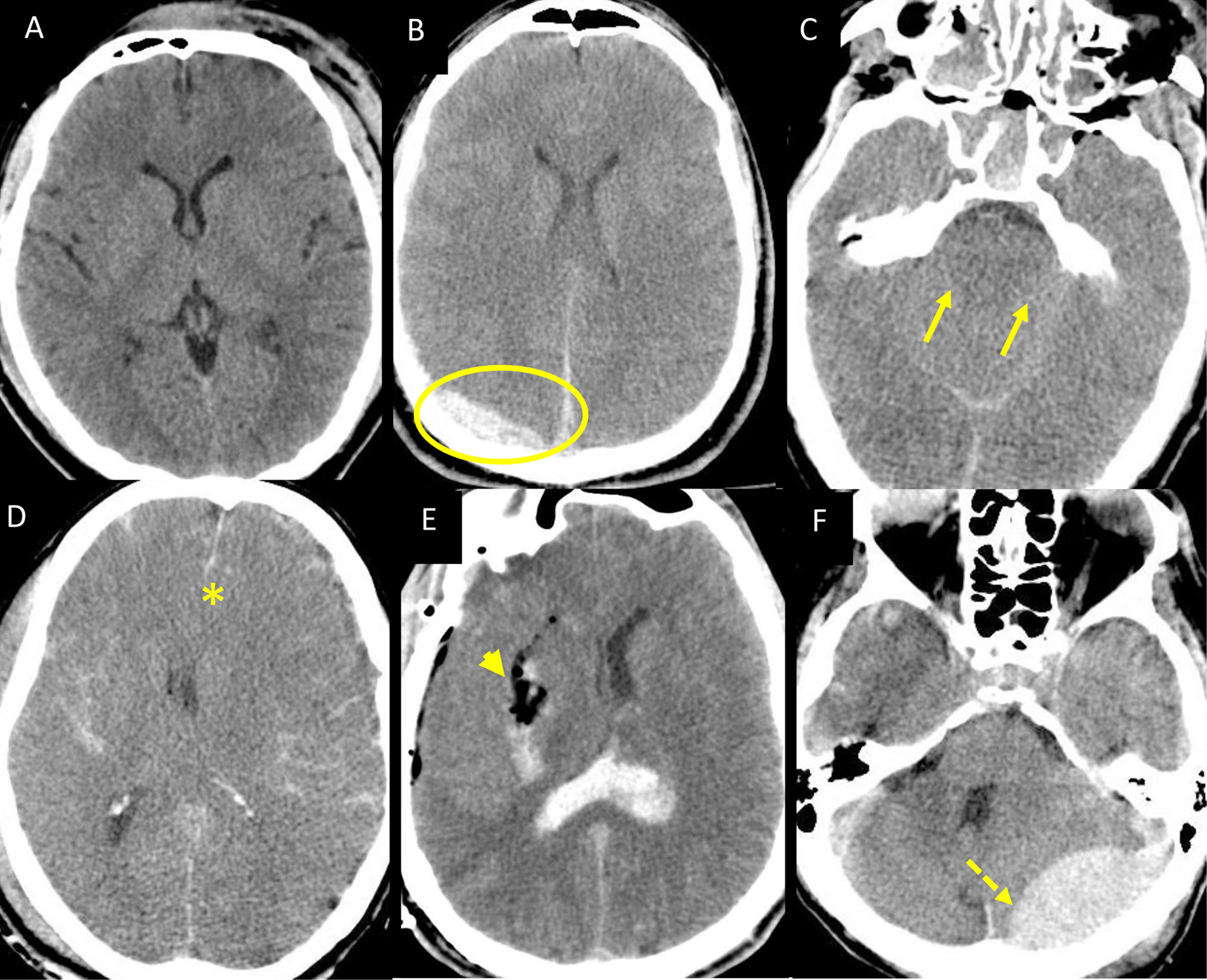
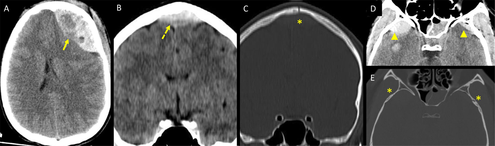
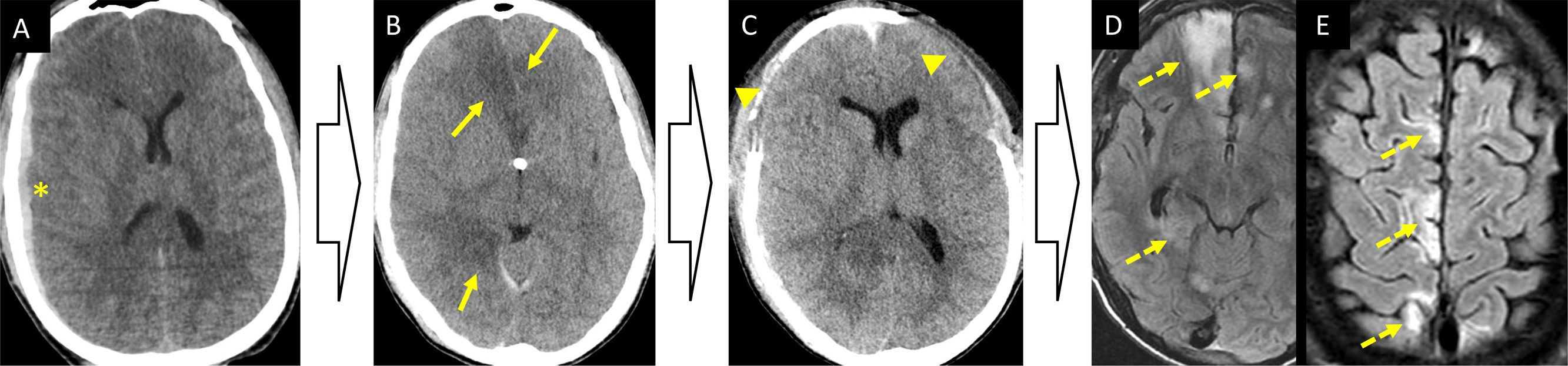
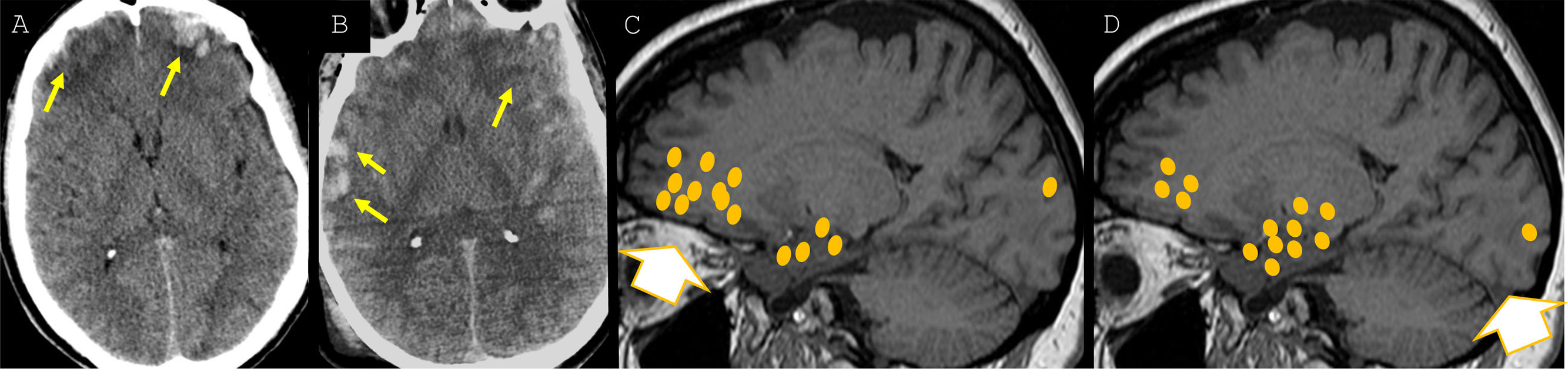
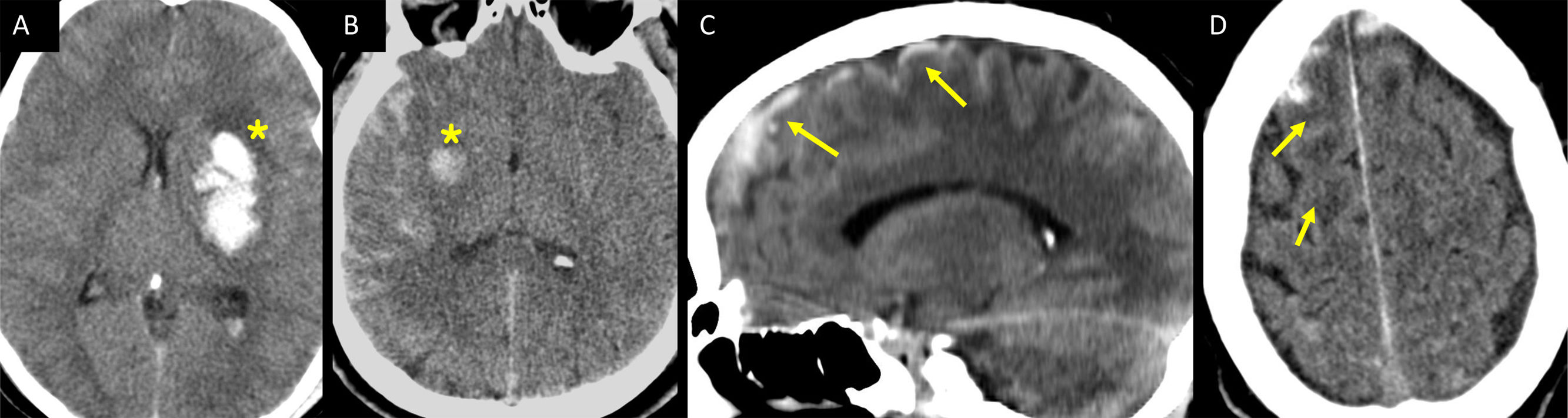
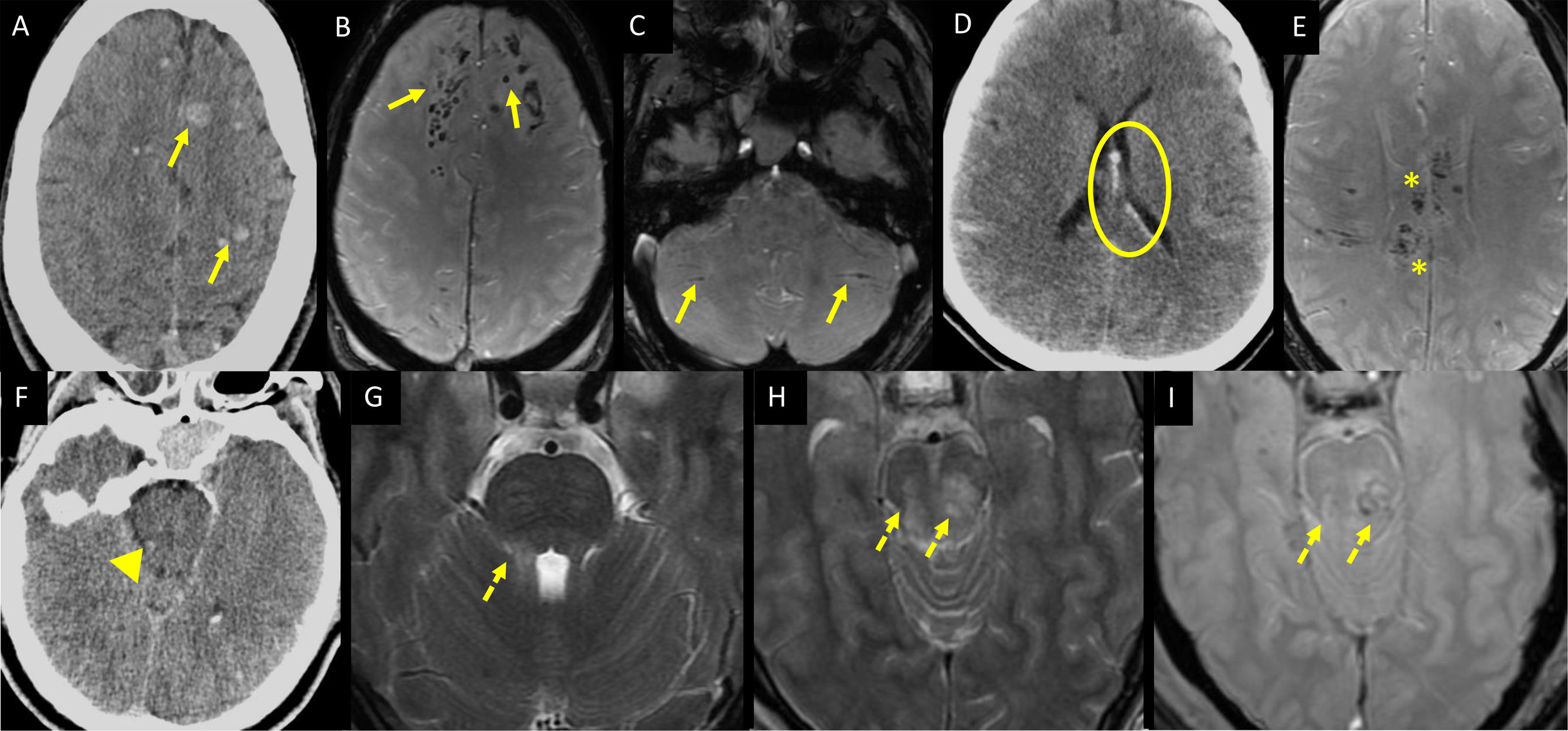
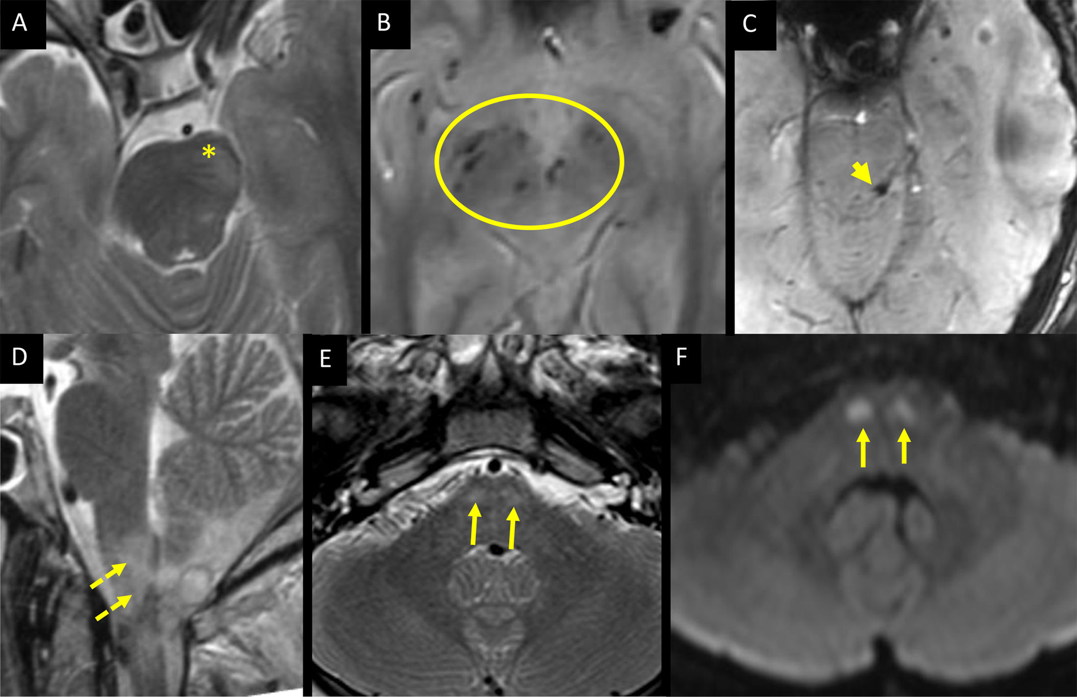
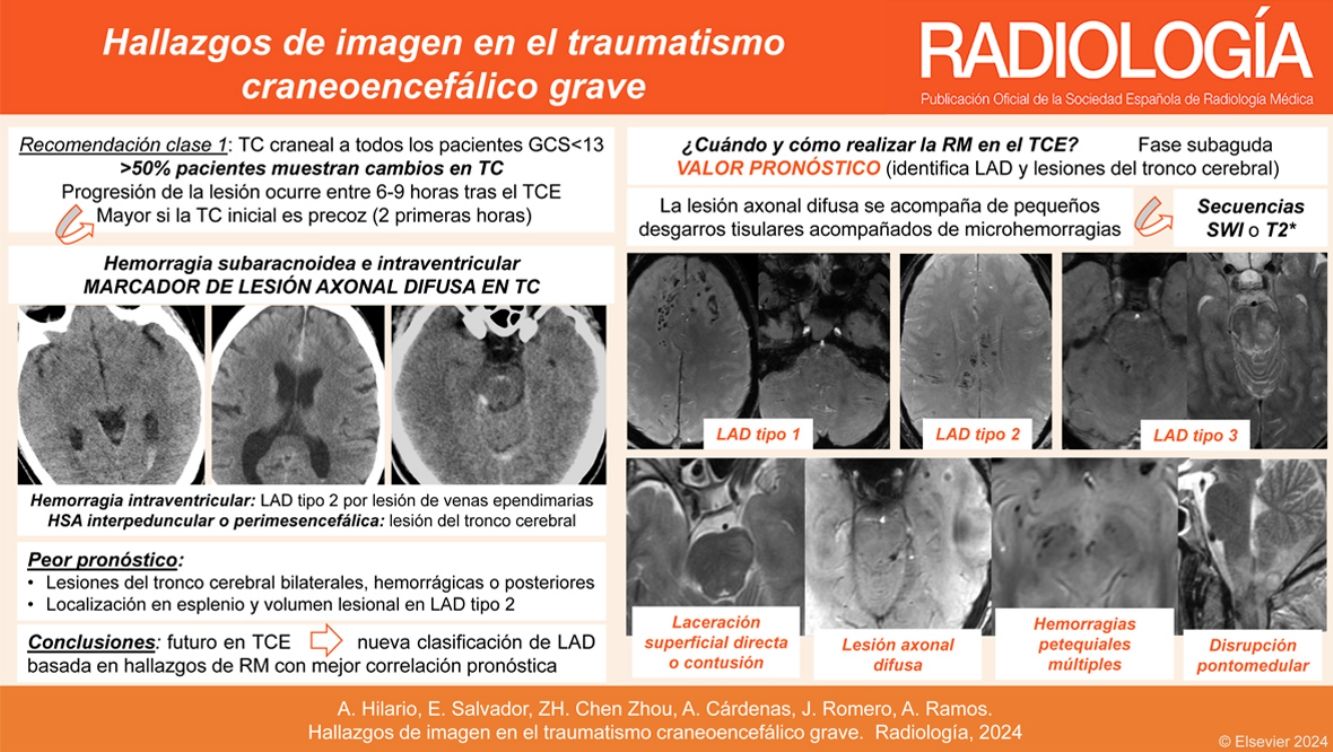
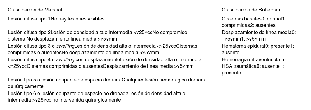
El traumatismo craneoencefálico (TCE) es la principal causa de morbimortalidad en los jóvenes. La clasificación de Marshall predice la mortalidad a 6 meses y divide a los pacientes con TCE grave en 6 grupos en función de los hallazgos de la tomografía computarizada (TC) en la fase aguda del trauma. La resonancia magnética (RM) tiene valor pronóstico, ya que detecta un 30% más de lesiones traumáticas, sobre todo lesiones del tronco cerebral y lesión axonal difusa. La lesión axonal difusa asienta en 3 áreas anatómicas diferentes, su gradación varía en función de la severidad, y la afectación cerebral se extiende de manera progresiva, en zonas más profundas cuanto mayor es el trauma. Las lesiones traumáticas del tronco cerebral que asocian peor pronóstico son aquellas de localización posterior, con afectación bilateral o hemorrágicas. Este artículo analiza el valor pronóstico de la TC y de la RM en la valoración del TCE grave, y describe las principales lesiones traumáticas intracraneales.

Palabras clave:
Hemorragia cerebral traumática
Hemorragia subaracnoidea
Lesión axonal difusa
pronóstico
Contusiones
Tomografía computarizada
Abstract

Traumatic brain injury (TBI) is the leading cause of morbidity and mortality in young patients. The Marshall classification predicts six-month mortality and divides severe TBI patients into six groups based on CT findings in the acute phase of trauma. MRI also has prognostic value because it detects 30% more traumatic lesions, especially brainstem injury and diffuse axonal injury. Diffuse axonal injury occurs in three different anatomical areas, graded according to severity, and the greater the trauma, the deeper the brain involvement extends. Traumatic brainstem injuries with the worst prognosis are those of posterior location, with bilateral or haemorrhagic involvement. This article analyses the prognostic value of CT and MRI in the assessment of severe TBI and describes the main intracranial traumatic injuries.

Keywords:
Traumatic brain haemorrhage
Subarachnoid haemorrhage
Diffuse axonal injury
Prognosis
Brain contusion
Computed tomography

Article

These are the options to access the full texts of the publication Radiología
Subscriber
Subscriber

If you already have your login data, please click here .

If you have forgotten your password you can you can recover it by clicking here and selecting the option “I have forgotten my password”
Subscribe
Subscribe to

Radiología

Purchase
Purchase article

Purchasing article the PDF version will be downloaded

Price 19.34 €

Purchase now
Contact
Phone for subscriptions and reporting of errors
From Monday to Friday from 9 a.m. to 6 p.m. (GMT + 1) except for the months of July and August which will be from 9 a.m. to 3 p.m.
Calls from Spain
932 415 960
Calls from outside Spain
+34 932 415 960
E-mail
Article options
es en pt

¿Es usted profesional sanitario apto para prescribir o dispensar medicamentos?

Are you a health professional able to prescribe or dispense drugs?

Você é um profissional de saúde habilitado a prescrever ou dispensar medicamentos