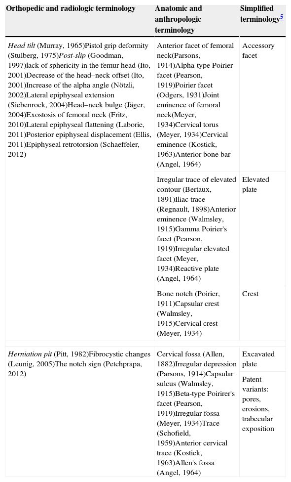
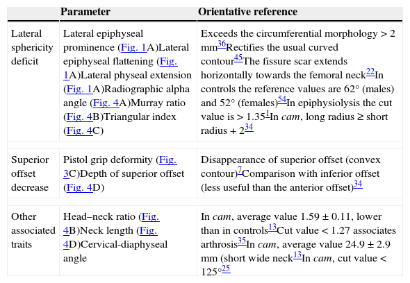
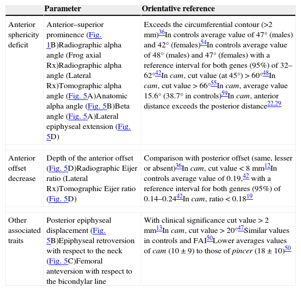
Interpreting imaging studies of a painful hip requires detailed knowledge of the regional anatomy. Some variants of the proximal femur, such as cam-type deformities, can course asymptomatically or cause femoroacetabular impingement. The principal numerical criterion for defining cam-type deformities, the alpha angle, has some limitations. In this article, we review the anatomic variants of the anterior aspect of the proximal femur, focusing on cam-type deformities. Using diagrams and multidetector CT images, we describe the parameters that are useful for characterizing these deformities in different imaging techniques. We also discuss the potential correspondence of imaging findings of cam-type deformities with the terms coined by anatomists and anthropologists to describe these phenomena.
Para interpretar correctamente los estudios radiológicos de una cadera dolorosa se requiere conocer detalladamente la anatomía regional. Algunas variantes del fémur proximal, como las deformidades tipo cam, pueden cursar de forma asintomática o causar un síndrome de choque femoroacetabular. El ángulo alfa, principal exponente numérico de estas deformidades, tiene algunas limitaciones. Nuestro objetivo es revisar las variantes anatómicas en la vertiente anterior del fémur proximal, centrando la atención en las deformidades tipo cam. Describimos los parámetros útiles para caracterizarla con métodos de imagen, utilizando diagramas e imágenes de tomografía computarizada multidetector. Exponemos además la correspondencia potencial de las deformidades tipo cam con términos descriptivos previamente acuñados por anatomistas y antropólogos.


