


To show the plain chest film findings in patients with confirmed infection with the new variant of the influenza A (H1N1) virus and to correlate these findings with the clinical history and evolution.
Materials and methodsWe reviewed the clinical histories and radiological studies in 99 patients infected with the new variant of H1N1 influenza who were admitted in two Hospitals in Cantabria, Spain. Plain chest film findings were classified according to their parenchymal pattern and the distribution of the lesions.
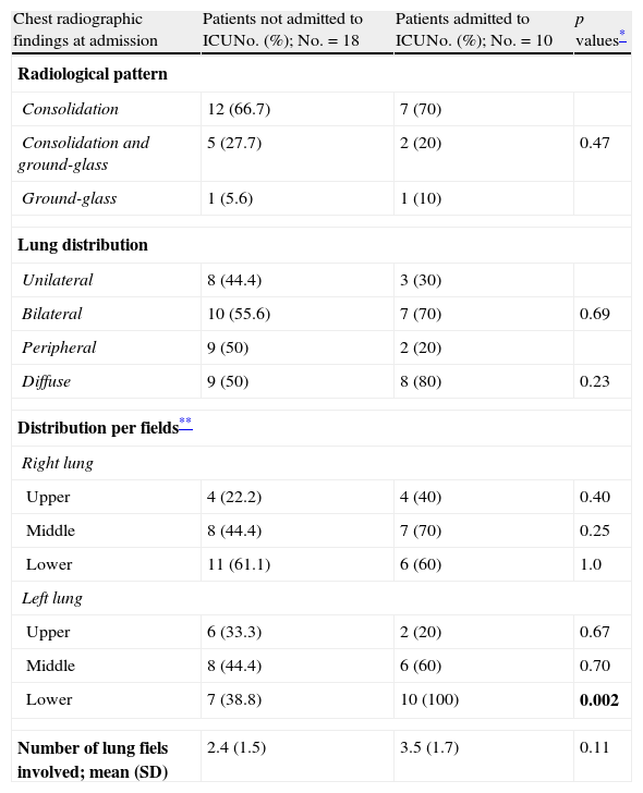
ResultsOf the 99 patients evaluated, 28 had changes on the plain chest film acquired at admission. In these 28 patients, the findings were: condensation in 19, condensation and ground-glass opacities in 7, and ground-glass opacities in 2; the distribution of the lesions was diffuse in 17 patients and bilateral in 17, with the lower and middle lobes being the most frequently affected. The lesions progressed in 13 patients, and the 7 patients who required mechanical ventilation had a higher frequency of diffuse lesion distribution and more lung fields affected on the plain chest field acquired at admission. Pathological findings on plain chest films were more common in males, in smokers, and in patients who presented with shortness of breath, pleuritic pain, and diarrhea (p<0.05).
ConclusionMost patients infected with the new variant of the H1N1 virus had no alterations on the plain chest film acquired on admission; when findings were present, the predominant pattern was diffuse, bilateral condensation mainly involving the bases of the lungs. Pleural effusion and hilar or mediastinal lymph node enlargement were uncommon.
Mostrar los hallazgos en la radiografía de tórax (RT) de pacientes con infección confirmada por la nueva variante del virus de la gripe A (H1N1) correlacionándolos con la historia y evolución clínica.
Material y métodosRevisión de la historia clínica y estudios radiológicos de 99 pacientes con infección por la nueva variante del virus de gripe A ingresados en dos hospitales del Servicio Cántabro de Salud. Los hallazgos en la RT fueron clasificados por el patrón parenquimatoso y la distribución de las lesiones.
ResultadosDe los 99 pacientes evaluados, 28 presentaron alteraciones en la RT realizada al ingresar. En estos 28 pacientes los hallazgos fueron: condensación en 19, condensación más vidrio deslustrado en 7 y vidrio deslustrado en dos; en 17 la distribución de las lesiones fue difusa, en 17 bilateral, y por campos los más afectados fueron el inferior y el medio. Trece pacientes experimentaron una progresión de las lesiones y los 7 que precisaron ventilación mecánica mostraron con mayor frecuencia en la RT del ingreso una distribución difusa de las lesiones y un mayor número de campos pulmonares afectos. Los pacientes con RT patológica fueron preferentemente varones, fumadores y presentaron disnea, dolor pleurítico y diarrea (p<0,05).
ConclusiónLa mayoría de los pacientes con infección por la nueva variante del virus de la gripe A no presentaron alteraciones en la RT del ingreso; sin embargo, cuando estaban presentes, el patrón predominante fue una condensación de distribución difusa, bilateral y con predominio en las bases. El derrame pleural y las adenopatías hiliares o mediastínicas fueron infrecuentes.

