To evaluate if the tumour perfusion at the initial MRI scan is a marker of prognosis for survival in patients diagnosed with High Grade Gliomas (HGG). To analyse the risk factors which influence on the mortality from HGG to quantify the overall survival to be expected in patients.
Patients and methodsThe patients diagnosed with HGG through a MRI scan in a third-level hospital between 2017 and 2019 were selected. Clinical and tumour variables were collected. The survival analysis was used to determine the association between the tumour perfusion and the survival time. The relation between the collected variables and the survival period was assessed through Wald’s statistical method, measuring the relationship via Cox’s regression model. Finally, the type of relationship that exists between the tumour perfusion and the survival was analysed through the Lineal Regression method.Those statistical analysis were carried out using the software SPSS v.17.
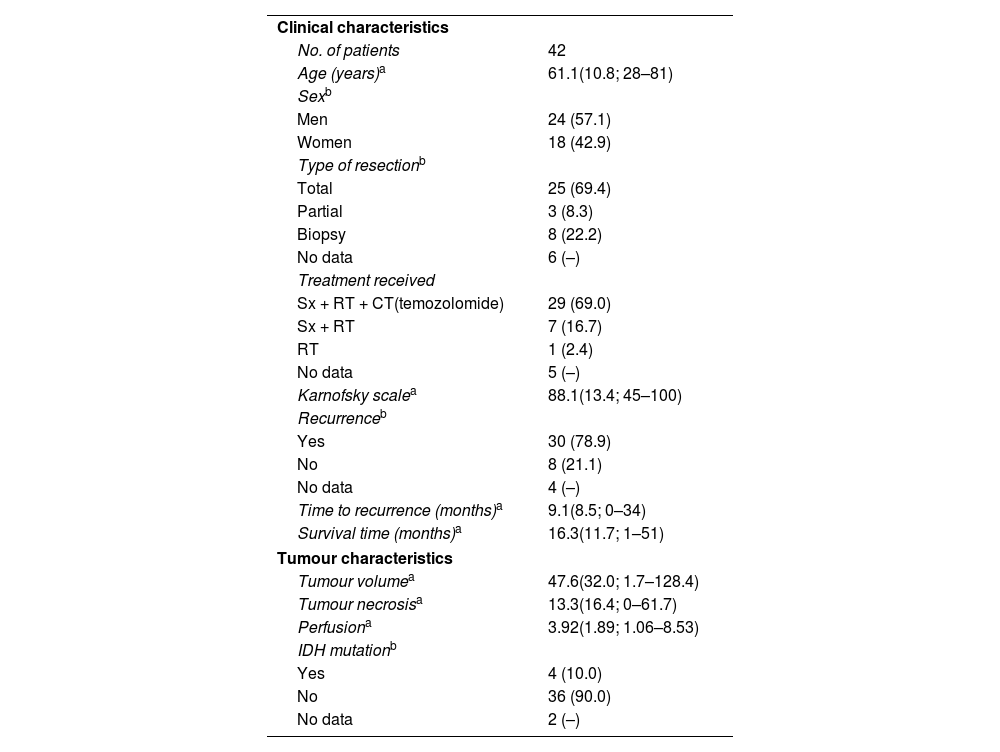
Results38 patients were included (average age: 61.1 years old). The general average survival period was 20.6 months. A relationship between the tumour perfusion at the MRI scan and the overall survival has been identified, in detail, a group with intratumor values of relative cerebral blood volume (rCBV)>3.0 has shown a significant decline in the average survival period with regard to the average survival period of the group with values <3.0 (14.6 months vs. 22.8 months, p = 0.046). It has also been proved that variables like Karnofsky’s scale and the response time since the intervention significantly influence on the survival period.
ConclusionsIt has become evident that the tumour perfusion via MRI scan has a prognostic value in the initial analysis of HGG. The average survival period of patients with rCBV less than or equal to 3.0 is significantly higher than those patients whose values are higher, which allows to be more precise with the prognosis of each patient.
Valorar si la perfusión tumoral en el estudio diagnóstico inicial de RM es un marcador pronóstico para la supervivencia en pacientes diagnosticados de gliomas de alto grado. Analizar los factores de riesgo que influyen en la mortalidad por gliomas de alto grado para poder cuantificar la supervivencia global esperada del paciente.
Pacientes y métodosSe seleccionaron las RM de todos los pacientes diagnosticados de glioma de alto grado en un hospital de tercer nivel entre los años 2017 y 2019. Se recogieron variables clínicas y tumorales. Se usó el análisis de supervivencia para determinar la asociación entre la perfusión tumoral y el tiempo de supervivencia. Se estudió la relación entre las variables recogidas y la supervivencia mediante el estadístico de Wald, cuantificando esta relación mediante la Regresión de Cox. Por último, se analizó el tipo de relación existente entre la perfusión tumoral y la supervivencia a través del estudio de Regresión Lineal. Estos análisis estadísticos se realizaron con el software SPSS v.17.
ResultadosSe incluyeron 38 pacientes (media de edad 61,1 años). La supervivencia media global fue de 20,6 meses. Se observó asociación entre la perfusión tumoral en la RM diagnóstica y la supervivencia global, mostrando el grupo con valores intratumorales de volumen sanguíneo cerebral relativo (rVSC) >3.0 una disminución significativa en el tiempo medio de supervivencia respecto al grupo con valores <3.0 (14,6 meses vs 22,8 meses, p = 0,046). También han demostrado influir significativamente en la supervivencia media variables como la escala de Karfnosky y el tiempo de recidiva desde la intervención.
ConclusionesSe ha evidenciado que la perfusión tumoral por RM tiene valor pronóstico en el estudio inicial de los gliomas de alto grado. La media de supervivencia de los pacientes con rVSC inferior o igual a 3.0 es significativamente mayor que en aquellos cuyo valor es superior, lo que permitiría una aproximación pronóstica más precisa en cada paciente en el momento del diagnóstico.


