Suplement “"Advances in Thoracic Radiology”
More infoTo assess the ability of an artificial intelligence software to detect pneumothorax in chest radiographs done after percutaneous transthoracic biopsy.
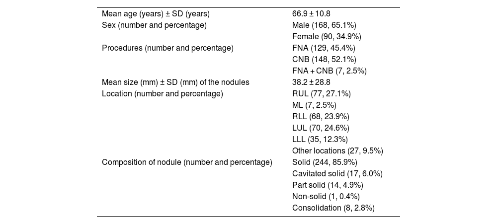
Material and methodsWe included retrospectively in our study adult patients who underwent CT-guided percutaneous transthoracic biopsies from lung, pleural or mediastinal lesions from June 2019 to June 2020, and who had a follow-up chest radiograph after the procedure. These chest radiographs were read to search the presence of pneumothorax independently by an expert thoracic radiologist and a radiodiagnosis resident, whose unified lecture was defined as the gold standard, and the result of each radiograph after interpretation by the artificial intelligence software was documented for posterior comparison with the gold standard.
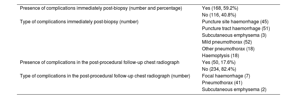
ResultsA total of 284 chest radiographs were included in the study and the incidence of pneumothorax was 14.4%. There were no discrepancies between the two readers’ interpretation of any of the postbiopsy chest radiographs. The artificial intelligence software was able to detect 41/41 of the present pneumothorax, implying a sensitivity of 100% and a negative predictive value of 100%, with a specificity of 79.4% and a positive predictive value of 45%. The accuracy was 82.4%, indicating that there is a high probability that an individual will be adequately classified by the software. It has also been documented that the presence of Port-a-cath is the cause of 8 of the 50 of false positives by the software.
ConclusionsThe software has detected 100% of cases of pneumothorax in the postbiopsy chest radiographs. A potential use of this software could be as a prioritisation tool, allowing radiologists not to read immediately (or even not to read) chest radiographs classified as non-pathological by the software, with the confidence that there are no pathological cases.
Evaluar la capacidad de detección de neumotórax por parte de un programa de inteligencia artificial (IA) en las radiografías de tórax de control posbiopsia percutánea transtorácica.
Material y métodosSe han incluido de forma retrospectiva los pacientes adultos sometidos a biopsia percutánea transtorácica de lesiones pulmonares, pleurales o mediastínicas guiada por TC desde junio de 2019 hasta junio de 2020 y que contaban con radiografía de tórax de control tras el procedimiento. Se procedió a la lectura independiente de las radiografías de tórax de control posprocedimiento por parte de un radiólogo torácico y un residente de radiodiagnóstico, cuya lectura unificada se definió como el gold estándar, y se anotó el resultado de cada radiografía de tórax tras la interpretación por un programa de IA para la detección de neumotórax.
ResultadosSe incluyeron 284 radiografías de tórax posprocedimiento, en las que la incidencia de neumotórax fue del 14,4%, sin existir discrepancias en el diagnóstico entre los dos lectores. El algoritmo de IA fue capaz de detectar 41/41 de los neumotórax presentes, suponiendo una sensibilidad del 100% y un valor predictivo negativo del 100%, a expensas de una especificidad del 79,4% y un valor predictivo positivo del 45%. La exactitud o valor global fue del 82,4%, indicando que hay una alta probabilidad de que un individuo sea clasificado adecuadamente por el modelo de IA. También se ha documentado que la presencia de Port-a-cath fue causa de 8 de los 50 falsos positivos.
ConclusionesEl programa ha detectado el 100% de casos de neumotórax en las radiografías de tórax de control. Un potencial uso de este software sería como herramienta de priorización, permitiendo postergar, o incluso obviar, la lectura de las radiografías de tórax clasificadas como no patológicas por el algoritmo con la garantía de que no haya casos patológicos.


