El acueducto vestibular dilatado (AVD) es la anomalía congénita más frecuentemente encontrada en técnicas de imagen en hipoacusia neurosensorial infantil. Nuestro objetivo es describir las características clínicas y audiológicas de los niños hipoacúsicos con hallazgo de AVD.
MétodosEstudio retrospectivo de 55 niños diagnosticados de AVD en el periodo 2000–2009. Se analizaron las pruebas audiológicas objetivas y/o subjetivas estándar realizadas según la edad de desarrollo de los niños. Se describen los hallazgos y concomitancias clínicas y audiológicas.
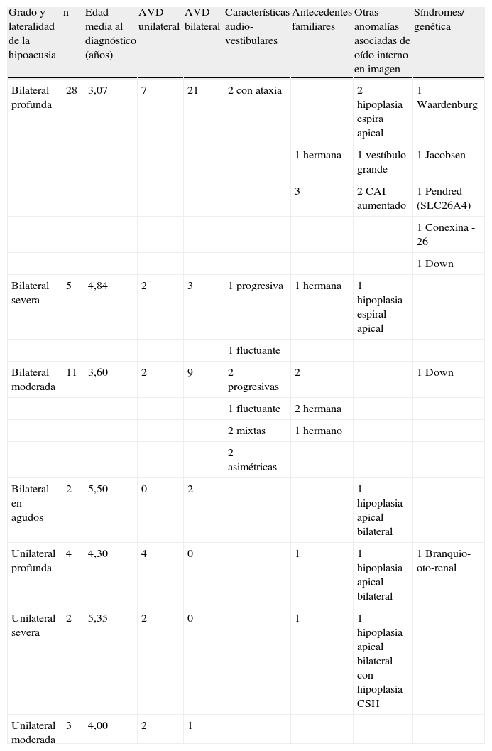
ResultadosTreinta y siete pacientes (67,27%) presentaban AVD bilateral y 18 (32,72%) unilateral. La hipoacusia era bilateral en 46 (83,63%) casos y unilateral en 9 (16,36%). La media de edad resultó 3,78 años. Presentaron hipocausa neurosensorial 53 (96,36%) casos (28 bilaterales y profundas), y 2 (3,63%) casos hipoacusia mixta. Tres casos fueron progresivos, 2 fluctuantes, 2 asimétricas y 2 presentaron síntomas vestibulares. Se evidenciaron otras anomalías radiológicas asociadas (6 hipoplasias cocleares, 2 conductos auditivos internos agrandados, 1 vestíbulo dilatado y 1 conducto semicircular horizontal hipoplásico), y 6 síndromes clínicos concomitantes (2 Down, 1 Jacobsen, 1 Pendred, 1 Waardenburg, 1 branquio-oto-renal). Un caso resultó positivo a la mutación GJB2. Se encontró historia familiar de hipoacusia en 12 (21,8%) casos.
ConclusiónLa presentación clínica de la hipoacusia infantil en el AVD se caracteriza por su variabilidad. Debe incluirse en el diagnóstico diferencial de la hipoacusia mixta. La asociación familiar y sindrómica del AVD deben considerarse en el estudio diagnóstico. Es necesario conocer la historia natural de la enfermedad con fines de información pronóstica a los padres.
Enlarged vestibular aqueduct (EVA) is the commonest congenital anomaly found with imaging techniques in paediatric sensorineural hearing loss (SNHL). Our aim was to describe clinical and audiological findings in paediatric hearing loss associated to EVA.
MethodsRetrospective review of 55 children with imaging-technique EVA findings from 2000 to 2009. Subjective and/or objective audiological tests were analysed and audiological findings related to clinical features were described.
ResultsThirty-seven patients (67.27%) showed bilateral EVA and 18 (32.72%) were unilateral. Hearing loss was bilateral in 46 (83.63%) patients and unilateral in 9 (16.36%). Mean age at diagnosis was 3.78 years. Fifty-three (96.36%) children showed SNHL (28 bilateral and profound), while 2 (3.63%) patients had mixed hearing loss. There were 3 cases of hearing loss progression, 2 fluctuations, 2 of them were asymmetric and 2 patients suffered from vestibular symptoms. Concomitant image findings were 6 cochlear hypoplasia, 2 enlarged internal auditory canals, 1 enlarged vestibule and 1 hypoplastic lateral semicircular canal. Six clinical syndromes were found (2 cases of Down's, and 1 each of Jacobsen, Pendred, Waardenburg and branchio-oto-renal). One child was positive for GJB2 mutation. Familial hearing loss was demonstrated on 12 (21.8%) cases.
ConclusionThe clinical picture of hearing loss associated to EVA is characterised by great variability. It should be included in the differential diagnosis of unexplained mixed hearing loss. Familial and syndromic findings have to be taken into consideration in the diagnostic evaluations of such patients. Knowledge about the natural history of this illness is needed so as to give parents prognostic information.
Artículo
Comprando el artículo el PDF del mismo podrá ser descargado
Precio 19,34 €
Comprar ahora


