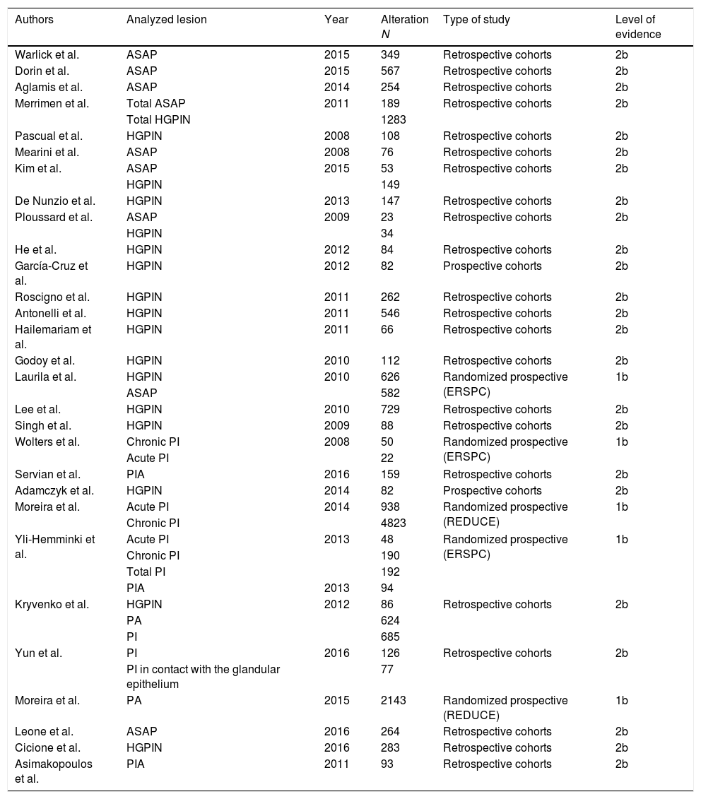
In cases of persistent suspicion of prostate cancer (PC), repeat prostate biopsies (PB) are frequently performed in spite of their low yield. In the context of a negative PB, there is a microscopic scenario (MS), which we define as the group of recognizable non-neoplastic lesions. While some of these lesions seem to have a protective effect, the existence of others increases the risk of PC detection in posterior PB. The objective of this systematic review is to identify the lesions that may belong to the MS of a negative PB and analyze the current evidence of their association with the risk of detecting PC in subsequent PBs.
Evidence acquisitionTwo independent reviewers conducted a literature search on Medline, Embase and Central Cochrane with the following search terms: small acinar proliferation, ASAP, prostatic intraepithelial neoplasia, HGPIN, adjacent small atypical glands, pinatyp, atrophy, proliferative inflammatory atrophy, pia, prostatic inflammation, prostatitis and prostate cancer. 1015 references were first identified, and 57 original articles were included in the study, following the PRISMA declaration and the PICO selection principles.
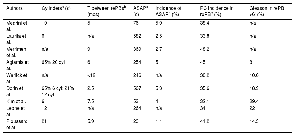
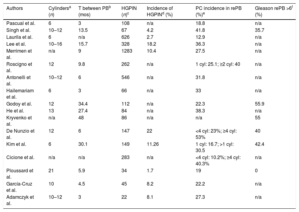
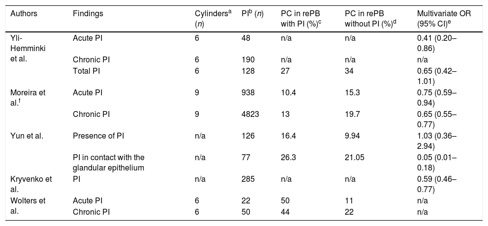
Evidence synthesisAtypical small acinar proliferation is associated with PC detection in repeat PB with rates ranging between 32 and 48%. High-grade prostatic intraepithelial neoplasia (HGPIN) is related to PC in 13–42% of cases. Studies show that HGPIN, when multifocal, is a significant independent risk factor for PC. Prostatic atrophy, inflammatory proliferative atrophy and prostatic inflammation seem to act as protective factors on the detection of PC in repeat PB. On the other hand, the risk of PC detection reduces significantly in male patients with multifocal HGPIN and coexistent PIA.
ConclusionsThe MS of a negative PB may include atypical small acinar proliferation, HGPIN, prostatic atrophy, inflammatory proliferative atrophy and prostatic inflammation lesions, since they all seem to be associated with the risk of PC detection in repeat PB. This review has led us to create the hypothesis that the MS of a negative PB might be a valuable and useful tool when considering repeat PB.
Las biopsias prostáticas (BP) de repetición, ante la persistencia de la sospecha de cáncer de próstata (CP), son frecuentes y su rendimiento bajo. En el contexto de una BP negativa existe un escenario microscópico (EM), que definimos como el conjunto de lesiones no neoplásicas identificable. La existencia de algunas de estas lesiones incrementa el riesgo de detección de CP en BP sucesivas, mientras que otras parecen tener un efecto protector. El objetivo de esta revisión sistemática es identificar el conjunto de lesiones que puede formar parte del EM de una BP negativa y analizar la evidencia actual de su asociación con el riesgo de detección de CP en BP sucesivas.
Adquisición de la evidenciaDos revisores independientes realizaron una búsqueda bibliográfica en Medline, Embase y Central Cochrane, con los términos de búsqueda: small acinar proliferation or ASAP or prostatic intraepithelial neoplasia or HGPIN or adjacent small atypical glands or pinatyp or atrophy or proliferative inflammatory atrophy or pia or prostatic inflammation or prostatitis and prostate cancer. Se identificaron 1.015 referencias y siguiendo los principios de la declaración PRISMA y de selección PICO, se identificaron 57 artículos originales válidos para esta revisión.
Síntesis de la evidenciaLa proliferación acinar atípica de célula pequeña se asocia a una tasa de detección de CP en BP sucesivas que oscila entre el 32 y 48%. La neoplasia intraepitelial prostática de alto grado (HGPIN) se asocia a CP entre el 13 y 42%, siendo su multifocalidad la que define el incremento en el riesgo de detección. La atrofia prostática, la atrofia proliferativa inflamatoria y la infamación prostática parecen tener un efecto protector sobre la detección de CP en BP sucesivas. Por otra parte, el riesgo de detección de CP en varones con HGPIN multifocal se reduce significativamente si coexiste atrofia proliferativa inflamatoria.
ConclusionesEl EM de una BP negativa puede estar compuesto por las lesiones de proliferación acinar atípica de célula pequeña, HGPIN, atrofia prostática, atrofia proliferativa inflamatoria e infamación prostática ya que todas parecen estar asociadas al riesgo de detección de CP en BP sucesivas. Esta revisión nos permite generar la hipótesis de que el EM de una BP negativa puede ser de utilidad en la decisión indicar BP de repetición.
Artículo
Comprando el artículo el PDF del mismo podrá ser descargado
Precio 19,34 €
Comprar ahora


