Introduction
Since Price's report in 1950,1 it has been recognized that monozygotic twins provide an opportunity to study genetic contribution in the development of cardiovascular malformations. The incidence of congenital heart defects among singletons is 6/1000 live births, compared to 17.2/1000 in monozygotic twins, while in dizygotic twins the incidence is approximately 8/1000.2 When both twins are affected, concordance is said to occur. Concordance incidence in dizygotic twins is 5%, compared to 25% in monozygotic twins.3 In this paper, two pairs of monozygotic twins with specific concordance of congenital heart defects are reported.
Case report 1
Monozygotic twins with pulmonary stenosis
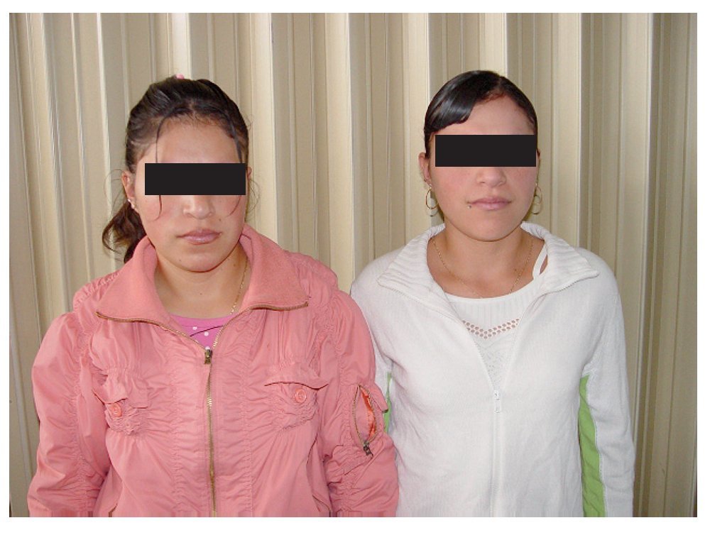
U.H. and E.H. were13 year-old monozygotic female twins; both girls had cardiac murmurs and were referred to our hospital. They were completely asymptomatic and their phenotypes were very similar (Figure 1A). Both of them had blood type B+; in a fluorescence hybridization test in situ the two girls were negative for chromosome 22q11 microdeletion. Echocardiographic examination showed a severe infundibular and pulmonary valve stenosis in both of them. Systolic gradient measured by Doppler was 140 mmHg in U.H., and 90 mmHg in E.H. During cardiac catheterization, U.H. had right ventricular pressures of 120/8 and 30/10 in the pulmonary artery, and E.H. had 200/14. Echocardiogram and selective angiogram showed severe dysplastic pulmonary valve in the right ventricle of both twins.
Figure 1A. Thirteen years-old monozygotic female twins with severe pulmonary valve stenosis.
In June 2006, the two patients underwent cardiac surgical correction with transannular patch under cardiopulmonary bypass. During surgery, severe pulmonary valve stenosis with hypoplastic annulus was found in both of them. In addition, U.H. presented infundibular stenosis which was removed. The patients recovered uneventfully from operation. Today, six years after the procedure, both patients are asymptomatic with normal development and a control echocardiogram revealed a residual gradient below 20 mmHg and minimal pulmonary insufficiency.
Case report 2
Monozygotic twins with Tetralogy of Fallot
J.T. and N. T. were 8 year-old male monozygotic twins, with almost identical phenotypes (Figure 1B). Both had blood O+; in a fluorescence hybridization test in situ the two boys were negative for microdeletion in chromosome 22q11. In both twins, Tetralogy of Fallot was diagnosed by echocardiogram, cardiac catheterization and angiocardiogram. In the first twin a Blalock-Taussig shunt had been performed. This patient had stress dyspnea and physical examination revealed cyanosis ++. Echocardiogram demonstrated Tetralogy of Fallot with an obstructive 60 mmHg gradient in the right ventricular outflow tract.
Figure 1B.Eight years-old monozygotic male twins with tetralogy of Fallot.
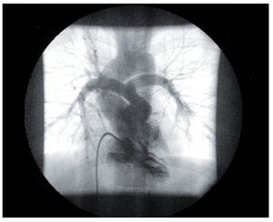
Cardiac catheterization confirmed the echocardiogram diagnosis and an additional stenosis was found at the origin of the left pulmonary artery (Figure 2a). The McGoon index was 1.3. In February 2008 surgical correction was performed with transannular patch, pulmonary valve prosthesis, and patches in left main coronary and pulmonary arteries; the ventricular septal defect was closed too. During surgery a laceration of the pulmonary artery with important bleeding occurred as a complication. In the intensive care unit, the patient developed pneumonia due to Sthaphylocococus aureus, with bad and long evolution. It was not possible to disconnect the patient from ventilator. Fifteen days later, this twin died because of septic shock.
Figure 2A.Angiogram of the main pulmonary artery in one of the twins with Tetralogy of Fallot. Severe pulmonary stenosis at the origin of the left pulmonary artery is documented.
In the second twin no previous shunt was done. During physical examination cyanosis ++ it was seen and arterial saturation was 82%. Echocardiography and angiocardiography showed favorable anatomy (Figure 2B). The McGoon index was 2.1. Total surgical correction was performed in January 28, 2008. He recovered uneventfully. Three months later, an echocardiogram revealed a maximum gradient of 22 mmHg and the patient presented no symptoms.
Figure 2B.Selective right ventriculogram in the second twin with Tetralogy of Fallot. Favorable anatomy is shown.
Discussion
Monozygotic twins are less frequent than dizygotic twins and account for approximately a third of total number of twins, but incidence of congenital anomalies in monozygotic twins is about 10%.4
Congenital heart disease concordance in monozygotic twins occurs at a frequency of 25%, which is high when compared to other congenital disorders. However, in previous literature usually it is not specified whether concordance refers to either the presence of the same cardiac defect in both twins, or to some cardiac malformation present in the two. We will refer to specific concordance in the former concept.
There are very few reports on specific concordance of congenital cardiac malformations. After detailed search of the literature (PubMed/Medline) only three reports of twins with tetralogy of Fallot were found. In one of the reports, both twins had pulmonary atresia and the same intracardiac defects, with anatomic differences in large vessels. Both patients were positive for microdeletion in chromosome 22q11.5 In France, a pair of twins had the same disorder, but they were negative for microdeletion in chromosome 22q11 (as in both of our cases); it was reported by Laugel et al in 2001 V.6 The first monozygotic pair of twins with Tetralogy of Fallot in India was reported by Patel AB et al in 2002.7 In these works, authors concluded that the differences found could not be explained on genetic grounds and suggested the influence of other factors in the developing embryo.
There are two other reports of specific concordance. In the first, the presence of persistent truncus arteriosus in both monozygotic twins, without DiGeorge phenotype syndrome and no deletion in chromosome 22q11 is described.8 The second report presents a successfully operated pair of twins with ventricular septal defect.9
This work, in which we have studied two pairs of identical twins, one with pulmonary valve stenosis and the other with Tetralogy of Fallot, adds to the few published cases of specific concordance. Just like in the previous reports, both twins had the same disorder, with minor differences, particularly in the severity of the lesion. In agreement with other authors, these differences could be explained by the influence of other factors probably acquired during the first stages of embryonic development.
Acknowledgements
Authors are thankful to Hector Alva PhD for his help in the preparation of the English manuscript.
Corresponding author:
Carlos Alva.
Congenital Heart Disease Department, Cardiology Hospital, Centro Médico Nacional Siglo XXI, Av.Cuauhtémoc 330, Col. Doctores, México City CP 06720, México.
Phone: 5255 5585 2443. E mail:echoca@yahoo.com
Received: December 12, 2008;
accepted September 23, 2009.


