Describir los cambios en el segmento anterior y en la presión intraocular (PIO) entre la iridotomía periférica láser (IPL) y la facoemulsificación en pacientes con sospecha de cierre angular primario (PACS) y cierre angular primario (PAC).
MétodoSe seleccionaron 47 ojos de 47 pacientes que presentaban un ángulo 0-II (Shaffer) en la gonioscopia y se excluyó a los pacientes con lesiones glaucomatosas. Según la esclerosis cristaliniana y la agudeza visual se separaron en 2 grupos: IPL (n=18) o facoemulsificación (n=29). Se realizó tonometría, gonioscopia, funduscopia y medidas de la cámara anterior (CA) con Pentacam antes de cada intervención, al mes y a los 3 meses.
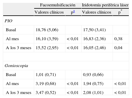
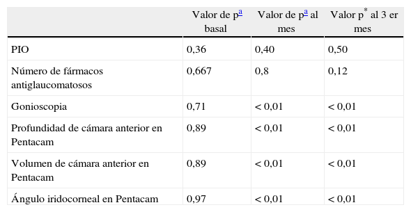
ResultadosLa facoemulsificación redujo la PIO al mes y a los 3 meses (p<0,01), mientras que la IPL redujo la PIO de forma estadísticamente significativa a los 3 meses (p<0,04; al mes p=0,38). La PIO fue 16,1 mmHg (DE: 3,59) en el grupo facoemulsificación versus 16,83 mmHg (DE: 2,36) en el grupo IPL al mes (p=0,4) y 15,52 (DE: 2.95) versus 16,05 (DE: 2,46) a los 3 meses (p=0,5). No se encontraron diferencias significativas en la media de fármacos antiglaucomatosos.
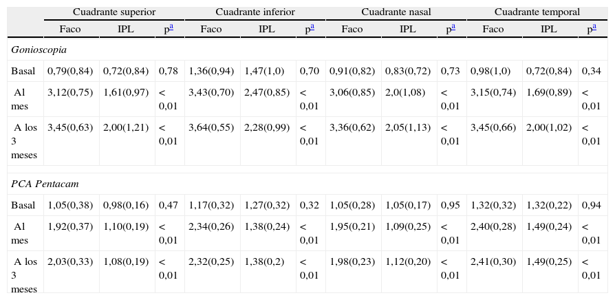
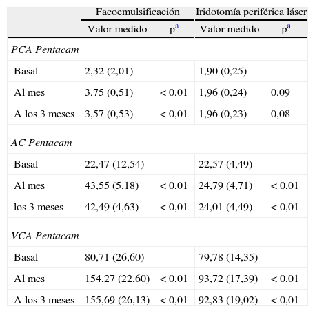
La apertura angular mediante gonioscopia fue mayor en el grupo de facoemulsificación (p<0,01), encontrándose la mayor diferencia en el cuadrante superior. La profundidad, el ángulo y el volumen de la CA obtenidos con Pentacam fueron superiores en el grupo de facoemulsificación (p<0,01).
ConclusionesTanto la IPL como la facoemulsificación son técnicas efectivas para prevenir el bloqueo pupilar en PAC, pero con la facoemulsificación se obtiene mayor amplitud del ángulo y de la CA de forma precoz.
A study was designed to determine and describe the changes induced in the anterior segment of the eye and the intraocular pressure (IOP) after laser peripheral iridotomy (LPI) versus phacoemulsification in primary angle closure suspects (PACS) and primary angle closure (PAC).
MethodsForty-seven eyes (47 patients) with Shaffer gonioscopy 0-II were included and split into 2 groups: cataract surgery (n=29) or LPI (n=18), depending on the lens sclerosis and visual acuity. Tonometry, gonioscopy, funduscopy, and automated measurements of the anterior chamber by Pentacam were performed before the intervention, and one and 3 months after the technique.
ResultsPhacoemulsification reduces IOP after one and 3 months (P<.01). LPI reduces IOP after 3 months (P<.04), and after one month (P<.38). IOP was 16.2mmHg (SD: 3.59) in the phacoemulsification group vs. 16.83mmHg (SD: 2.36) in the LPI group after one month (P=.4), and 15.52 (SD: 2.95) vs. 16.05 (SD: 2.46) in the third month (P=.5). There were no significant differences in the antiglaucoma drugs.
Shaffer gonioscopy grading was greater in the phacoemulsification group vs. in the LPI group one and 3 months after the intervention (P=.01). The highest difference between both techniques was found in the superior quadrant. The anterior chamber depth, angle and volume by Pentacam were wider in the phacoemulsification group after one and 3 months (P<.01).
ConclusionsAlthough phacoemulsification and LPI could both be effective techniques in the prevention of pupillary block in PAC, faster and greater amplitude of the angle and the anterior chamber can be obtained after phacoemulsification than after LPI.
Artículo
Comprando el artículo el PDF del mismo podrá ser descargado
Precio 19,34 €
Comprar ahora


