< 25 tenía osteoporosis. Conclusiones. La prevalencia de osteoporosis en nuestras pacientes fue mayor que en otros estudios (30%). Insistimos en la necesidad de realizar el cribado de la osteoporosis con UOC en atención primaria. Sugerimos que los programas de educación comunitaria deberían comenzar en edades más tempranas para identificar los factores que contribuyen a mantener la densidad mineral ósea en las mujeres posmenopáusicas.
Introduction
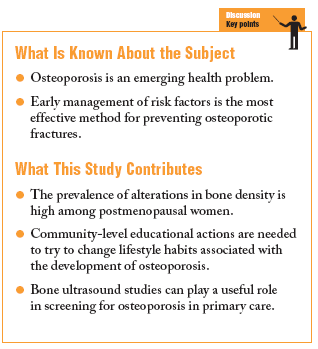
Osteoporosis is defined as a skeletal disease characterized by diminished bone strength that predisposes a person to an increased risk of fractures. In epidemiological terms it affects 35% of all women older than 50 years, with the percentage rising to 52% in women more than 70 years old.1
Because of the steady aging of our society, osteoporosis can be considered an emerging health problem.2 It is important to determine the factors involved in this, as the most effective way to prevent osteoporotic fractures is by early management.3
Measuring bone mineral density (BMD) is the best way to confirm or rule out a diagnosis of osteoporosis.4 Dual absorption x-ray absorptiometry (DEXA) is the most widely used diagnostic technique.
Predictions of the risk of fractures are most accurate when BMD is measured directly in the bone tissues involved most often (spinal column and hip). However, measurements in peripheral bone are technically simpler. Among the methods for measuring bone mass in peripheral bone, ultrasound has shown an association with the prevalence (cross-sectional studies) and risk of fractures (prospective studies). This method provides an indication of the risk of fracture (especially hip fractures) that is independent from BMD, and is currently recommended as a rapid alternative for bone mass measurements that does not involve irradiation.5
Quantitative bone ultrasound examination or broadband ultrasonic attenuation (BUA) has been shown to have the same predictive value for vertebral column fractures as DEXA for the spinal columna and hip (OR, 2.2; 95% CI, 1.7-2.9 per standard deviation in the spinal column; OR, 1.7; 95% CI, 1.3-2.1 per standard deviation in the hip).6,7
The prevalence of osteoporosis is probably higher than reported figures indicate, and the present study was done to determine the prevalence of alterations in BMD in postmenopausal women with bone ultrasound studies.
Patients and Methods
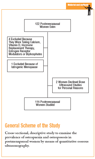
This descriptive, cross-sectional prevalence study was carried out at the Salvador Allende Primary Care Health Center in Valencia (eastern Spain). We selected all postmenopausal women served by our center who were seen during September, October, and November 2003. As exclusion criteria we used iatrogenic menopause and use of calcium, vitamin D, hormone replacement therapy, estrogen receptor modulators or biphosphonates.
Bone ultrasound studies were done in the right calcaneus with a Norland McCue CUBA Clinical ultrasound sonometer (Norland Medical Systems) and the results were recorded as BUA. The findings for this study were reported as osteoporosis, osteopenia, or normal bone density on the basis of the t-score according to WHO criteria. Osteoporosis was recorded when bone mass figures were more than 2.5 standard deviations (SD) below peak bone mass, i.e., the maximum value for bone mass in young women. Osteopenia was recorded when bone mass was between 1 and 2.5 SD below peak bone mass.
The sample size needed, as calculated with the Epi-info program for a precision of 95%, was 86 patients (P<.05; alpha level, .05) assuming a prevalence of BMD alterations of 49%.
The variables studied here were age in years, weight in kilograms, height in meters, ultrasound bone densitometry findings, smoking habit (more than 10 cigarettes/day during the previous year), use of diuretics, and years of amenorrhea.
Laboratory values and pathological findings were recorded from the patient's medical record held at the health center.
Mean age of the population served by our center was 39.6 years. The population in our catchment area was considered mostly mature (Friz index, 71) and regressive, i.e., the percent population older than 50 years was greater than the percent population between 15 and 50 years of age.
All data were processed and analyzed with the SPSS (version 11.0 for Windows). Descriptive statistics for each variable were reported as the mean and SD for quantitative variables, and as percentages for qualitative variables.
Results
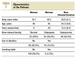
The characteristics of our patients are summarized in Table 1. Of the 115 postmenopausal women studied here, 58 had osteoporosis and 34 had osteopenia. The prevalence of osteoporosis was 50.4% (95% CI, 45.7%55%), and the prevalence of osteopenia was 29.6% (95% CI, 25.3%33.8%). Thus 80% (95% CI, 76.3%83.7%) of the women in this study had some BMD alteration.
Of the 58 women with osteoporosis, the 55-60 year old age group accounted for 17.2% of this group and the 70-75 year old age group accounted for 27.6%. Of the 34 postmenopausal women with osteopenia, 32.4% were 55-60 years old. Most of the postmenopausal women with osteoporosis (82.8%) were less than 75 years old.
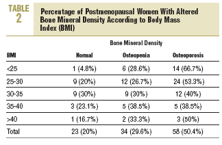
Two thirds (66.7%) of the postmenopausal women with a body mass index less than 25 had osteoporosis. Table 2 shows the percentage figures for the frequency of alterations in BMD according to body mass index.
According to the criteria of the Agència d'Avaluació de Tecnología Mèdica11 and the Sociedad Española de Medicina de Familia y Comunitaria4 for the indication for densitometry, this diagnostic test is indicated for 64 (55.6%) of the patients in the present study. Of the 51 (44.4%) women for whom densitometry was not indicated, 21 (41.2%) were found on ultrasound examination to have osteoporosis.
Discussion
The prevalence of postmenopausal osteoporosis in the present study is higher than in other reports.8 Díez Curiel et al9 estimated the prevalence of osteoporosis by age group in Spanish women on the basis of densitometric findings. The prevalence of osteoporosis according to measurements in lumbar vertebrae was 24.29% in women aged 60 to 69 years, and rose to 40% in women aged 70 to 79 years. The prevalence in women older than 50 years of age was 22.8%. In comparison to the present study, the difference for each of these age groups is statistically significant. We found osteoporosis in 45.5% of our patients aged 60-69 years, 65.6% of those aged 70-79 years, and 48.1% of the women older than 50 years.
Two thirds (66.7%) of the postmenopausal women with osteoporosis had a normal body mass index. Díez Pérez et al10 evaluated the prevalence of risk factors for osteoporosis in women older than 65 years. They considered body weight lower than 57 kg to be a risk factor for osteoporosis, with a prevalence of 14.6% (95% CI, 13.6%15.5%). In the present study 13% of the women weighed less than 57 kg.
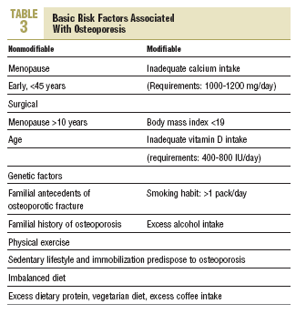
In view of our results, we suggest that community-level educational programs about this widely prevalent disorder should begin at an earlier age in order to identify modifiable risk factors (Table 3) associated with the appearance of osteoporosis.12 Such programs should also aim to identify patients who may require early treatment.13
We believe that quantitative bone ultrasound studies, despite their limitations, are a potentially useful tool for osteoporosis screening in primary care for postmenopausal women. However, uniform diagnostic criteria for osteoporosis are still not available for these techniques. As an added word of caution, ultrasound techniques and should not be used to monitor response to treatment5 as the peripheral skeleton responds to scanning with small increases in bone density that are within the margin of error of precision of these devices.


