


Secondary basilar invagination or basilar impression is an anomaly at the craniovertebral junction where the odontoid process prolapses into the foramen magnum with the risk of compressing adjacent structures and obstructing the proper flow of cerebrospinal fluid (CSF). The incidence is less than 1% in the general population and occurs mainly in the first three decades of life when it is associated with malformations of the neuroaxis. In older age, the main aetiologies are diseases that alter bone mineral density. The clinical course is usually progressive and the most common symptoms are asthenia, cervical pain and restricted movement, but also dysphonia, dyspnoea and dysphagia. It is a progressive disease which, if left untreated, can cause severe neurological damage and death. We report the case of a 79-year-old woman with osteoporosis and progressive dysphagia leading to severe malnutrition, which conditioned the decision not to intervene due to the high perioperative risk.
La invaginación basilar secundaria o impresión basilar es una anomalía de la unión craneovertebral en la que la apófisis odontoides prolapsa en el foramen magnum con el riesgo de comprimir estructuras adyacentes y obstruir el correcto flujo del líquido cefalorraquídeo. Presenta una incidencia inferior al 1% en la población general y se produce principalmente en las 3 primeras décadas de la vida, cuando se asocia a malformaciones del neuroeje. En edades avanzadas, las principales etiologías son enfermedades que alteran la densidad mineral ósea. La evolución clínica suele ser progresiva y los síntomas más frecuentes son: astenia, dolor cervical y restricción de movimientos, si bien también disfonía, disnea y disfagia. Se trata de una enfermedad progresiva que, si no se trata, puede causar daños neurológicos graves y la muerte. Presentamos el caso de una mujer de 79 años con osteoporosis y disfagia progresiva que derivó en desnutrición grave, lo que condicionó la decisión de no intervenirla por el alto riesgo perioperatorio.
Basilar impression is an alteration of the craniovertebral junction where the odontoid process prolapses into the foramen magnum with the associated neurological and life-threatening risks. The terms basilar invagination and basilar impression are often incorrectly used as synonyms in the craniovertebral junction literature, but basilar impression refers to the acquired form of basilar invagination resulting from softening of the bone at the base of the skull. It is a rare condition in the elderly, caused by diseases that alter bone mineral density, mainly rheumatoid arthritis, Paget's disease, osteomalacia, bone tumours and infections, hyperparathyroidism and osteogenesis imperfecta.1–3
We report the case of a patient whose main clinical symptom was dysphagia and who was diagnosed with a basilar impression.
The patient was a 79-year-old woman whose reason for consulting was progressive asthenia and dysphagia. Her medical personal history included euthyroid multinodular disease since 2021 and osteoporosis since 2018 treated with denosumab, calcium and vitamin D. She had no known cardiovascular risk factors, harmful lifestyle habits or surgical procedures. She had no cognitive impairment and lived at home, but needed help with basic activities and used a walking stick. Her usual treatment consisted of denosumab 60mg subcutaneously every 6 months, chondroitin sulphate 800mg daily, cholecalciferol/calcium pidolate 400/600mg daily, esomeprazole 20mg daily, lorazepam 1mg daily and paracetamol 1g/8h.
In the anamnesis the patient reported that her symptoms had started 2–3 years ago and had worsened in the last 6 months. She complained of dysphonia, a sensation of instability with some falls without cranioencephalic trauma and progressive fine motor clumsiness. She reported a weight loss of 20kg in the last 2–3 years due to hyporexia and mixed dysphagia with foreign body sensation in the oropharynx, without infectious processes suggesting bronchoaspiration. Due to the patient's clinical condition and in order to complete the study, it was decided to admit the patient to hospital.
Physical examination revealed severe cachexia with generalised muscle wasting. There were no abnormalities on cardiorespiratory auscultation or abdominal examination, no lower limb oedema or signs of venous pathology. Neurological examination showed dysphonia with nasal voice and generalised symmetrical vivid osteotendinous reflexes, preserved muscle tone and strength without fasciculations with significant generalised atrophy, no changes in speech, eye movements, pupils, campimetry, facial musculature or other lower cranial nerve pairs with symmetric elevation of the palatine pillars and normal position of the lingula, no altered sensitivity, no dysmetria of the limbs and no gait ataxia, but she required two supports to walk.
Morpho-functional assessment was performed by the Endocrinology and Nutrition Department at the time of admission: height 143cm, usual weight 50kg, current weight 30kg, weight loss 40% in 3 years, ideal weight 47kg, corrected weight 41.4kg and BMI 14.6kg/m2, hand grip strength 8kg, calf circumference (CC) adjusted to BMI of 30cm, corrected basal energy expenditure of 951kcal, total energy expenditure (corrected with a stress factor of 1.2) of 1142kcal, protein requirement of 56–70g The 24-h intake record of the shredded diet was 30% (900kcal and 40g of protein).
With the diagnosis of severe malnutrition based on the GLIM criteria, mixed dysphagia and high risk of refeeding syndrome, additional tests were performed to aid in the aetiological diagnosis. Blood tests showed prealbumin of 17mg/dl, retinol binding protein (RBP) of 2.5mg/dl and normocytic normochromic anaemia with Hb 10.1g/dl. Other parameters, including renal, hepatic and lipid profiles, vitamins B12, B1 and D, coagulation, thyroid hormones, autoimmune profile and tumour markers were in the normal range; and an endoscopy and a cervico-thoracic-abdominal computed tomography (CT) scan ruled out structural and tumour pathologies.
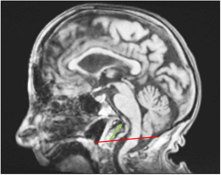
Finally, cranio-cervical magnetic resonance imaging (MRI) was performed, which showed craniometric changes at the cranio-cervical junction, with marked basilar invagination (Fig. 1), with impression of the medulla oblongata and bulbo-medullary junction, without signal intensity changes.
 5mm, defining a marked basilar invagination, imprinting the medulla oblongata and the medullary-bulbar junction.' title='Sagittal cranio-cervical MRI. Chamberlain's line (red mark) is exceeded by the odontoid process (green mark) by >5mm, defining a marked basilar invagination, imprinting the medulla oblongata and the medullary-bulbar junction.'/>
The patient was assessed by the Neurosurgery Department who, in agreement with the patient's family and the patient herself, declined surgery due to the high perioperative risk.
Management and evidenceBasilar impression is an acquired condition resulting from a change in bone tissue at the craniovertebral junction. Diagnosis is based on clinical and imaging studies demonstrating protrusion of the odontoid process into the foramen magnum beyond normal limits established by radiographic reference lines (Fig. 2). The most common signs and symptoms are asthenia, cervical pain and limited movement, and symptoms secondary to medulla oblongata and cervical spinal cord compression and CSF flow obstruction. An aetiological study should be performed, including diseases that cause changes in bone mineral density, mainly ruling out rheumatoid arthritis, Paget's disease, osteomalacia and osteoporosis, hyperparathyroidism, osteogenesis imperfecta, Hurler syndrome, and skull base tumours and infections.1–3
 5mm above this line. 2 McGregor's line between HP and the lowest point of occiput, positive diagnosis if odontoid process protrudes >2.5mm above line. 3 McRae's line between basion and opisthion should be above the odontoid process.' title='Reference lines. 1 Chamberlain's line between hard palate (HP) and opisthion (OP). Basilar impression is present if the odontoid process protrudes >5mm above this line. 2 McGregor's line between HP and the lowest point of occiput, positive diagnosis if odontoid process protrudes >2.5mm above line. 3 McRae's line between basion and opisthion should be above the odontoid process.'/>
Reference lines. 1 Chamberlain's line between hard palate (HP) and opisthion (OP). Basilar impression is present if the odontoid process protrudes >5mm above this line. 2 McGregor's line between HP and the lowest point of occiput, positive diagnosis if odontoid process protrudes >2.5mm above line. 3 McRae's line between basion and opisthion should be above the odontoid process.
This is a progressive disease and treatment is based on correction of the cranio-cervical junction with neurosurgical management and treatment of the underlying aetiology. The type of surgical intervention must be individualised and depends on certain characteristics of the patient, the presence or absence of cranial nerve deficits, C1-C2 mobility, imaging findings, being often complicated by abnormalities such as cranial settling, autofusion of cervical vertebrae, platybasia, and syringomyelia, and the coexistence of other medical conditions that increase perioperative complications. Due to the mechanical and anatomical complexities, surgical treatment involves varying combinations of anterior or posterior decompression with or without traction or fusion. Common approaches to decompression include endonasal and transoral ventral approaches (transoral atlantoaxial reduction plate (TARP) fixation) or occipital cervical posterior decompression and fusion.3,4
In our case, the patient could not be operated on because of her severe malnutrition, which makes the surgical risk too high, and her dependent baseline standard of living. Treatment focused on management of oropharyngeal dysphagia with clinical assessment of safety and efficacy of swallowing and detection of silent aspirations at the bedside. A modified swallowing assessment (MSA)5–7 was performed with negative results, and a fibreoptic endoscopic evaluation of swallowing (FEES) was attempted, but it was not possible to perform a complete evaluation due to the protrusion of the posterior pharyngeal wall from the cavum to the hypopharynx, which did not allow the passage of the fibrescope; it was possible to visualise the vocal cords which were mobile without lesions, with an adequate glottic lumen and no salivary retention. A diet of blended solids, free water in small volumes and hypercaloric and hyperproteic oral nutrition supplements were prescribed with good tolerance and no direct or indirect signs of bronchoaspiration. In addition, to avoid refeeding syndrome,8 thiamine and a multivitamin supplement were prescribed while she was in the hospital. The patient was discharged from the hospital with nutritional recommendations and treatment, maintaining the previous osteoporosis treatment, and was followed up by the nutrition service.
After six months, the patient underwent a nutritional re-evaluation with significant weight recovery (weight 51.8kg) and improvement in analytical and functional parameters (prealbumin 22.9mg/dl, RBP 4.7mg/dl, hand grip strength 12kg, CC 32cm). Symptoms had improved, with less asthenia and good oral tolerance, enabling a better quality of life, but the rest of the symptoms persisted.
Areas of uncertaintyOne of the things we were concerned about was the presence of cranial nerve deficits such as dysphagia and dysphonia due to brainstem compression. The literature reports that compression of the spinal cord and medulla oblongata can lead to severe neurological injury, and persistent compression can lead to respiratory compromise with high mortality rates. When these symptoms are present, neurosurgical decompression is required.3,4 A review of the literature shows that patients with oropharyngeal disease or a small oral space, as in our case, were preferentially treated with occipitocervical fixation, although this has a significantly higher intraoperative blood loss, longer operative time and lower rates of bone fusion at 3 and 6 months postoperatively than TARP.9 In this case, when the perioperative risks were assessed, they were too high to be taken. Furthermore, it has also been reported that preoperative nutritional support must be provided to patients with compromised nutritional status, and that failure to do so may result in wound dehiscence and even non-fusion.1,3,9,10
Another concern was what to do in cases where intervention was not possible. Sasun et al.11 published the case of a patient with basilar impression due to degenerative spondylolisthesis, with clinical symptoms of proximal upper limb weakness and torticollis. In this case, rehabilitation was chosen with improvement in symptoms, allowing independent living. There are no published cases of conservative management with clinical manifestations of cranial nerve deficits and their outcomes, neither there are cases published of basilar invagination isolated secondary to osteoporosis.
GuidelinesOn the basis of the literature reviewed, there are no well-established studies that clearly delineate a treatment algorithm for this condition. Based on the review of the literature, intervention should be considered in all patients due to its progressive course, but is of particular importance in those with high cervico-medullary and brainstem compression.1–4,9,10 Throughout the management triage process, careful preoperative planning and perioperative risk assessments should be undertaken. Treatment of symptoms and the treatment of the underlying aetiology must be carried out. In our case the patient's assessment was in accordance with the GLIM guidelines for malnutrition12 and guidelines for the risk of refeeding syndrome,13 the nutritional treatment was carried out in accordance with the ESPEN guideline,14 dysphagia was managed following the guidelines of the European Society for Swallowing Disorders,15,16 and to treat osteoporosis the European guidance for the diagnosis and management of osteoporosis in postmenopausal women were followed.17
Conclusions and recommendationsIn conclusion, basilar impression is a progressive disease that can lead to severe neurological damage and even death if left untreated.1–4,9,10 This case shows that dysphagia can be a leading symptom and its management is essential to ensure proper nutritional status, reduce perioperative risk and allow for better recovery.
Conflict of interestThe authors declare that they have no conflict of interest.
