


Although the most common clinical form of infection by Mycoplasma pneumoniae (M. pneumoniae) is respiratory, this microorganism can cause extrapulmonary manifestations, the most serious of which is neurological1. We report here a case with anisocoria and optic neuritis.
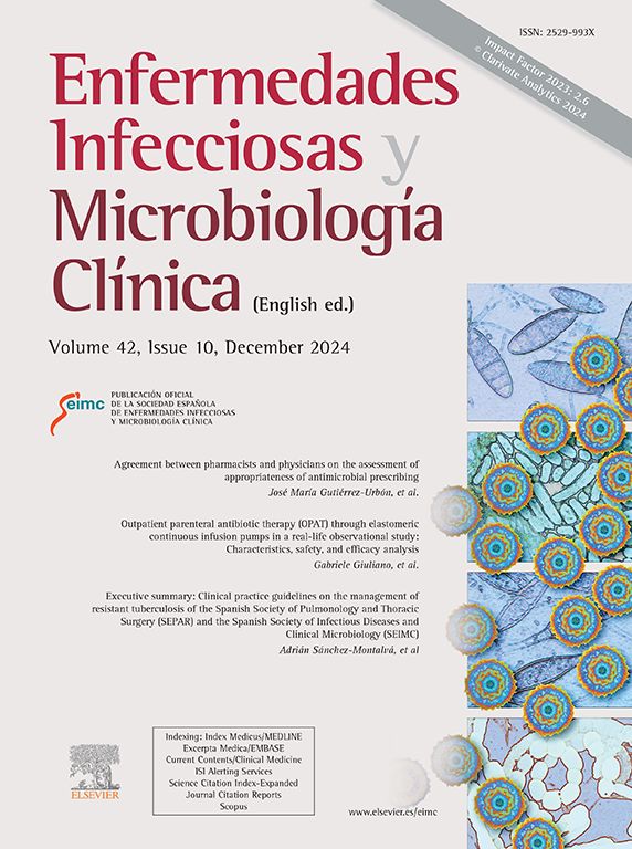
This was a 32-year-old man with no previous medical history who had a two-week history of productive cough, dyspnoea, low-grade fever, nasal congestion and left hearing loss. Chest X-ray (Fig. 1, upper) revealed an infiltrate in the right upper lobe. Legionella pneumophila and pneumococcal urinary antigen tests were negative and treatment was started with amoxicillin/clavulanic acid 875/125mg/8h.
 and brain MRI of the patient (lower image).")
Two days later, the patient had not improved and attended Accident & Emergency; he had tachypnoea, with a baseline saturation of 88%, which rose to 95% with nasal cannula at 2 lpm, HR 46 bpm, blood pressure 124/68 and rhonchi in the upper right lung field.
Tests revealed leucocytosis with neutrophilia, coagulopathy (INR 1.3) and C-reactive protein of 103mg/l, and a repeat chest X-ray showed no changes. Treatment was escalated to ceftriaxone 2g/24h with levofloxacin 500mg/24h and the patient was admitted to internal medicine.
On day three of admission he developed anisocoria, with greater mydriasis in his left eye. CT scan of the brain showed no acute intracranial findings. Pilocarpine eye drop test showed involvement of the left third cranial nerve.
CT angiogram of the head ruled out vascular injury and MRI ruled out cavernous sinus pathology, showing right maxillary sinusitis and left ethmoid sinus retention cyst (Fig. 1, lower). After 24h, the anisocoria resolved spontaneously.
Multiplex PCR was carried out on nasopharyngeal exudate and sputum, which was positive for M. pneumoniae and negative for coronavirus, MERS-CoV, rhinovirus/enterovirus, influenza and parainfluenza virus, metapneumovirus, adenovirus and respiratory syncytial virus. Serum was also positive for M. pneumoniae IgM. HIV, syphilis and hepatotropic virus serologies were all negative. Azithromycin 500mg/24h for seven days was prescribed.
Four days later, the patient consulted once more due to loss of visual acuity in his left eye. Visual field testing revealed a diffuse loss of sensitivity with a superior altitudinal visual field defect in the left eye and visual acuity of 0.5. Lumbar puncture ruled out infection of the central nervous system. As parainfectious retrobulbar optic neuritis was suspected, he was started on corticosteroid therapy and made a full recovery.
At subsequent check-ups, the electroencephalogram, cervical spine magnetic resonance imaging and brain magnetic resonance angiography were normal, and the follow-up lumbar puncture showed no oligoclonal bands. Blood aquaporin 4 antibodies and myelin oligodendrocyte glycoprotein antibodies were negative, but he had a mild sustained IgG2 deficiency. After seven weeks, he continued to be IgM positive, with IgG seroconversion against M. pneumoniae (signal 1.41).
The incidence of central nervous system complications due to Mycoplasma spp. has not been established; it ranges from 1% to 7% in hospitalised patients, with the mortality rate as high as 10%1. Over half of cases are found in patients between 6 and 21 years of age2, but it also occurs in adults. Parainfectious neuritis usually occurs bilaterally and is more active in children3, and up to 20% may have no prior respiratory infection4.
There are several mechanisms involved in the neurological complications, none of which is exclusive. There may be direct cell damage following haematogenous spread of the microorganism reaching the central nervous system. When the bacteria damages cells, the innate immune response is activated, attracting different cytokines such as IL 18, which activates helper T cells 1 and 2, or IL 8, which attracts neutrophils. It can be mediated by immune complexes due to antigenic elements, which bind to the patient's macrophages and monocytes, initiating immune reactions due to the antigenic similarities between M. pneumoniae and brain parenchyma antigens5. Vascular damage is also possible6.
The incidence of Mycoplasma spp. infections is increasing in patients with hypogammaglobulinaemia, which indicates compromise of humoral immunity in the pathogenesis of these cases7. Antibodies inhibit the growth of M. pneumoniae in vitro, preventing proliferation on the surface of colonised mucosa8, without affecting neutrophil opsonisation9.
Our patient had optic neuritis as a rare complication in M. pneumoniae infections. It is likely that partial IgG2 deficiency facilitated infection and cell mimicry led to the neurological symptoms. We need to be aware of the association between hypogammaglobulinaemia and the risk of M. pneumoniae infection, as these patients can suffer from more severe and prolonged illnesses and develop more complications10.
