Valorar la utilidad de la técnica positron emission tomography (PET, «tomografía por emisión de positrones») con 18F-fluorodesoxiglucosa (18F-FDG) combinada con tomografía computarizada (TC) en la localización del tumor primario en pacientes diagnosticados de tumor de origen desconocido (TOD).
Pacientes y métodoSe analizaron retrospectivamente los estudios PET-TC con 18F-FDG realizados, entre noviembre de 2006 y noviembre de 2010, a pacientes con el diagnóstico de TOD para búsqueda de tumor primario. A todos los pacientes se les realizó un estudio estándar PET-TC, 50-60 minutos después de la administración intravenosa de 296-370MBq de 18F-FDG. Se valoraron los estudios PET-TC en patológicos, con/sin identificación del tumor primario, y no patológicos. El diagnóstico final se estableció mediante confirmación histológica y/o seguimiento clínico/radiológico superior a 6 meses.
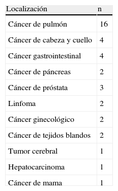
ResultadosSetenta y cuatro pacientes fueron estudiados (59 varones, 15 mujeres), con un intervalo de edad de 41-89 años. En 38 (51%) pacientes la PET-TC determinó correctamente el origen del tumor primario, realizándose confirmación histológica en 8 casos sobre el mismo. En 4 pacientes la PET-TC mostró un resultado falso positivo.
ConclusiónLa técnica PET-TC permitió identificar el 51% de los tumores primarios en nuestra muestra de pacientes.
We determined the utility of the 18F-fluorodeoxyglucose (18F-FDG) positron emission tomography (PET)-computerized tomography (CT) in the localization of the primary tumor in patients with tumor of unknown origin (TUO).
Patients and method18F-FDG PET-CT scans, performed between November 2006 and November 2010, in search for the primary tumor in patients with TUO, were retrospectively evaluated. Patients underwent a standard PET-CT, 50-60minutes after intravenous injection of 296-370MBq 18F-FDG. PET-CT studies were assessed as pathological, with/without identification of the primary tumour and no pathological. Final diagnosis was established by histological confirmation and/or clinical/radiologic follow-up longer than 6 months.
ResultsWe studied 74 patients (59 males, 15 females), with ages ranging from 41-89 years. In 38 (51%) patients the PET-CT assessed the correct origin of the primary tumour. In 8 cases, a histological confirmation in the primary lesion was obtained. In 4 patients the PET-CT showed a false positive result.
ConclusionPET-CT scanning identified 51% of the primary sites in our sample of patients.
Artículo
Comprando el artículo el PDF del mismo podrá ser descargado
Precio 19,34 €
Comprar ahora


