


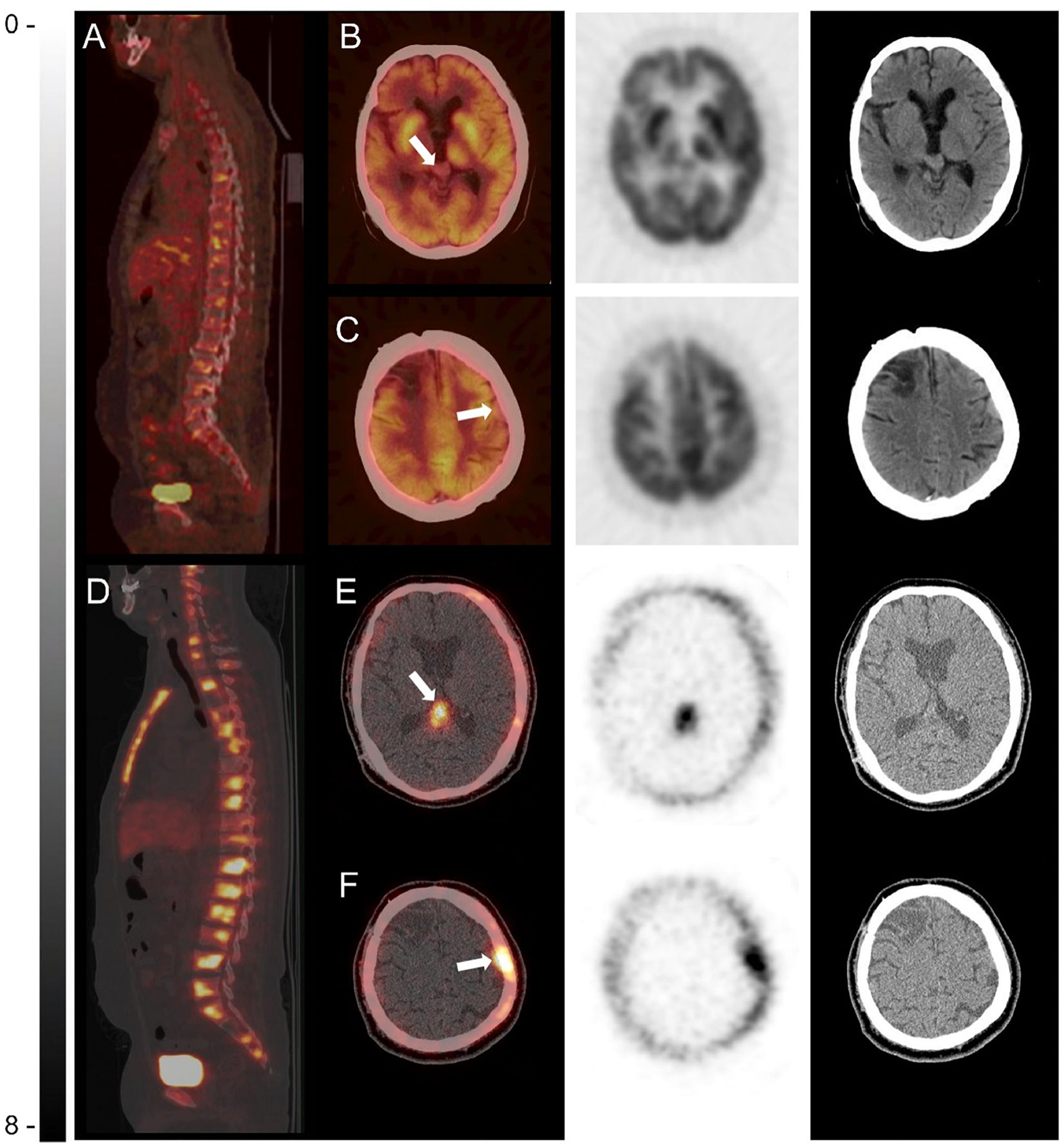
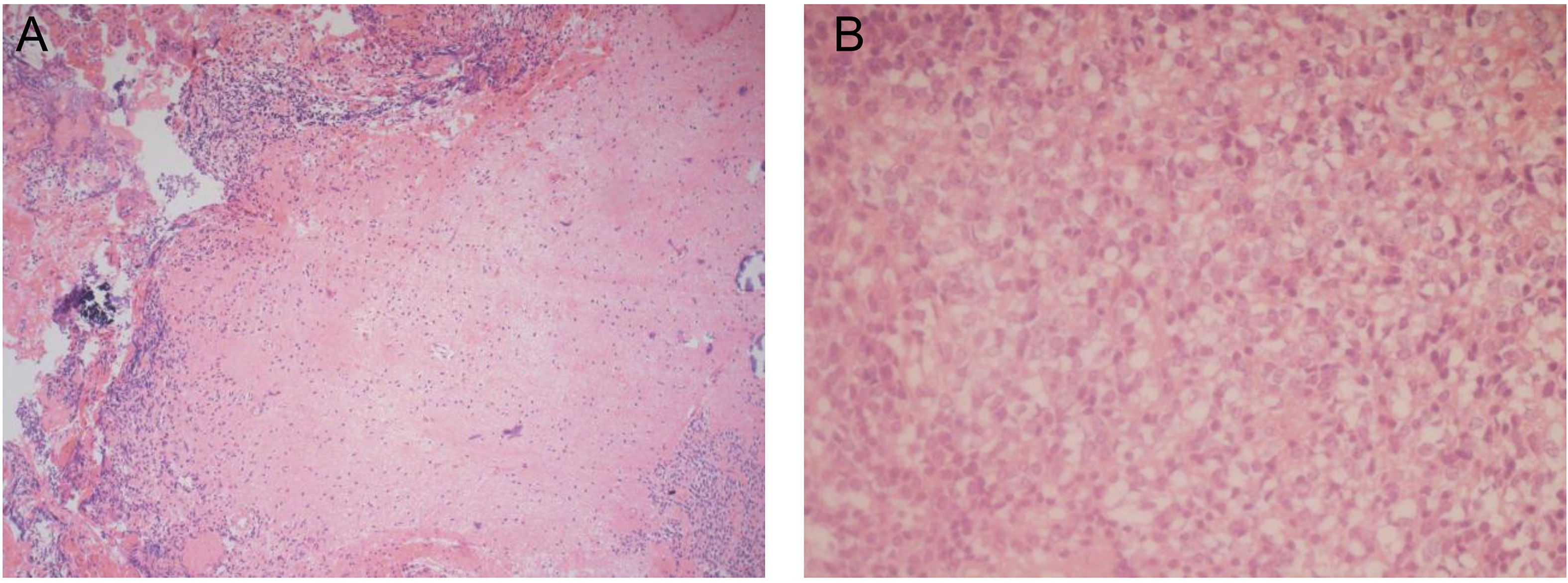
A 45-year-old man with a history of pinealoma and radical pinealectomy 15 years ago. Recently, the patient went to hospital for treatment with dizziness, weakness and fever. 18F-FDG PET/CT in local hospital demonstrated pinealoma recurrence with systemic bone metastases (Fig. 1A and B, the white arrow shows the current lesion in the pineal region), the left frontoparietal lesion (Fig. 1C, the area indicated by the white arrow) was misinterpreted as meningioma due to no abnormality in glucose metabolism. 68Ga-DOTATOC PET/CT performed in our hospital showed increased metabolism of pineal region (Fig. 1E, the region indicated by the white arrow is consistent in 1B), left frontoparietal lesion (Fig. 1F, the region indicated by the white arrow is consistent in 1C) and multiple bones (Fig. 1D), which were diagnosed as pinealoma recurrence with intracranial and extracranial metastases. Finally, combined with the previous pathological sections of pinealoma (Fig. 2A) and results of bone marrow biopsy (Fig. 2B), it was confirmed as pineal parenchymal tumors of intermediate differentiation (PPTIDs) recurrence with intracranial and extracranial metastases by multi-disciplinary team consultation.


PPTIDs is a rare lesions, and extraneural metastasis is even more rare[1]. The 2016 World Health Organization (WHO) Classification of Central Nervous System Tumors recognized three pineal parenchymal tumors (PPTs): pineocytomas, pineoblastomas, and PPTIDs grade II–III. The WHO grade II–III PPTIDs represent between 20% and 62% of PPTs [2,3]. In the present case, there was a history of pinealoma resection and this time with multiple systemic metastases. PET/CT shows that the glucose metabolism of left frontoparietal lesion is not consistent with other lesions, which makes the diagnosis difficult. Further octreotide imaging shows a good consistency between the primary focus and the metastatic focus, providing very useful diagnostic information. It shows the important value of octreotide imaging in the diagnosis of neuroendocrine tumors.
Authors' contributionsHP and ZY: preparation of manuscript and retrieval of images; ZY: involved in the initial workup and management; ZXS and HP: oversaw preparation of manuscript and decided on final submission.
Competing interestsNone declared.
FundingThe authors have not declared a specific grant for this research from any funding agency in the public, commercial or not-for-profit sectors.
