metricas
covid
Buscar en
Medicina Clínica Práctica
Toda la web
Inicio Medicina Clínica Práctica Toxic epidermolysis after diclofenac
Información de la revista
Vol. 6. Núm. 1.
(enero - marzo 2023)
Compartir
Compartir
Descargar PDF
Más opciones de artículo
Vol. 6. Núm. 1.
(enero - marzo 2023)
Images in medicine
Open Access
Toxic epidermolysis after diclofenac
Necrólisis epidérmica tóxica por diclofenaco
Visitas
537
Vasco Nevesa,
Autor para correspondencia
vbenj@icloud.com

Corresponding author.
, Maria Inês Gonçalvesb, Maria Pachecoa, Renato Gonçalvesb, Gonçalo Mirandab, Ana Moreiraa, Pedro Venturaa
a Serviço de Medicina Interna, Hospital Sousa Martins – ULS Guarda, Av. Rainha D. Amélia, Guarda, Portugal
b Serviço de Medicina Interna, Centro Hospitalar Universitário Cova da Beira, Quinta do Alvito, 6220-251 Covilhã, Portugal
Este artículo ha recibido

Under a Creative Commons license
Información del artículo
Texto completo
Descargar PDF
Estadísticas
Figuras (3)
Mostrar másMostrar menos
Texto completo

Lyell Syndrome is a rare and severe mucocutaneus adverse reaction mostly common induced by medication. It is related to Stevens-Johnson syndrome and both are collectively known as epidermal necrolysis. Classification is based on the affected total body area, with Lyell syndrome skin detachment affecting more than 30% of the total body area, and therefore is a more severe condition.

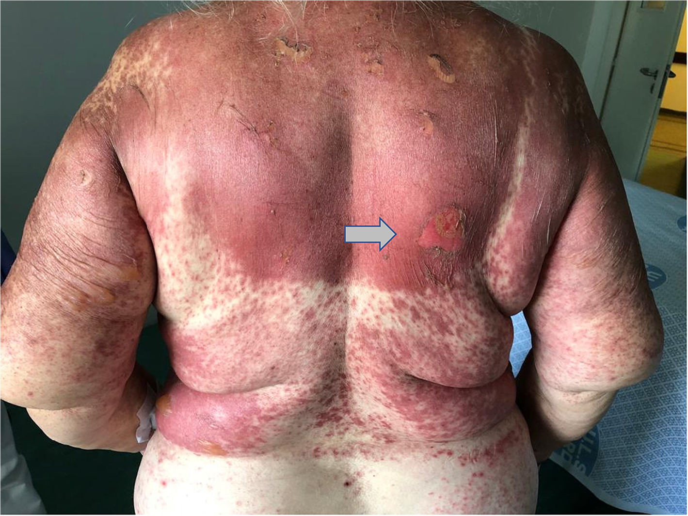
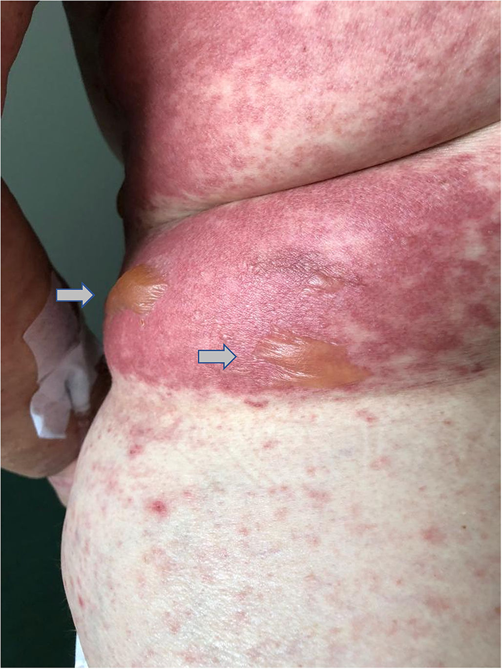
This case reports a 75 year-old woman whom was prescribed diclofenac after having fallen at home. One day after starting diclofenac, the patient showed up to the emergency department complaining of multiple skin lesions (that started in the right arm). Clinical examination revealed a generalized disseminated mucocutaneous reaction, characterized by confluent macular erythematous skin lesions (Fig. 1), associated with blisters, namely in the arms, posterior trunk and feet (Fig. 2 and 3). The patient started immunoglobulin associated with corticosteroid and anti-histamines, along with skin care. Over time, more and more blisters appeared, affecting more that 30% of total body area, becoming very difficult to control patient pain.

Fig. 1.

Dorsal area involvement. Coalescent macular erythematous skin lesions with ruptured blister (arrow).

(0,24MB).
Fig. 2.

Blisters present in the right flank (arrows), surrounded by macular erythematous lesions.

(0,19MB).
Fig. 3.

Patient feet with bulky blisters in its toes.

(0,13MB).

Medical team decided to transfer the patient to a Burn Unit, so that the best wound care could be provided. Later, the patient developed a respiratory complication, namely pneumonia with severe ARDS and sadly died.

Copyright © 2022. The Author(s)
Opciones de artículo
es en pt

¿Es usted profesional sanitario apto para prescribir o dispensar medicamentos?

Are you a health professional able to prescribe or dispense drugs?

Você é um profissional de saúde habilitado a prescrever ou dispensar medicamentos

Quizás le interese:
10.1016/j.mcpsp.2020.100179
No mostrar más