Definir el impacto diagnóstico de la PET/TC con 18F-FDG en función de las características clínicas del síndrome paraneoplásico neurológico (SPN).
Material y métodosEstudio retrospectivo multicéntrico y longitudinal de pacientes con sospecha de SPN.
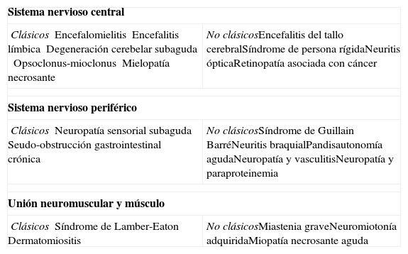
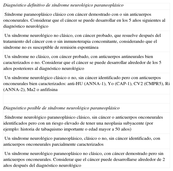
El cuadro clínico se clasificó en síndrome clásico (SC) o no clásico (SNC). Tras el seguimiento se estableció el diagnóstico de SPN definitivo o posible. Los cuadros que no encajaron en ninguna de las categorías previas se catalogaron como no clasificables. Se analizó el estado de los anticuerpos onconeuronales.
La PET/TC se clasificó en positiva o negativa para la detección de malignidad. Se determinó la relación entre los hallazgos PET/TC y el diagnóstico final. Se analizaron las diferencias entre variables (Chi cuadrado de Pearson) y la relación entre el resultado de la PET/TC y el diagnóstico definitivo.
ResultadosSe analizaron 64 pacientes. El 30% de los cuadros clínicos se catalogaron como SC y el 42% como SNC. Tras el seguimiento el 20% se clasificó en SPN posible y el 16% en definitivo. El 13% de los pacientes tenía anticuerpos onconeuronales positivos.
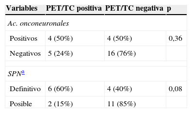
El hecho de poseer un SPN definitivo se relacionó con un resultado positivo de la PET/TC (p=0,08). Se demostró relación significativa entre la positividad de los anticuerpos y el diagnóstico final de proceso neoplásico (p=0,04).
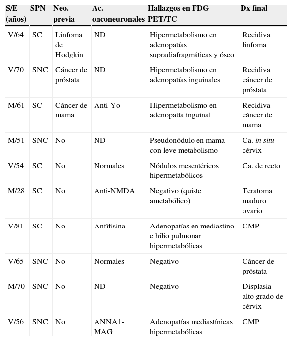
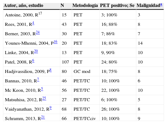
La PET/TC fue eficaz en la correcta localización tumoral en 5/7 casos con cáncer invasivo.
ConclusionesLa PET-TC mostró un mayor porcentaje de resultados positivos en pacientes con diagnóstico de SPN definitivo. A pesar de la baja prevalencia de malignidad en nuestra serie, la PET/TC detectó malignidad en una significativa proporción de pacientes con cáncer invasivo.
This study aimed to determine the diagnostic impact of 18F-FDG PET/CT based on the clinical features of paraneoplastic neurological syndrome (PNS).
Material and methodsMulticenter retrospective and longitudinal study of patients with suspicion of PNS.
The clinical picture was classified into classic (CS) and non-classic syndrome (NCS). After the follow-up, the definitive or possible diagnosis of PNS was established. The pictures that did not match any of the previous criteria were categorized as non-classifiable. The state of the onco-neural antibodies was studied.
The PET/CT was classified as positive or negative for the detection of malignancy. The relationship between PET/CT findings and the final diagnosis was determined. The differences between variables (Pearson test X2) and the relationship between the results of the PET/CT and the final diagnosis were analyzed.
ResultsA total of 64 patients were analyzed, classifying 30% as CS and 42% as NCS. After the follow-up, 20% and 16% of subjects were diagnosed as possible and definitive PNS, respectively. Positive onco-neural antibodies were found in 13% of the patients.
A definitive diagnosis of PNS was associated with a positive PET/CT (P=.08). A significant relation between antibodies expression and final diagnosis of neoplasia (P=.04) was demonstrated.
The PET/CT correctly localized malignancy in 5/7 cases of invasive cancer.
ConclusionsThe PET/CT showed a higher percentage of positive results in patients with definitive diagnosis of PNS. Despite the low prevalence of malignancy in our series, the PET/CT detected malignancy in a significant proportion of patients with invasive cancer.
Artículo
Comprando el artículo el PDF del mismo podrá ser descargado
Precio 19,34 €
Comprar ahora


