La lectura automatizada de citología basada en ThinPrep® es una evidente ayuda para el citotécnico y para el patólogo, al identificar campos de interés para ser evaluados. Algunos autores han demostrado la utilidad de la citología líquida ThinPrep® como sustitución del citospin en citología de orina, pero existe muy escasa información acerca de su utilidad.
ObjetivoEvaluar la utilidad de la lectura automática de citología, en citología de orina.
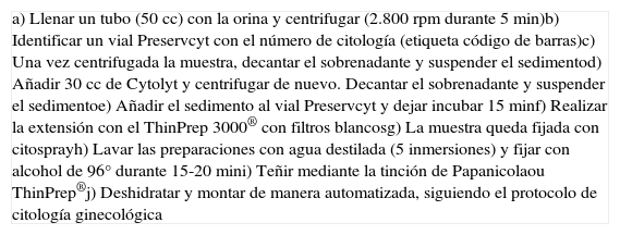
Material y métodosEstudio retrospectivo de 180 extensiones de orina. Procesamiento según protocolo con plataforma ThinPrep®, tinción automatizada con teñidor Leica®, lectura manual de la extensión y diagnóstico. Lectura automatizada de citología utilizando Imager® y posterior revisión por los citotécnicos.
ResultadosLos resultados fueron clasificados como positivos, negativos, atipia o polioma.
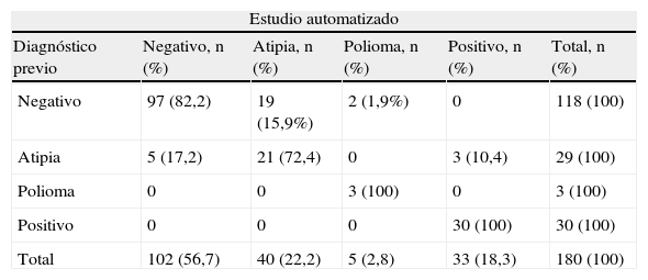
La coincidencia diagnóstica fue del 83,9% (Kappa; IC95%: 0,713 [0,619-0,807]). Todos los casos positivos fueron detectados por el sistema automático. De los 118 casos interpretados previamente como negativos, 97 (82%) fueron reinterpretados como tales. Ningún caso informado previamente como negativo fue interpretado como positivo.
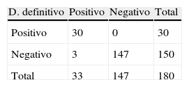
La sensibilidad y el valor predictivo negativo del Imager® para la detección de carcinoma en esta serie son del 100%. La especificidad es del 98%, y el valor predictivo positivo, del 91%.
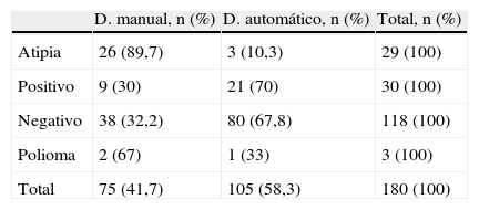
El citotécnico revisó 75 de las 180 laminillas (41,7%), la mayoría por atipias (89,6%) y negativos (32,3%).
ConclusionesEl uso del Imager® como pre-cribado podría ayudar en la interpretación de la citología de orina. El porcentaje de casos que no precisan de revisión completa podría aumentar con la experiencia del citotécnico.
ThinPrep® Imager system reading method of liquid-based cytology is currently a very helpful tool for both the cytotechnologist and the pathologist. Some authors have shown the usefulness of liquid-based cytology ThinPrep® as a replacement of the citospin in urine cytology. However, there is very little information about its usefulness.
ObjectiveTo evaluate the utility of reading urine cytology using ThinPrep® Imager System.
Material and methodsA retrospective study of 180 urine cytology smears was carried out. Processing was done according to ThinPrep® platform protocol, automated staining with stainer Leica® and manual reading of the slide and diagnosis, followed by automated reading of cytology using Imager®. Review by cytotechnologists.
ResultsThe results were classified as positive, negative, atypia or polioma.
Diagnostic coincidence was 83.9% (Kappa; 95%IC, 0.713 [0,619-0,807]). All positive cases were detected by the automated system. Of the 118 cases previously interpreted as negative, 97 (82%) were reinterpreted as such. None previously reported as negative were interpreted as positive in the study with Imager®.
Sensitivity as well as negative predictive value of the Imager® for the detection of carcinoma in this series are 100%. The specificity is 98% and the positive predictive value is 91%.
The cytotechnologist reviewed 75/180 slides (41.7%), mostly with atypia (89.6%) or negative (32,3%).
ConclusionsThe use of the Imager® as a pre-screening method could help in the interpretation of urine cytology and a useful tool for the cytotechnologist. The percentage of cases that do not require complete revision could increase with the experience of the cytotechnologist.
Artículo
Comprando el artículo el PDF del mismo podrá ser descargado
Precio 19,34 €
Comprar ahora


