


Dermatophytic infections are a worldwide public health problem. In Nigeria, children of nomadic herdsmen are thought to be more at risk because of their early and continuous exposure to different kinds of animals. However, little is known about the level of infection in these children in southeastern Nigeria or elsewhere within the West African sub-region.
ObjectiveThis study investigated dermatophytic infections among children of nomadic herdsmen living in isolated camps in southeastern Nigeria from October 2008 to May 2009.
Methods390 children aged between 4 and 17 years with clinically suggestive lesions were sampled. Isolates were identified by microscopy, culture or both. Biochemical tests and sequencing of the ITS2 region of the ribosomal DNA were used to identify isolates with atypical morphology.
Results162 children were confirmed to be infected. Ten different species of dermatophytes were recovered with Trichophyton mentagrophytes showing the highest incidence. There was a significant difference (P < 0.05) in the frequency of isolates recovered among the different age groups screened. No significant (P > 0.05) observations were found according to gender or location of the two camps investigated.
ConclusionThis study suggests that tinea capitis is the predominant clinical type of dermatophytosis among children exposed to different kinds of animals based on parental nomadic lifestyle. It also dominated in children with mixed infections in different anatomical sites. The high prevalence of 41.25% suggests that animal to child transmission may be very common.
Las infecciones por dermatofitos son un problema de salud pública en todo el mundo. En Nigeria, los hijos de los pastores tienen mayores probabilidades de sufrirlas debido a su exposición temprana y continua a diversos animales. Sin embargo, poco se sabe sobre el nivel de infección en estos niños en el sureste de Nigeria o de cualquier otra parte del oeste de África.
ObjetivosEste estudio investigó las infecciones por dermatofitos entre los hijos de pastores nómadas que viven en campamentos aislados en el sureste de Nigeria. El periodo de estudio abarcó desde octubre de 2008 hasta mayo de 2009.
Métodos390 niños de edades comprendidas entre los 4 y 17 años, con lesiones sugestivas de dermatofitosis, fueron incluidos en la muestra. Los aislamientos fueron identificados por microscopía, cultivo o por ambas técnicas. Para la identificación de aislamientos con morfología atípica, se utilizaron pruebas bioquímicas y la secuenciación de la región ITS2 del ADN ribosómico.
ResultadosSe aislaron un total de 10 especies de dermatofitos en 162 niños, con Trichophyton mentagrophytes con el mayor número de aislamientos. Hubo una diferencia significativa (p < 0,05) en la frecuencia de aislamientos entre los diferentes grupos de edad estudiados. La ubicación de los campamentos estudiados o el sexo de los niños no fueron variables significativas (p > 0,05).
ConclusiónEste estudio sugiere que la tiña de la cabeza es la dermatofitosis predominante en los niños expuestos al contacto con animales donde los padres tienen un estilo de vida nómada. En niños con infecciones mixtas en diferentes localizaciones anatómicas la tiña de la cabeza fue también la de mayor importancia. La alta prevalencia del 41,25% sugiere que los animales son transmisores habituales de los agentes etiológicos.
Dermatophytes, including Trichophyton, Microsporum and Epidermophyton, cause superficial fungal infections of the skin and are a public health problem.11 An increase in the incidence of dermatophytoses has been noted worldwide, especially in developing countries such as Nigeria,5-7,9,12 among the elderly and in immunocompromised patients.14 Our group and others have carried out several surveys in Nigeria on the clinical and etiological aspects of dermatophytoses.7,11,12,14 In Nigeria (shown in Fig. 1) and surrounding neighboring countries of Chad, Ghana, Niger and Benin Republic, nomadism – that is races or tribes without fixed place of abode – is common among the predominantly Hausa/Fulani kindred. Since they rear animals such as cows, camels, sheep and goats, they usually move from place to place according to the availability of pasture or food supplies required by the animals under their care. The animals are kept for use as source of meat, milk and sometimes blood, and their hides and hair are used for clothing, tents and other equipment. The children of nomads are thereby exposed to a variety of risks and infections. In recent times, the life pattern of nomads has been changing. They typically migrate from the predominantly dry northern Nigeria and neighboring countries to the southern part of Nigeria, where they live in camps. To date, there have been no studies among the children of nomads aimed at investigating dermatophyte infections, and this information is important since these children interact with persons outside their community, who visit nomadic camps to transact business or work and/or play with these children, potentially leading to the spread of dermatophyte infections, and possibly contributing to increasing infections in Nigeria, which is already known to be endemic for dermatophytoses.12 The present study was conducted to investigate the incidence of dermatophytoses, their causative agents and their distribution according to age group, gender and body sites amongst children of northern herdsmen living in southeast Nigeria.
Materials and methodsStudy background and population.")
The studied areas are part of southeastern Nigeria. They were the Awka and Enugu nomadic camps. In these areas the nomadic Fulanis/Hausas are usually found. They rear some domestic animals such as goats, sheep, and camels, among others. Most of the children sampled in the study, particularly the males, rarely attend school as they follow their fathers in herding.
Pre-survey contact and mobilizationA preliminary survey and consent approvals were first carried out in the camps/bush encampments, in order to ensure maximum co-operation including sampling of siblings. This was successfully carried out through the consent of the heads of encampments, who mobilized participants for sample collection. All children aged 4–16 years, whose parents had been informed and who gave consent, were included in the study. A questionnaire was administered for each child and included questions about age, sex, animal contact and other questions relevant to the study. Each child with clinically suggestive lesions was examined for any superficial fungal infection, and samples were taken from relevant lesions. The clinical examination and sample collection were performed between October 2008 and May 2009. A total of 390 children in the specified age group were included, of which 205 were boys and 185 girls.
Specimen collection/study protocolSpecimen collection and study protocol adopted were as described in previous investigations with some modifications.11,12 Briefly, the affected skin was cleaned with alcohol and the advancing border of the lesion was scraped with the blunt edge of a sterile disposable scalpel. Hairs and scales were plucked with sterile tweezers. For transport of specimens, clean, dry presterilized paper envelopes were used. Samples were transported to the laboratory and processed within 24h. Portions of specimens were treated with 10% KOH for microscopic identification of typical hyphae or arthroconidia at ×100–400 magnifications. For the cultures, Dermasel agar (Oxoid, UK) slants, supplemented with cycloheximide, 0.4 mg/l, chloramphenicol 0.05mg/l and gentamicin 0.16mg/l, was used as a standard substrate. Cultures were incubated for 4–6 weeks at 30°C. Identification of dermatophyte species was performed by macro- and micro-morphological examination of colonies and by biochemical methods.3 In the cases of isolates that had atypical morphology and/or biochemical test results, sequencing of rDNA internal transcribed spacer region 2 (ITS 2) was performed.18
Statistical analysisStatistical analysis was done using the statistical package SPSS version 10.0 for windows.
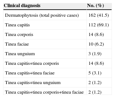
ResultsOut of the 390 children sampled, 162 (41.5%) had dermatophytoses. Tinea capitis was the predominant clinical type with a total number of 112 (69.1%) cases. It also dominated in children with mixed infections in different anatomical sites, having been found to be present in all cases of mixed infections examined (23, 14.2%) (Table 1). For the mixed infections, there were 14 (8.6%) cases of tinea capitis and tinea corporis, 5 (3.1%) of tinea capitis and tinea faciae and 2 (1.2%) of tinea capitis/tinea unguium. Only 2 (1.2%) cases were mixed infections that involved more than two clinical types. There were also 14 (8.6%) cases of tinea corporis, 10 (6.2%) tinea faciae and 3 (1.9%) of tinea unguium infections (Table 1). One hundred and fifteen nomadic children out of the 162 with clinically diagnosed lesions in Enugu and Awka camps, 115 were positive for dermatophytic infection by both microscopy and culture, 28 by microscopy alone and 19 by culture alone. The number of children therefore with lesions positive by microscopy or culture or both was 162, giving an incidence of 41.5% with respect to the total number of children investigated 390.
Clinical presentation in 390 children of nomadic herdsmen screened for dermatophytoses.
| Clinical diagnosis | No. (%) |
| Dermatophytosis (total positive cases) | 162 (41.5) |
| Tinea capitis | 112 (69.1) |
| Tinea corporis | 14 (8.6) |
| Tinea faciae | 10 (6.2) |
| Tinea unguium | 3 (1.9) |
| Tinea capitis+tinea corporis | 14 (8.6) |
| Tinea capitis+tinea faciae | 5 (3.1) |
| Tinea capitis+tinea unguium | 2 (1.2) |
| Tinea capitis+tinea corporis+tinea faciae | 2 (1.2) |
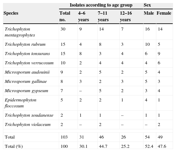
The incidence of dermatophytosis was significantly higher (P<0.05) in children aged 7–11 years, followed by children aged 4–6 years and children aged 11–16 years (Table 2). There was no significant difference (P>0.05) either in the general occurrence of dermatophytes in the two camps studied (data not shown) or among the two sex groups studied. A total of 103 isolates representing 10 species, namely Trichophyton mentagrophytes, Trichophyton rubrum, Trichophyton tonsurans, Trichophyton verrucosum, Microsporum audouinii, Microsporum gallinae, Microsporum gypseum, Microsporum canis, Trichophyton soudanense and Trichophyton violaceum, were recovered. The distribution of the different species according to age groups and sex is shown in Table 2. T. mentagrophytes was the most common etiological agent (29.1%) of dermatophytes recovered by culture were from children aged 7–11 years followed by those from 4 to 6 years and the least.
Distribution of dermatophytes according to age group and sex among the children of nomads sampled in the study.
| Isolates according to age group | Sex | |||||
| Species | Total no. | 4–6 years | 7–11 years | 12–16 years | Male | Female |
| Trichophyton mentagrophytes | 30 | 9 | 14 | 7 | 16 | 14 |
| Trichophyton rubrum | 15 | 4 | 8 | 3 | 10 | 5 |
| Trichophyton tonsurans | 15 | 8 | 3 | 4 | 6 | 9 |
| Trichophyton verrucosum | 10 | 2 | 4 | 4 | 4 | 6 |
| Microsporum audouinii | 9 | 2 | 5 | 2 | 5 | 4 |
| Microsporum gallinae | 8 | 3 | 2 | 3 | 5 | 3 |
| Microsporum gypseum | 7 | – | 5 | 2 | 3 | 4 |
| Epidermophyton floccosum | 5 | 2 | 2 | 1 | 4 | 1 |
| Trichophyton soudanense | 2 | 1 | 1 | – | 1 | 1 |
| Trichophyton violaceum | 2 | – | 2 | – | – | 2 |
| Total | 103 | 31 | 46 | 26 | 54 | 49 |
| Total (%) | 100 | 30.1 | 44.7 | 25.2 | 52.4 | 47.6 |
The present study has confirmed that tinea capitis is the predominant clinical type of dermatophytosis among children of herdsmen living in nomadic camps in southeastern Nigeria. In the past, several authors have reported tinea capitis as an important clinical problem widely distributed throughout the world, especially among children.2,4,10–12,17 In Nigeria, the incidence of dermatophytoses has been increasing in the last decade. The spectrum of the species and their frequency are known to vary from one geographical area of the country to another, based on several factors.11,12 In addition to geography, other possible factors such as locality, socioeconomic status, age, gender, animal contact and communal lifestyle are also important factors for the transmission of dermatophytes.11,12 This study aimed to specifically elucidate the role of animal contact in dermatophyte etiology and transmission in children exposed to different kinds of animals based on parental nomadic lifestyle.
There was a difference between the number of positive samples by microscopy and actual number of isolates recovered by culture in the study. This could be due to the nonviability of arthroconidia and hyphae which are recognized microscopically.
The dermatophytes recovered were either antropophilic, geophilic or zoophilic species. However, the recovery of 15 isolates of anthropophilic species (T. tonsurans) and seven geophilic isolates (M. gypseum) deserves mention. The finding of T. tonsurans as the second most occurring species with T. rubrum is not surprising because T. tonsurans was previously reported as the predominant species among children in the Anambra state,12 where one of the nomadic camps investigated is located. T. soudanense and E. floccosum were recovered as etiological agents. These are not typically animal-acquired dermatophytes and it is possible that these children acquired them from other sources. However, there are reports of previous isolation of both species from animals.13
The high frequency of T.mentagrophytes, and the relatively high frequency of T. rubrum, T. verrucosum, M. audouniii and M. gallinae is understable: first, animals have been implicated in the transmission of these species and second, rearing of animals is the major occupation of the parents of these children. In addition, some of the male children of nomads participate in rearing of the animals. Some authors1 also reported that T. mentagrophytes was responsible for 13.7% of similar cases in Italy. The recovery of seven isolates of M. gypseum in the study would appear to dispute the view of Philpot15 that this organism rarely occurs as an etiological agent in Africa. However, this finding is in agreement with previous data on dermatophytes recovered from animals in northern Nigeria.13 It is probably possible that this species has re-emerged and clinicians should be aware of this species as a potential infectious agent. The ranking of T. rubrum and T. verrucosum as the second and third most frequent in this study, respectively, is not surprising. T. verrucosum has been predominantly recovered from cattle by previous investigators,16 and T. verrucosum from infected skin and hair, remaining viable and infective for a period of 15 months up to 4 years, thus providing an opportunity for the continuous infection of new animals introduced in the flock.8 The same authors also observed in a different study that children got T. verrucosum infection from debris on clothing worn by others in a dairy barn.13 Furthermore, it is normal to suspect that the origin of the recovered dermatophytes is from the animals, but it may be difficult to suggest where the animals got them. It is possible that the animals got infected in the Nigeria environment or from other neighboring countries in West Africa. The animal trade between Nigeria and countries like Niger, Benin, Ghana and Benin Republic could be important transmission routes. Unfortunately, not much is currently done to control the movement of these animals by the authorities of these countries at the borders. This raises a lot of concern considering the apparent free movement of persons within the West African sub-region. In the literature, information is scarce on documented cases of a similar profile of dermatophytosis amongst children in the neighboring West African countries. One way of protecting these children from infections by dermatophytes and other agents is by ensuring that government agencies regulate and monitor these nomadic camps.
In most animals, symptoms of mycoses are sometimes too discrete or recognized too late and are often left untreated, leading to transfer of infections to humans. In other cases, mycotic diseases in animals persist asymptomatically without evident clinical signs and thus become a reservoir for transmission. Routine check on animals and treatment of infected ones is also very essential. Furthermore, a supposed collaborative effort by governments of neighboring countries in the West African sub-region will certainly prove useful.

