


Presentamos el caso de un hombre de 75 años con adenocarcinoma sigmoide metastásico con compromise único del lóbulo hepático izquierdo. Post sigmoidectomía y metastectomía hepática, fue derivado para cintigrafía hepatobiliar (HBS por sus siglas en inglés), dado que presentaba una fuga biliar persistente a través de sus drenajes. Las exploraciones se realizaron con imágenes dinámicas y estáticas.
La HBS es un examen ampliamente utilizado para el diagnóstico de interrupción de las vías biliares. A pesar de brindar importante información, rara vez puede demostrar específicamente el lugar de lesión. En este caso, se pudo identificar, de manera excepcional, la fuga de bilis en la porción anterior del octavo segmento hepático.
We present the case of a 75-year-old man with metastatic sigmoid adenocarcinoma with sole left hepatic lobe lesion. After sigmoidectomy and liver metastectomy, he was referred for hepatobiliary scintigraphy (HBS), due to persistent bile leakage. The explorations were carried out with dynamic and static images.
HBS is widely used to diagnose and provide important information about biliary disruption. In this case, we were able to identify the unusual source of bile leakage in the subcapsular anterior portion of the eighth hepatic segment.
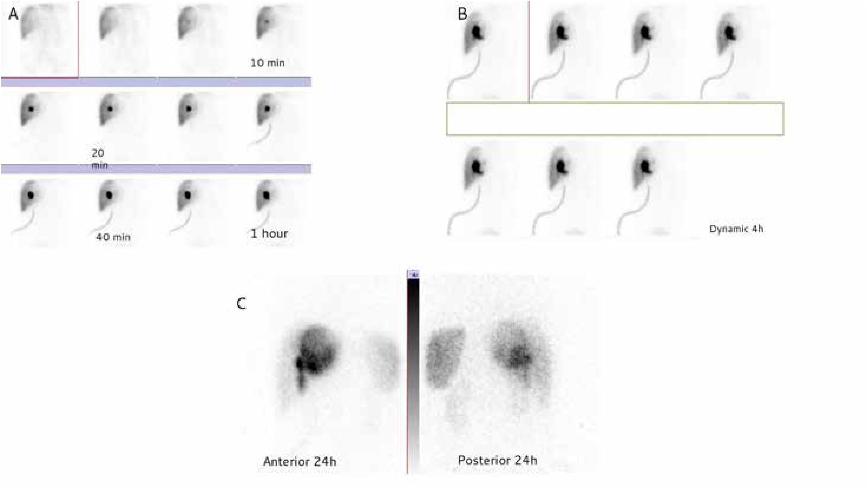
We present the case of a 75 y/o man with a metastatic sigmoid adenocarcinoma with solitary left liver lobe involvement. He was referred post-sigmoidectomy and hepatic metastatectomy for an hepatobiliary scintigraphy (HBS) due to persistent biliary leakage through the tubes, starting from 3 weeks after surgery. After an IV injection of 5 mCi of Tc-99m-BrIDA, scanning was performed dynamically every 1minute for 60minutes (Figure 1A) and delayed dynamic images were also obtained every hour up to 4hours (Figure 1B). After the standard dynamic images, 24-hour delayed static (Figure 1C) and SPECT/CT images (Figure 2) were taken. The scan showed normal and uniform hepatic uptake with no significant defects but abnormal progressive accumulation at the center of the right hepatic lobe is seen beginning at 10minutes of imaging. The intrahepatic and extrahepatic ducts were not seen even on delayed images. The scan showed excretion of the radiotracer through the draining tube beginning at 23minutes of the study without intestinal activity even after 24hours.

 as well as in the medial part of the right liver lobe (adjacent to the surgical drainage catheter insertion (pink arrow)). There is also increased tracer accumulation in the left upper abdominal quadrant (sub capsular region of the spleen (blue arrow)) which was due to bilomas.")
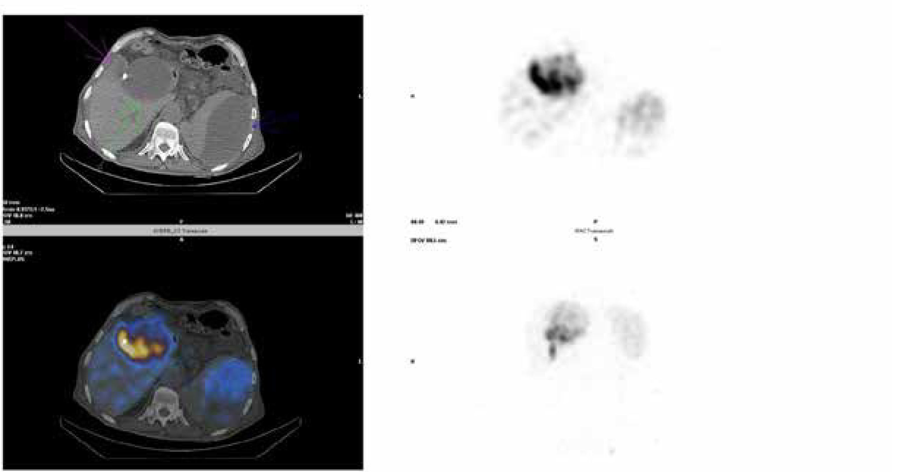
BrIDA SPECT/CT.
The SPECT/CT images depict abnormal tracer accumulation in the liver; sub capsularly in the region of segment VIII (green arrow) as well as in the medial part of the right liver lobe (adjacent to the surgical drainage catheter insertion (pink arrow)). There is also increased tracer accumulation in the left upper abdominal quadrant (sub capsular region of the spleen (blue arrow)) which was due to bilomas.
The SPECT/CT images confirmed a well-defined hypodense intrahepatic collection with abnormal tracer accumulation (subcapsularly in the region of VIII segment (green arrow)) and medial part of the right liver lobe (adjacent to the surgical drainage catheter insertion (pink arrow)) as well as ill-defined increased tracer accumulation in left upper quadrant of the abdomen (splenic area) (blue arrow) which were due to bilomas (Figure 2).
HBS is widely used to diagnose biliary disruption1. While it can provide important information, HBS commonly shows bile leaks as a persistent collection of tracer outside the biliary system and cannot further specify leak location2–4. In this case, we were able to identify the unusual source of bile leakage in the sub-capsular anterior portion of the VIII hepatic segment.
Conflict of interestThe authors have no conflict of interest to declare.
Ethical considerationsThe authors ensure that the work described has been carried out in accordance with “The Code of Ethics of the World Medical Association” (Declaration of Helsinki) for experiments involving humans. The manuscript is in line with the Recommendations for the Conduct, Reporting, Editing and Publication of Scholarly Work in Medical Journals and aim for the inclusion of representative human populations (sex, age and ethnicity) as per those recommendations.
Written patient consent was taken for the use of their anonymized images.