


Female patient 13 years of age with severe TMJ dysfunction who presented symptoms of pain and generalized muscular hypertonicity, vertigo, difficulty for walking, limitation of mandibular movements, weak chewing and maximum oral opening of 17mm. She was diagnosed as a skeletal Class II due toretrognathia and micrognathia. She had a convex profile severe upper and lower anterior dental crowding. Treatment began with neuromuscular reprogramming with a splint for muscular relaxation, thermotherapy with moist heat, muscle relaxants and afterwards, orthodontic treatment. The results of the treatment were satisfactory since the patient experienced improvement in her physical and emotional health. Correct masticatory function was restored giving the patient a better quality of life and allowing her to resume her normal daily activities.
Paciente femenino de 13 años de edad, con severa disfunción craneomandibular. Presenta sintomatología dolorosa e hipertonicidad muscular generalizada, vértigo, dificultad para caminar, limitación de movimientos mandibulares, masticación débil y apertura bucal máxima de 17mm clase II esqueletal por retrognatismo mandibular, micrognatismo, perfile convexo, severo apiñamiento dental anterior superior e inferior. Los objetivos del tratamiento fueron lograr una oclusión estable, mejorar el tono muscular y la función craneomandibular. Disminuir en lo posible el dolor, obtener una apertura bucal adecuada. El tratamiento inició con reprogramación neuromuscular con férula de miorrelajación, termoterapia con calor húmedo, relajantes musculares y posteriormente tratamiento ortodóntico. Los resultados del tratamiento fueron satisfactorios, ya que la paciente experimentó mejoría en su salud, física y emocional. Se recuperó la correcta función masticatoria, brindándole a la paciente una mejor calidad de vida, reintegrándose a sus actividades cotidianas.
Craniomandibular dysfunction (CMD) is a complex and multifactorial disorder that may cause 2 types of alterations: local and systemic.
One of the most frequent etiologies is stress. The more susceptible structures are: muscles, the temporomandibular joint (TMJ), teeth and their supporting structures.
The body reacts to the stressing factor making demands for readjustment or adaptation. The magnitude of such adaptation will depend on the amount of stress. Muscles experience pain upon palpation and during mandibular movements. The patient describes it as a limitation of movement with associated pain, tenderness, joint pain, clicking and wear on the teeth.
CMD is a multifactorial disease that must be treated multi-disciplinarily.
A correct diagnosis will lead to a successful treatment. There are different ways of treating CMD.
Diagnosis of the craniomandibular dysfunction should be carried out by means of a thorough evaluation of the stomatognathic system, which includes basically a complete medical history and a thorough physical examination which may be supplemented with imaging tests.
The main signs and symptoms of TMJ disorders are associated with a disruption of the disc-condyle complex.
Epidemiological studies suggest that approximately 30% of the general population shows some sign of functional alteration of the masticatory system.
Craniomandibular dysfunction etiologyStress, muscle hyperactivity, premature contact points, bruxism and trauma.
Craniomandibular dysfunction signs and symptomsPain, myofacial pain syndrome, trigger points, articular sounds(clicking and crackling), periauricular pain, dizziness and vertigo, limitation of mandibular movements, malocclusion, alterations at joint level (morphologic alterations, adhesions, dislocation, inflammatory disorders of the TMJ, inflammatory disorders of associated structures) and decreased visual acuity.
Craniomandibular dysfunction diagnosisClinical chart, physical exam, functional analysis, radiographic diagnosis and diagnostic aids (videos, photographs, study models).
Craniomandibular dysfunction treatmentTreatment of the CDM is classified into 2 types:
- 1.
Final: It is aimed to control or eliminate etiologic factors.
- 2.
Supportive: They are therapeutic to modify symptoms.
Another way for classifying CDM treatment is the following: palliative therapy, natural resolution, therapy related to the cause, specific therapy and rehabilitation.
Treatment is performed by means of a myorelaxation splint, medications, physical therapy, thermotherapy, transcutaneous electrical nerve stimulation (TENS), type ABotox, ultrasound, laser, psychotherapy, orthodontic appliances and surgical treatment.
CASE REPORTFemale patient of 13 years of age with very strong painful symptoms and generalized muscular hypertonicity, vertigo, inability to walk, limitation of mandibular movements, weak chewing and maximum oral opening of 17mm (Figures 1 and 2). She was referred from Neurology, and her reason for consultation was to eliminate the pain. Cefalometrically, she presented a skeletal class II due to retrognathism, micrognathia, a tendency to horizontal growth, bi-proclinationand dentoalveolar bi-protrusion. Facial analysis showed a convex profile and bi-prochelia. She suffered from bilateral TMJ pain as well as widespread muscle pain.


Functionally, the patient performed abnormal and limited opening and closing movements and there was a generalized muscle contracture, tongue thrusting, lip suction and weak chewing. In relation to her dental features, she had a left and right nonassessable canine class, severe upper and lower anterior crowding, increased overbite and overjet, deviated dental midlines and upper and lower nonideal archform (Figures 3 and 4).
TREATMENT PLAN

Therapy with muscle relaxants and thermotherapy with moist heat.
- 1.
Neuromuscular reprogramming with myo-relaxation splint.
- 2.
Upper and lower first premolar extractions.
- 3.
0.022” Roth system.
- 4.
Phase I. Initial aligning and leveling 0.014”-0.016” NiTi archwires.
- 5.
Phase II-1. Second and third order movements 0.016” x 0.016”NiTi to 0.019”x 0.025” SS archwires.
- 6.
Phase II-2: retraction of the anterior segment with 0.019” x 0.025” SS DKL archwires.
- 7.
At this stage, the possibility of using mini-implants for upper anchorage would be re-assessed.
- 8.
Posterior space closure.
- 9.
Phase III: consolidation and stabilization 0.019” x 0.025”0,021”x 0.025”SS archwires.
- 10.
Phase IV: occlusal settling.
- 11.
Retention.
First Robaxisal, a muscle relaxant, was prescribed: 1every 8hours for 15 days, coupled with hot -water bags and massage of the face and neck muscles 3 times a day as palliative therapy. An upper myorelaxation splint was placed (Figure 5) and the patient¿s appointments were scheduled every 15 days for review of the contact points and wear on the splint, which were smoothed. The appliance was worn for 3 months, time required to determine if the problem was caused by the malocclusion. When the mouth opening, the chewing and the pain improved it was confirmed that the treatment would be orthodontic and that the problem was not caused by a systemic problem such as juvenile rheumatism thus treatment was begun.

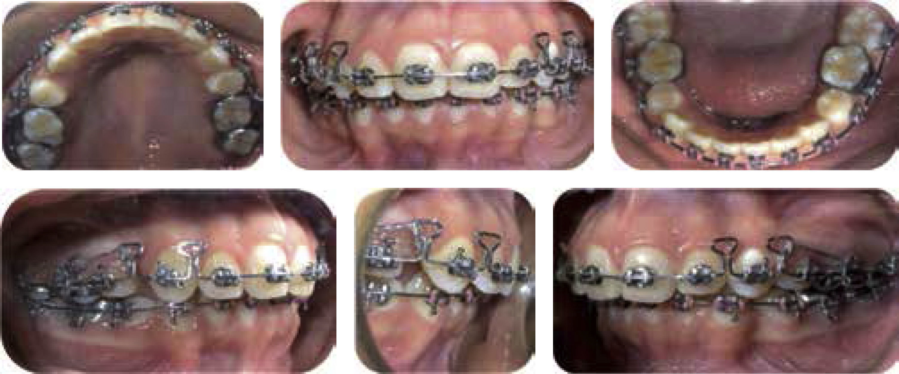
Upper and lower first premolar extractions were performed. Molar bands and 0.022 slot Roth brackets were placed on the upper and lower arch. Phase 1consisted in initial aligning and leveling with 0.014 NiTi archwires. Phase II, the arches were coordinated through light second and third order movements. Anchorage was obtained through mini-implants. Retraction of the anterior segment was done with a DKL archwire and all spaces were closed. Consolidation, stabilization and retention (Figures 6 to 9).


The facial and intraoral photographs, the X-rays and the functional analysis show completely satisfactory results since the axial axis of the teeth was corrected and dentoalveolar biprotrusion decreased. The profile improved by decreasing the biprocheilia. Craniomandibular function was improved by eliminating the widespread muscle pain. An opening and closing pattern without deviation was attained, obtaining a maximum opening of 38mm. Oral habits were corrected and adequate masticatory function was recovered. Bilateral molar and canine class I was obtained. Dental crowding was eliminated and the overbite, overjet and dental midlines were corrected. Arch shape was improved (Figures 10 to 12).


All treatment objectives were achieved demonstrating that in cases such these, if they are properly diagnosed and treated, patient's health may be restored.
CONCLUSIONSCraniomandibular dysfunctions are each day more frequent in our population, affecting more women than men and with an increasing incidence in children and teenagers.
This disease is multifactorial therefore it should be treated multi-disciplinarily in order to obtain an integral result that involves facial, articular, functional, dental, aesthetic, psychological, social, and emotional changes.
There are currently different alternatives to deal with the CMD so it is important to be trained to implement the most appropriate for each patient and in this way manage pain and relieve the patient's suffering.
The main challenge for the orthodontist in CMD cases is the diagnosis in order to determine the extent to which the malocclusion is affecting the craniomandibular problem.
Graduate.
This article can be read in its full version in the following page: http://www.medigraphic.com/ortodoncia.