


The aim of this study was to estimate the prevalence and conditions of supernumerary teeth not associated to syndromes in the University of Latin America Orthodontics Department Campus Valle in Mexico City.
Material and methodsA retrospective study was made using 1038 panoramic radiographs, 622 females and 416 males, ages between 9, and 57 years old in a period of time of 20 months. Considering age, sex, classification of the supernumerary tooth and location. The variables of gender, number, and age, were specified in rates as well as the Mean, and standard deviation respectively.
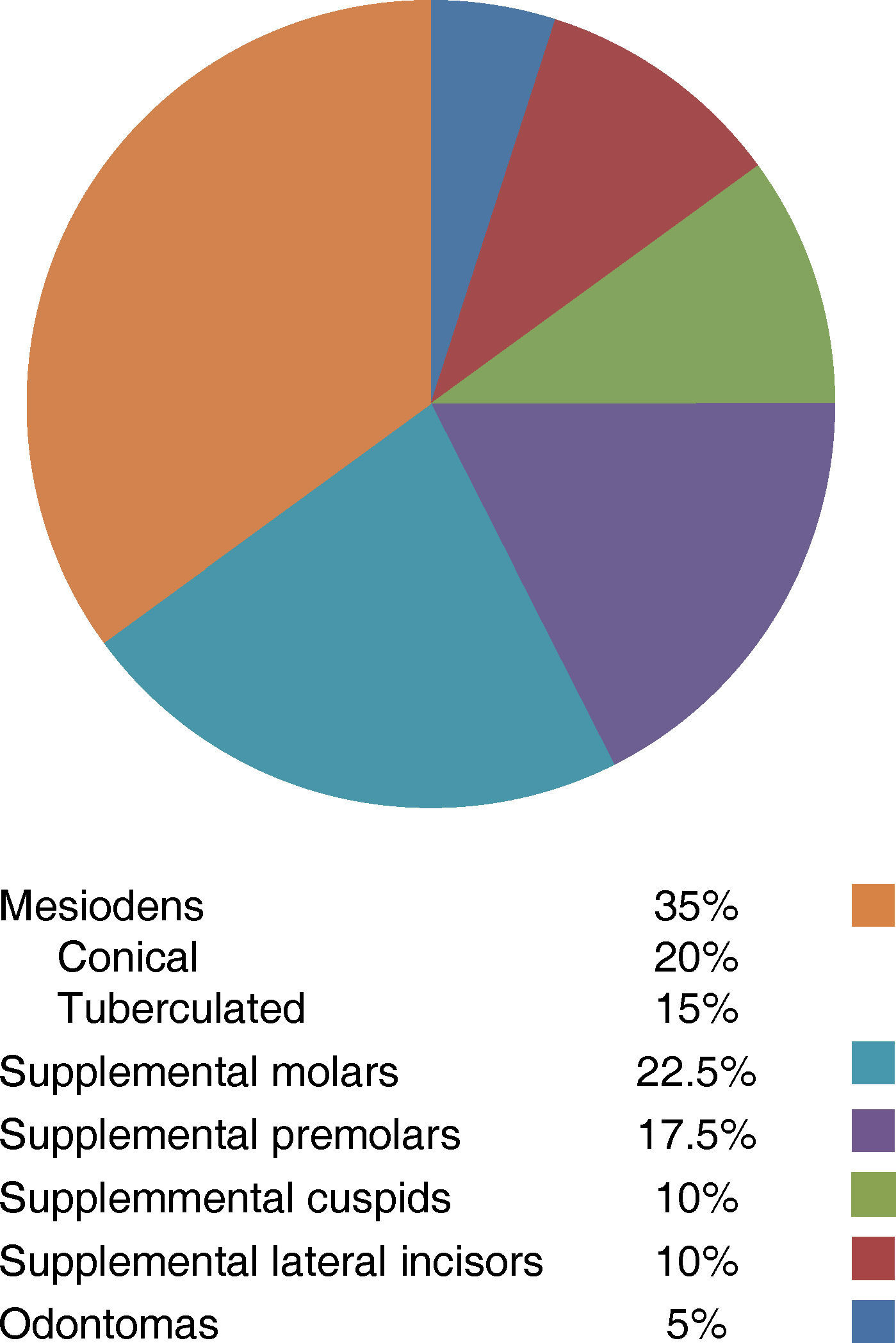
Results40 supernumerary teeth were found in 29 patients (2.8%), 17 in males, 12 in females. In accordance to their classification, the next rates were found: mesiodens 35%, conical 20% and tuberculated 15%, supplemental molars 22.5%, supplemental premolars 17.5%, supplemental cuspids and lateral incisors 10% each. With respect to their location, there is no statistically significant difference between maxilla and mandible (p= 0.168).
ConclusionsIt seems that the mesiodens are the most frequent supernumerary teeth, being the upper maxilla the most affected in males, although females had more supernumerary teeth.
El propósito de este estudio fue determinar la prevalencia y condición de los dientes supernumerarios no asociados a síndromes en el Departamento de Ortodoncia de la Universidad Latinoamericana, Campus Valle en México D.F.
Material y métodosSe realizó un estudio retrospectivo utilizando 1,038 radiografías panorámicas de 622 mujeres y 416 hombres, con edades comprendidas entre los 9 y 57 años de edad en un periodo de 20 meses. Se contemplaron la edad y el sexo del paciente, la clasificación del diente supernumerario y la ubicación. Las variables de género, número y edad se resumieron con porcentajes y la media y desviación estándar respectivamente.
ResultadosSe encontraron 40 dientes supernumerarios en 29 pacientes (2.8%); de éstos, 17 fueron hombres y 12 mujeres. En los hombres se encontró el 55% de dientes supernumerarios, mientras que en las mujeres el 45% del total. De acuerdo a su clasificación se observaron los siguientes porcentajes: mesiodens 35% entre cónico 20% y tuberculado 15%, molares suplementarios 22.5%, premolares suplementarios 17.5%, caninos e incisivos laterales suplementarios 10% cada uno. Respecto a su ubicación no se encontró gran diferencia entre el maxilar y la mandíbula (p = 0.168).
ConclusionesAl parecer, el mesiodens es el diente supernumerario que se presenta con mayor frecuencia, siendo el maxilar superior el más afectado en el sexo masculino; sin embargo, las mujeres presentaron mayor cantidad de dientes supernumerarios.
Supernumerary teeth are a dental anomaly that consists of an increase in the number of teeth of the normal formulae in the Primary or Permanent dentition.1 They may be present in any region of the dental arch with predominance of the anterior part of the Maxilla and be simple, or multiple, unilateral, or bilateral. The multiple hyperodontia can be related to cleidocranial disostosis, Gardner syndrome, Ellis Van Creveld syndrome, among others. The etiology of supernumerary teeth is supported by several theories: The Dichotomy theory describes that the dental laminae divides during the early development giving place to a normal sized tooth and a smaller one, one with normal shape, and the other amorphous.
The Genetics theory supports that inheritance plays a main role in the appearance of this anomaly. The Filogenetic theory or the atavism talks of a regression of the extinct ancestral tissues of the mammals. It has been suggested that with evolution, the number of teeth tends to disappear, while morphology becomes more complex, although this hasn’t been proved yet. The Hyperactivity theory is highly accepted and describes that once the permanent tooth is formed, the cells of the dental laminae degenerate and this proliferation produces supernumerary teeth.2 Nonsyndromic supernumerary teeth are not common although the damage they can cause to neighboring teeth and tissues is sometimes severe. They participate in the development of malocclusions by provoking dental displacements, cyst formations occasionally when they do not erupt and radicular resorption of neighboring teeth due to unusual positioning.
MATERIAL AND METHODSFor this study all patients files with clinical history and panoramic X-rays who entered the clinic of University of Latin America Orthodontics Department Campus Valle in Mexico City, between March 2011, and October 2012, were included. 1,038 panoramic X-rays and clinical histories of 622 females and 416 males, in ages between 9, and 57 years old, were reviewed in a period of 20 months. Each X-ray was reviewed and discussed by the panel in a negatoscope with a 7X lens. Differing opinions were solved by consensus between dentists with specialties in Orthodontics or Periodontics by request of the main examiner. All X-rays belonged to patients without syndromes, healthy and without congenital diseases, facts corroborated in their clinical histories. After examination, the next facts were registered: Patient's name, sex, age, absence or presence of supernumerary teeth, classification, and location.
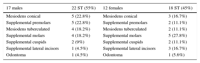
RESULTS40 supernumerary teeth were found in 29 patients, which represents 2.8% of the total sample. From the total of patients with supernumerary teeth, 17 were males (58.6%) and 12 females (41.4%). The distribution by gender is shown on table I below.
Supernumerary teeth distribution by gender.
| 17 males | 22 ST (55%) | 12 females | 18 ST (45%) |
|---|---|---|---|
| Mesiodens conical | 5 (22.8%) | Mesiodens conical | 3 (16.7%) |
| Supplemental premolars | 5 (22.8%) | Supplemental premolars | 2 (11.1%) |
| Mesiodens tuberculated | 4 (18.2%) | Mesiodens tuberculated | 2 (11.1%) |
| Supplemental molars | 4 (18.2%) | Supplemental molars | 5 (27.8%) |
| Supplemental cuspids | 2 (9%) | Supplemental cuspids | 2 (11.1%) |
| Supplemental lateral incisors | 1 (4.5%) | Supplemental lateral incisors | 3 (16.7%) |
| Odontoma | 1 (4.5%) | Odontoma | 1 (5.6%) |
As indicated in the sectors diagram, the mesiodens was the most frequent supernumerary tooth; the patients with mesiodens (n = 12) represent the 41.4% of the patients with supernumerary teeth: eight males (66.7%) and 4 females (33.3%). The age range was between 10, and 20 years with a mean of 14.7 years (SD 3.5 years). In this study, 14 mesiodens were found; 10 cases were shown isolated and two cases in pairs. Nine mesiodens were observed in males (64.3%) and five in females (35.7%). According to their morphology, eight conicals were found (57%), five in males, three in females, and six tuberculated (43%); four in males and two in females, according to Garvey's classification.3
The supplemental premolars of the sample were seven cases, and seven teeth, (24.1% of the total patients with ST), five males (71.4%), and two females (28.6%), ages between 12, and 20 years with a mean of 14.7 years (SD 3.3 years) and all the cases were found in the mandible, five in the left side, and two in the right.
The supplemental molars were five cases, and nine teeth (17.2% of the patients with ST), three females (60%), and two males (40%), ages between 12, and 17 years, with a mean of 15.2 years (SD 1.9 years). Of the nine supplemental molars, two cases were females (15 and 16 years) located between the first and second molars, upper left and lower right, with roots totally formed. The other three cases were seven fourth molars without root development, two males (16 and 17 years) and one female (12 years). There were four cases of supplemental lateral incisors in four patients (13.8% of the total patients with ST), three in females (75%) and one in males (25%), ages between 13, and 17 years old with a mean of 15 years (SD 1.8 years), three located in the upper maxilla, two in the right side and one in the left and only one in the lower left side. Supplemental cuspids were found in three patients with four teeth (10.3%), two males (66.7%), and one female (33.3%) which had two lower right cuspids; the other two cuspids were also found in the same lower location, none in the upper maxilla (Figure 1). The sample ages were between 12 to 21 years with a mean of 16.3 years (SD 4.5 years).

Odontomas were classified according to Howard4 finding only two cases (6.9%) of 29 total. One located in the upper maxilla between central and the lateral incisor, which was discovered in a 12 year-old male classified as a composite odontoma, and another in an 18 year-old female located alongside the lateral incisor to the distal side, which was subsequently sent to pathology for examination.
DISCUSSIONThere are numerous reports of the prevalence of supernumerary teeth which vary according to the type of population studied and in most cases these reports were made in infant populations.5 Amongst Latin American studies that include adults, we found one performed by Salcido-García in Mexican patients.6 In Caucasian patients, the prevalence varies from 1.05 to 2.1%.5,7–9 The lowest prevalence we found with a sample of 1,751 patients in a study conducted in Iran10 with patient's ages between 9, and 27 years. The results of the present study revealed a prevalence of 2.8% similar to those found by Davis11 in Hong Kong, China, 2.7%, and Esenlik E et al.12 in Isparta and Ankara, Turkey, of 2.7%. Regarding gender, males were more affected. This fact coincides with all the other reviewed articles. However, some studies reveal that the rate in males is more than twice as much than females.7,9,11,13 In the current study, supernumerary teeth were found slightly more frequently in males than females in a 1.4: 1 ratio coinciding with studies in Turkey, India and Mexico6,12,13 however, related to the number of supernumerary teeth found in males and females, we observed the females more affected in a 1.3: 1.5 ratio. We agree that the area corresponding to the midline in the upper maxilla is the one that presents the highest number of supernumerary teeth.6,7,12
Mesiodens were observed in 12 patients like the study of Salcido-García,6 although the frequency was observed this way: supplemental molars, premolars, cuspids, and lateral incisors and finally the odontomas. Salcido-García6 reported in second place premolars, lateral incisors and ultimately the molars found with the 9.7%.
Our results agree with the frequency reported by MT Garvey,3 and Menardia-Pejuan.14
In the studies reviewed from Turkey12 and São Paulo Brazil15 no supplemental molars are found and results related to distribution were different from this study. Odontomas were not taken into consideration because they are considered neoplasias. Another interesting fact is a study of Simões15 with a slightly larger sample than this study, found 30 patients with supernumerary teeth, 29 in the upper arch and, 1 in the mandible.
Schmuckli9 reported 44 supernumerary teeth, 5 in the Mandible while 16 were observed in the lower arch.
CONCLUSIONSFactors involved in finding supernumerary teeth such as age, classification, type of population, size of the sample, influence the prevalence of supernumerary teeth. These factors must be documented and recorded carefully in order to achieve an accurate correlation between them and the occurrence of supernumerary teeth.
This article can be read in its full version in the following page: http://www.medigraphic.com/ortodoncia