Various clinical and radiologic variables impact the neurologic prognosis of patients with ischemic cerebrovascular accidents. About 30% of ischemic cerebrovascular accidents are caused by proximal obstruction of the anterior circulation; in these cases, systemic thrombolysis is of limited usefulness. CT angiography is indicated in candidates for endovascular treatment. Various radiologic factors, including the grade of leptomeningeal collateral circulation, as well as the length, density, and extension of the thrombus, have been identified as predictors of neurologic prognosis after anterior ischemic cerebrovascular accidents due to proximal vascular obstruction. Final infarct volume correlations with mortality and long-term functional outcome in these patients. This study aimed to determine the best predictors of final infarct volume on CT angiography in patients with ischemic cerebral accidents due to proximal occlusion.
Materials and methodsThis retrospective observational study included adults with ischemic cerebrovascular accidents due to obstruction of the anterior circulation diagnosed by CT angiography in the period comprising June 2009 through December 2019. We measured the length and density of the thrombus in unenhanced CT images, and we used the clot burden score to record the grade of leptomeningeal collateral circulation and the extension of the thrombus. Then we measured the final infarct volume on follow-up CT and analyzed the correlations among these radiologic factors in the infarct volume.
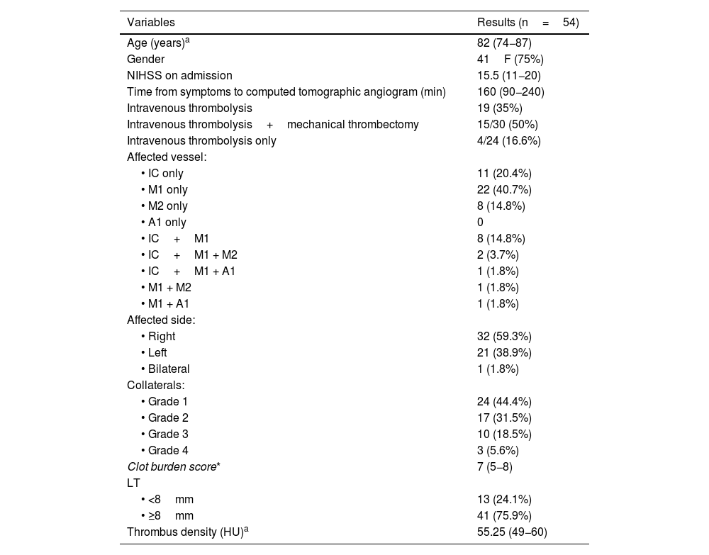
ResultsWe included 54 patients [mean age, 82 y; 41 (75%) women] with ischemic cerebrovascular accidents due to proximal occlusion. About 60% of the cerebrovascular accidents affected the right cerebral hemisphere, and the most commonly affected vessel was the M1 segment of the medial cerebral artery (40.7%). Final infarct volume correlated with the grade of leptomeningeal collateral circulation (p=0.03) and with the clot burden score (p=0.01). Neither the length nor the density of the thrombus correlated with final infarct volume.
ConclusionThe final infarct volume can be estimated on the initial CT angiogram. Nevertheless, we found no useful predictive factors in unenhanced CT images. The best independent radiologic predictors of the final infarct volume are the grade of collateral circulation and the clot burden score, especially in patients who did not undergo mechanical thrombectomy, because mechanical thrombectomy improves outcomes. These factors are important for decision making in the management of patients with ischemic cerebrovascular accidents due to proximal occlusion.
Múltiples variables clínicas y radiológicas están involucradas en el pronóstico neurológico de los pacientes con accidente cerebrovascular (ACV) isquémico. Alrededor del 30% de los ACV isquémicos son causados por la obstrucción vascular proximal del circuito anterior; en estos casos, la utilidad de la trombólisis sistémica es limitada. La angiotomografía está indicada en los pacientes que pueden ser candidatos a tratamiento endovascular. Diferentes factores radiológicos como el grado de colaterales leptomeníngeas, o el largo, densidad o extensión del trombo, fueron descritos como predictores del pronóstico neurológico tras un ACV isquémico con compromiso vascular proximal. El volumen final del infarto cerebral se correlaciona con la mortalidad y el grado funcional a largo plazo de estos pacientes. El propósito de este estudio es determinar los mejores predictores radiológicos del volumen final del infarto cerebral en pacientes con ACV isquémico con compromiso proximal, utilizando angiotomografía.
Materiales y métodosRealizamos un estudio observacional retrospectivo. Incluimos pacientes adultos con ACV isquémico causado por la obstrucción de un vaso proximal, diagnosticados mediante angiotomografía en el período de junio de 2009 a diciembre de 2019. Medimos la densidad y el largo del trombo en la adquisición sin contraste, registramos el grado de colaterales leptomeníngeas y la extensión del trombo utilizando el clot burden score. Luego medimos el volumen final del infarto en una tomografía de control y analizamos el grado de correlación entre estos factores radiológicos en el volumen infartado.
ResultadosIncluimos 54 pacientes con ACV isquémico por compromiso vascular proximal; 41 (75%) fueron mujeres. La mediana de edad fue de 82 años. Alrededor del 60% de los ACV comprometieron el hemisferio derecho y el vaso más afectado fue el segmento M1 de la arteria cerebral media (40,7%). Encontramos una asociación entre el grado de colaterales leptomeníngeas (p=0,03) y el clot burden score (p=0,01) con el volumen final del infarto. Tanto el largo como la densidad del trombo no se correlacionaron con el volumen final del infarto.
ConclusiónEs posible estimar el volumen final infartado en la angiotomografía realizada al momento del evento isquémico. Sin embargo, no se hallaron factores predictivos útiles en la tomografía computarizada sin contraste. Los mejores predictores radiológicos del volumen final infartado como factores independientes son el grado de vasos colaterales y la escala de extensión del trombo o clot burden score, en especial en los pacientes que no fueron sometidos a trombectomía mecánica, ya que este tratamiento modifica positivamente la evolución natural de la enfermedad. Estos factores son de importancia para la toma de decisiones en el manejo de los pacientes con ACV isquémico con compromiso vascular proximal.


