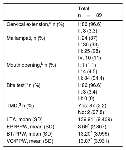
To establish a correlation between 4 measurements made on preoperative computed axial tomography and the presence of difficult airway, as well as its clinical prediction in patients undergoing otorhinolaryngological surgery.
Materials and methodsA retrospective, observational study was carried out using the information gathered from the clinical notes of 104 patients undergoing general anaesthesia and endotracheal intubation for oncological otorhinolaryngological surgery over a period of 36 months. Based on the findings in the preoperative imaging tests, a multivariate logistic regression analysis was performed, where the dependent variable was the presence of extreme grades of visualisation of the glottis visualisation (Cormack III–IV) or the presence of predictors of difficult intubation (Mallampati III–IV). This resulted in a total of 4 tomographic and clinical factors of difficult airway being introduced in this model.
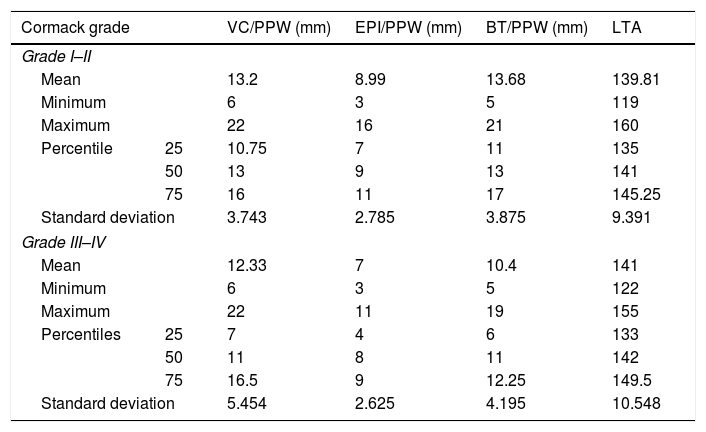
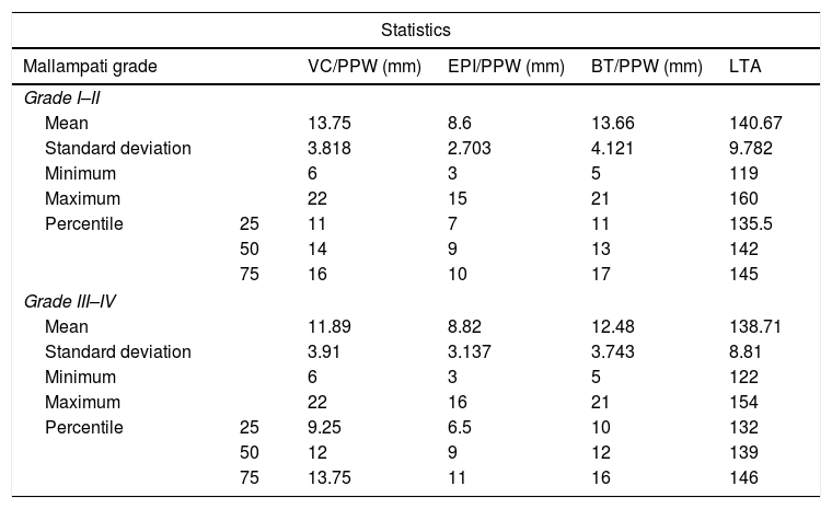
ResultsIn the Cormack III–IV group, the results were not statistically significant in the multivariate model when compared to the tomography predictors, distance from epiglottis to posterior pharyngeal wall (95% CI; 0.030–2.31, p<0.05), and the distance from the base of the tongue to the posterior pharyngeal wall (95% CI; 0.018–1.37, p<0.05). In the Mallampati III–IV group, in the multivariate model only the distance from the vocal cords to the posterior pharyngeal wall showed clinically significant results (95% CI; 0.104–8.53, p<0.05).
ConclusionsIn the approach to the airway, reliance on predictors is based on physical examination to anticipate situations that put oxygenation and ventilation of the patients at risk. There are still insufficient data to recommend imaging tests in this area, however it seems that in the future they may be added to the diagnostic performance of physical examination as predictors of difficult airway.
Establecer una correlación entre 4 mediciones realizadas en la tomografía axial computarizada preoperatoria y la presencia de vía aérea difícil, y con la predicción clínica de la misma, en pacientes intervenidos mediante cirugía otorrinolaringológica.
Material y métodosSe realizó un estudio observacional, retrospectivo, usando como fuente de información las historias clínicas de 104 pacientes intervenidos bajo anestesia general e intubación endotraqueal por enfermedad oncológica durante un periodo de 36 meses. Sobre la base de los hallazgos obtenidos en las pruebas de imagen preoperatorias se realiza un análisis de regresión logística multivariante, donde las variables dependientes son grados extremos de visualización de la glotis (Cormack III-IV) o la presencia de predictores de intubación dificultosa (Mallampati III-IV). Se introdujeron en dicho modelo un total de 4 factores tomográficos y clínicos de vía aérea difícil.
ResultadosEn el grupo Cormack III-IV, en el modelo multivariante los resultados no fueron estadísticamente significativos cuando se comparaban con los predictores tomográficos (p>0,05; IC 95% distancia de la epiglotis a la pared faríngea posterior 0,030–2,31; distancia de la base de la lengua a la pared faríngea posterior 0,018-1,37). En el grupo Mallampati III-IV, en el modelo multivariante únicamente la distancia de las cuerdas vocales a la pared faríngea posterior muestra resultados clínicamente significativos (p<0,05; IC 95% 0,104-8,53).
ConclusionesEn el abordaje de la vía aérea actualmente nos podemos apoyar en los predictores correspondientes al examen físico para adelantarnos a situaciones que pongan en riesgo la oxigenación y la ventilación de nuestros pacientes. Aunque aún los datos son insuficientes para recomendar las pruebas de imagen en este ámbito, parece que en un futuro pueden sumarse al examen físico para aumentar el rendimiento diagnóstico.


