


Analizar el grosor coroideo macular (GCM) en la neuropatía óptica isquémica anterior no arterítica (NOIA-NA).
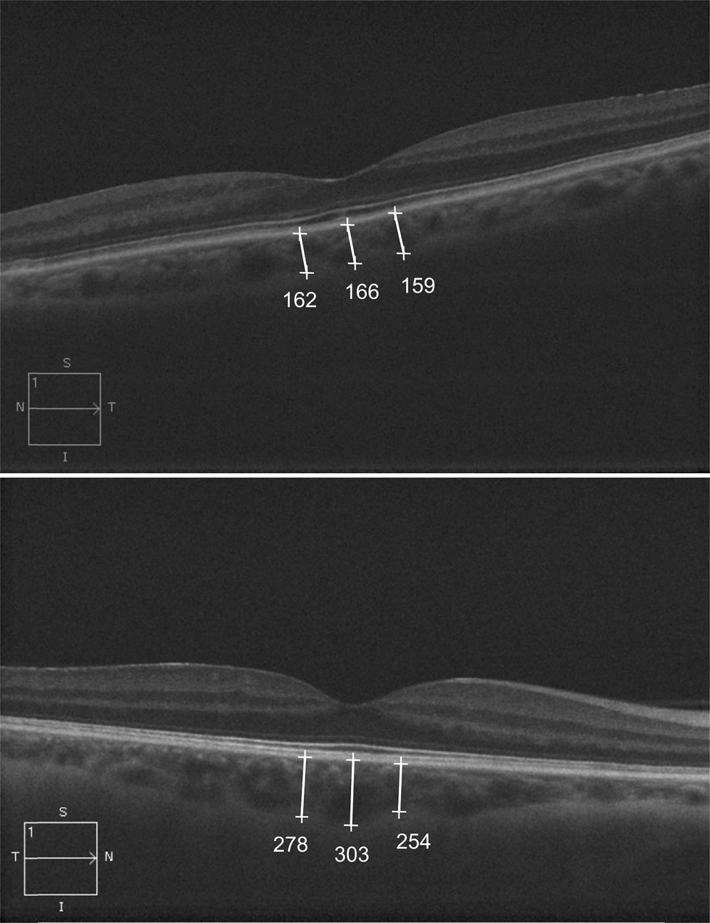
Material y métodosUn total de 22 pacientes diagnosticados de NOIA-NA (22 ojos) y 42 sujetos sanos (42 ojos) fueron estudiados usando tomografía de coherencia óptica con técnica Enhanced Depth Imaging (EDI-OCT). Se realizó un escáner de una línea horizontal centrado en la fóvea 3 meses después del inicio de NOIA-NA. Se tomaron 3 medidas desde la parte posterior del epitelio pigmentario hasta la unión esclerocoroidea a intervalos de 500μm en las 1.500μm centrales de la mácula. Los resultados fueron analizados estadísticamente comparando la media de GCM entre grupos y correlacionando el GCM con otros parámetros oculares y sistémicos.
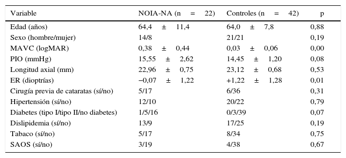
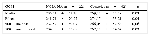
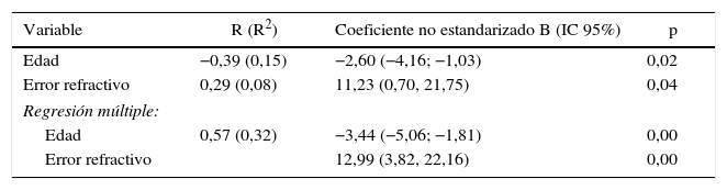
ResultadosExcepto en el error refractivo (p=0,01), no hubo diferencias significativas en longitud axial (p=0,53), edad (p=0,88) ni en otros parámetros oculares ni epidemiológicos entre grupos. La media de GCM en la NOIA-NA y en el grupo control fue 236,21±63,29μm y 269,13±52,28, respectivamente. La media del GCM fue significativamente más delgada en ojos con NOIA-NA que en sanos (p=0,03). El adelgazamiento del GCM estuvo asociado con el diagnóstico de NOIA-NA después de ajustar por error refractivo (p=0,04).
ConclusionesLos ojos afectos con NOIA-NA mostraron un GCM significativamente más adelgazado que en sujetos sanos, después de ajustar por error refractivo.
To analyse macular choroidal thickness (MCT) in non-arteritic ischaemic optic neuropathy (NAION).
Materials and methodsAn analysis was made on 22 patients diagnosed with NAION (22 eyes) and 42 healthy controls (42 eyes) using enhanced-depth imaging of spectral-domain optical coherence tomography. A horizontal raster scan centred on the fovea was obtained per eye 3 months after the onset of NAION. Three measurements of MCT were obtained from the posterior edge of the retinal pigment epithelium to the choroid-sclera junction at 500μm intervals. Statistical analysis was used to compare the mean MCT and to correlate MCT with other ocular and systemic parameters.
ResultsExcept for refractive error (P=.01), there were no statistically significant differences between both groups in axial length (P=.53), age (P=.88) and other epidemiological and ocular parameters. Mean MCT in NAION eyes and control group was 236.21±63.29μm and 269.13±52.28, respectively. Mean MCT was significantly thinner in NAION eyes than in healthy eyes (P=.03). Thinner MCT, adjusted for refractive error, was associated with the diagnosis of NAION (P=.04).
ConclusionsEyes affected by NAION showed significantly thinner MCT compared with healthy control eyes after adjusting for refractive error.
