La ecocardiografía transesofágica permite una adecuada monitorización de la hemodinamia intraoperatoria. Un parámetro frecuentemente utilizado es la fracción de eyección medido con método de Simpson. Con el advenimiento de Doppler tisular y la medición de onda s’, que corresponde a la velocidad de la perfusión del tejido miocárdico durante la sístole, podría estimarse la fracción de eyección de manera más rápida y fácil durante una cirugía.
ObjetivosComparar la fracción de eyección calculada con método de Simpson con las mediciones intraoperatorias de la velocidad de la onda s’ medida con Doppler tisular.
Material y métodoSe estudiaron pacientes afectos de patología cardiovascular crónica sometidos a cirugía cardiaca y no cardiaca electiva. Se excluyeron pacientes en ritmo no sinusal y con enfermedad mitral. Se midió en 4 y 2 cámaras volumen de fin de diástole y volumen de fin de sístole para calcular la fracción de eyección con método de Simpson. Este grupo fue dividido en aquéllos con fracción de eyección normal (> 50%) y un segundo grupo con fracción de eyección disminuida (< 49%) Luego se utilizó Doppler tisular de anillo mitral para medir la velocidad de la onda s’. Para estimar la fracción de eyección con s’ se utilizó la fórmula: FE = 5,5xs’+8.
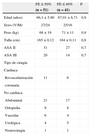
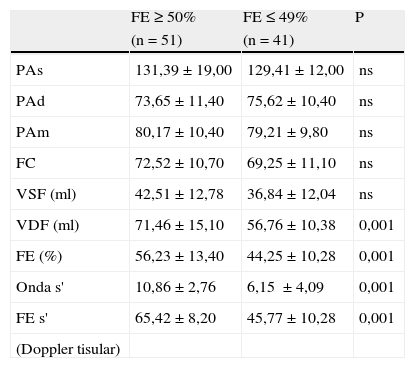
ResultadosFueron estudiados 92 pacientes, 51 (55%) casos con fracción de eyección normal y 41 (45%) disminuida. El grupo con fracción de eyección < 49% tuvo una buena correlación con la calculada mediante la velocidad de la onda s’ del Doppler tisular (r = 0,91, p < 0,01). En cambio en el grupo con fracción de eyección normal esta correlación fue menor (r=0,61, p > 0,5).
ConclusiónLa estimación de la fracción de eyección con Doppler tisular es una técnica fácil y reproducible. Su utilidad podría ser mayor especialmente en aquellos pacientes que tienen un ventrículo izquierdo alterado.
Transesophageal echocardiography is appropriate for intraoperative monitoring of hemodynamics. The parameter often estimated is ejection fraction (EF) by means of Simpson’s rule. With the advent of tissue Doppler imaging and measurement of the systolic (S) wave, corresponding to the rate of myocardial perfusion during the systole, it is possible to estimate the EF more easily and rapidly during surgery.
ObjectiveTo compare EF estimates obtained by Simpson’s rule to those based on intraoperative tissue Doppler measurements of S-wave velocity (S').
Material and methodsPatients with chronic cardiovascular disease undergoing scheduled cardiac and noncardiac surgery were studied. Patients in nonsinus rhythm and with mitral valve disease were excluded. To apply Simpson's rule for calculating the EF, we measured end-diastolic volume in 4- and 2-chamber views. The group was divided into patients with normal (≥50%) and diminished (≤49%) ejection fraction. Tissue Doppler imaging of the mitral annulus was then used to measure S'. Ejection fraction was calculated according to the formula EF = 5.5x S' +8.
ResultsNinety-two patients were studied; in 51 (55%) the EF was normal and in 41 (45%) it was reduced. In patients whose EF was ≤49% according to Simpson’s rule, the correlation between that measurement and EF based on tissue Doppler estimate of S' was good. The correlation was lower, however, in the group with normal EF (r=0.61; P>0.5).
ConclusionsEF is easy to estimate with tissue Doppler imaging and the procedure is reproducible. This approach is probably more useful in patients with left ventricular dysfunction.


