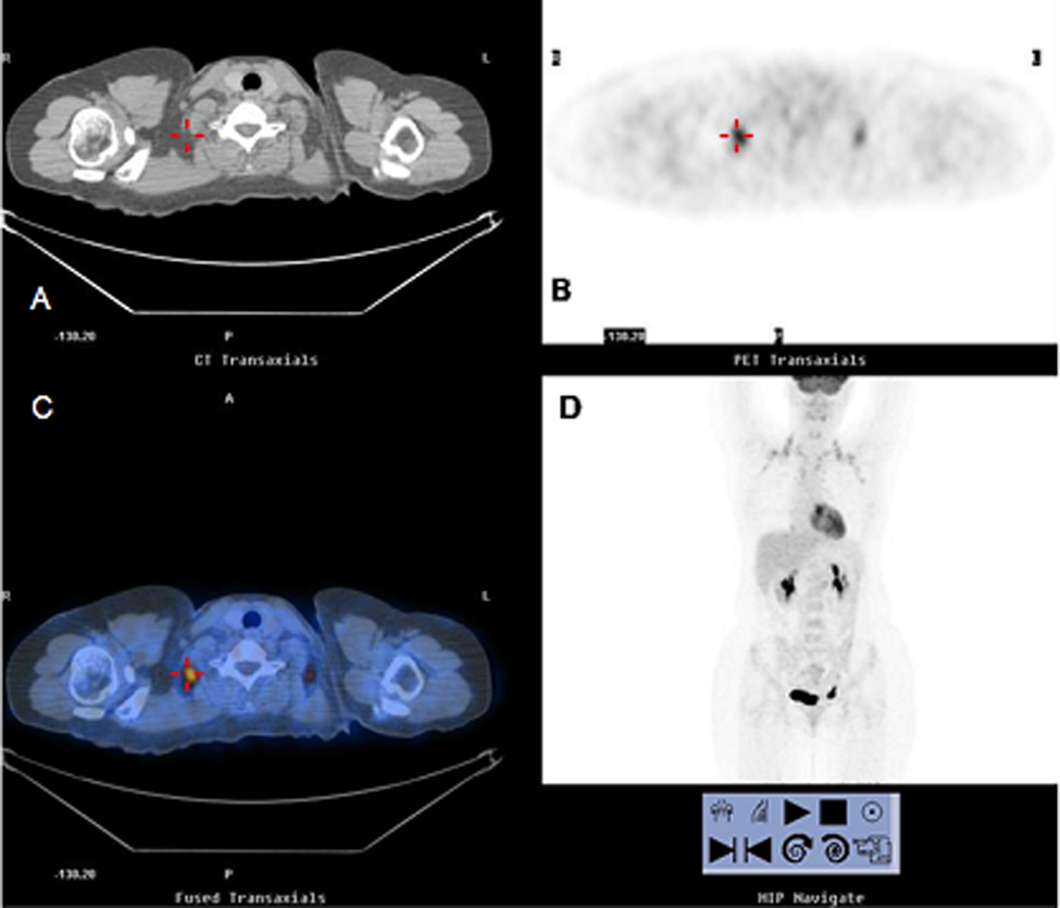
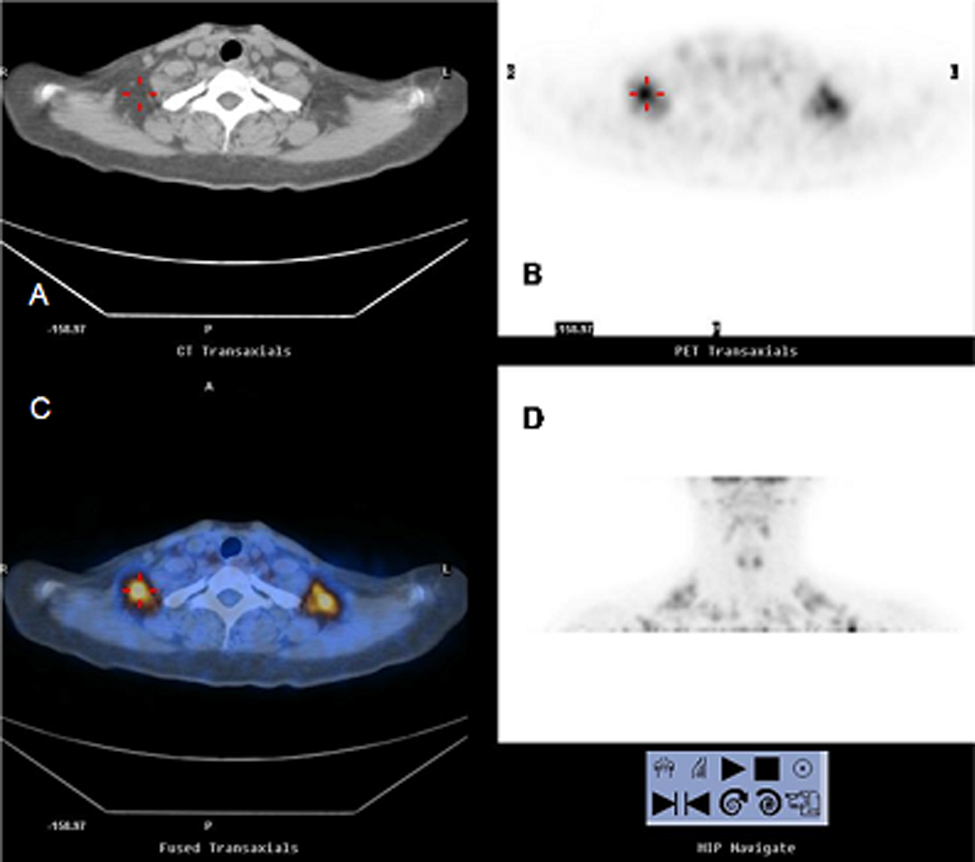
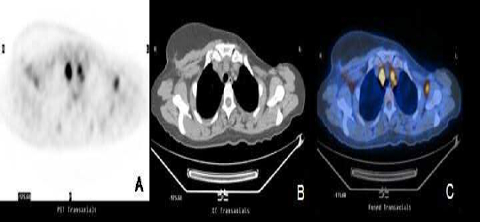
Brown adipose tissue (BAT) is a potential source of false-positive findings on [18F] FDG PET. In this report, we have discussed the 18F-FDG uptake mechanisms in BAT and have aimed to determine if dual time point PET imaging helps to differentiate BAT from malignant lesions.
MethodsPatients with dual-time-point PET/CT scans were reviewed retrospectively and 31 cases (11 males, 20 females, age: 28.6±9.7) having hypermetabolic BAT were included for this study. 18F-FDG uptake in BAT was quantitatively analyzed by maximum standardized uptake values (SUVmax), and average percent change in SUVmax of BAT between early and delayed images was calculated.
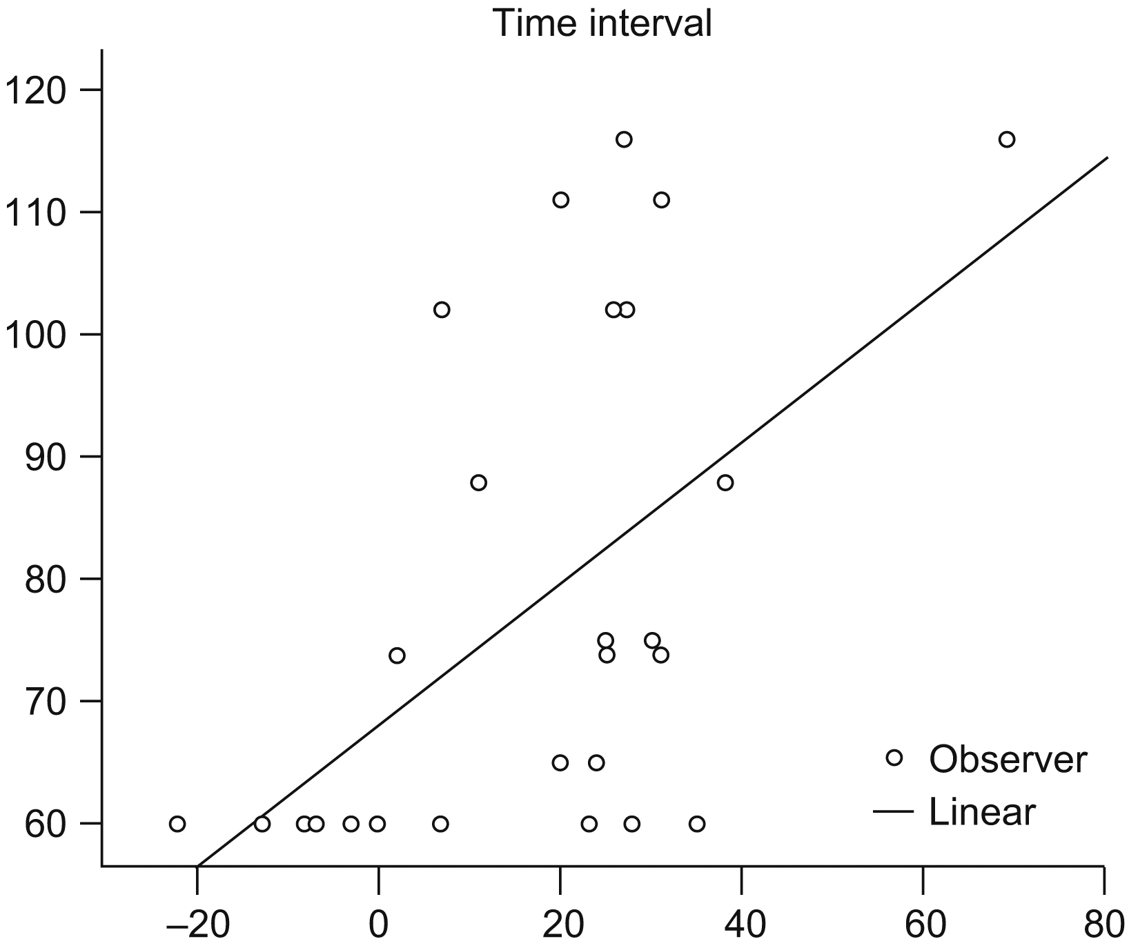
ResultsCompared to the initial scans, 18F-FDG uptakes in BAT in delayed images were higher in 26 of the patients, and lower in one patient. In terms of body regions, 18F-FDG uptake increased in 80.6%, remained unchanged in 5.5% and decreased in 13.9% of the body regions. Mean percent change in SUVmax, including all BAT regions, was 19.8±19.1% while the mean percent increase was calculated as 69±25% in regions where progressive accumulation was observed. The increase in SUVmax correlated with the time interval between the two scans.
ConclusionPhysiologic 18F-FDG uptake in BAT increases over time and may mimic the behavior of malignant lesions on dual time point PET imaging. Without the exact anatomic definition of the CT scan, false positive interpretation of PET data may be possible in cases with atypical BAT.
El tejido adiposo marrón (TAM) es un fuerte potencial de falsos-positivos en el 18F-FDG PET. En este informe, comentamos los mecanismos de captación del 18F-FDG en TAM e intentamos averiguar si la adquisición de imágenes con el PET en dos fases ayuda a diferenciar el TAM de las lesiones malignas.
MétodosLos pacientes sometidos a estudios PET-TAC en dos fases fueron estudiados retrospectivamente y se incluyeron 31 casos (11 hombres, 20 mujeres, edad: 28,6±9,7) con TAM hipermetabólico en el estudio. La captación de 18F-FDG en el TAM fue analizada cuantitativamente, utilizando los valores estandarizados de captación máxima (SUVmax), y se calculó el cambio medio porcentual en el SUVmax de TAM entre imágenes tempranas y retardadas.
ResultadosComparadas con los estudios iniciales, las captaciones de 18F-FDG en TAM en las imágenes retardadas fueron más altas en 26 de los pacientes y más baja en uno. En cuanto a las regiones del cuerpo, la captación de 18F-FDG aumentó 80,6 %, no cambió en 5,5 % y disminuyó en el 13,9% de las regiones del cuerpo. El cambio medio porcentual en SUVmax, incluyendo todas las regiones del TAM, fue de 19,8 %±19,1, mientras que el aumento medio porcentual calculado fue de 69±25% en las regiones en que se observó acumulación progresiva. El aumento en SUVmax correlacionó con el intervalo de tiempo entre las dos fases.
ConclusiónLa captación fisiológica de 18F-FDG en el TAM aumenta con el tiempo y puede simular el comportamiento de lesiones malignas en el PET en dos fases de tiempo. Sin tener la definición anatómica exacta del estudio TAC, puede haber una interpretación de falso-positivo de los datos del PET en casos con un TAM atípica.
Artículo
Comprando el artículo el PDF del mismo podrá ser descargado
Precio 19,34 €
Comprar ahora


