Introduction
Clinicopathological correlation studies focusing on the various clinical forms of meningococcal disease are scarce. A review of the Brazilian and international literature shows few such studies, particularly in recent years, and those that exist are generally restricted to case reports or pathological analyses of only one organ, such as the skin, adrenal glands, or heart. Although focal or diffuse myocarditis can be considered a classically reported finding in necropsy studies of meningococcal septicemia1-3, clinicopathological correlation analyses are quite rare. In one such study, among 27 cases of myocarditis detected at necropsy, the authors4 found only one clinical diagnosis of heart failure. According to some authors3,5, left ventricular failure due to myocarditis was probably the main cause of death in patients with meningococcal septicemia, in contrast to another group of researchers6 who identified multiple organ failure as the principal cause of death. According to various authors2,3,7,8, neither focal nor diffuse adrenal hemorrhage was correlated with manifestations of shock, and evaluations associating pathological findings and clinical forms of meningococcal disease are even less frequent. In the light of this scarcity of clinicopathological correlation studies and their potential contribution in terms of improving prognosis based on early clinical interventions, we proposed to review the necropsies performed in a series of meningococcal disease cases, comparing the results to the clinical and laboratory data available at the source, according to the reclassification of the corresponding clinical forms and cause of death.
Methods
The pathological analysis was a descriptive study conducted at the Antônio Pedro University Hospital in Rio de Janeiro from 1971 to 1996, based on a review of all cases treated with a clinical diagnosis or clinical suspicion of meningococcal disease (MD) at the Department of Infectious and Parasitic Diseases and which were submitted to necropsy at the Department of Pathology.
Inclusion of clinical cases followed the criteria adopted by the National Commission for Meningitis Control of the Brazilian Ministry of Health9, accepted by international literature10, defining as a confirmed case of meningococcal disease the presence of one the following criteria: a) fever of abrupt onset and purpuric skin manifestations, without meningitis/meningoencephalitis, and with or without microbiological confirmation, designated as meningococcal disease with septicemia (MD-S); b) fever of abrupt onset, with purpuric and/or maculopapular eruption and meningitis/meningoencephalitis, and with or without microbiological confirmation, designated as meningococcal disease with meningitis/meningoencephalitis and septicemia (MD-MS); and c) purulent meningitis/meningoencephalitis with microbiological confirmation, designated as meningococcal disease with meningitis/meningoencephalitis alone (MD-M). Microbiological confirmation included one of the following positive results: a) isolation of Neisseria meningitidis from cerebrospinal fluid (CSF) culture, b) identification of gramnegative diplococci by CSF Gram stain, or c) detection of meningococcal capsular antigens in CSF by means of latex agglutination. In the MD-M and MD-MS clinical forms, CSF had to contain more than 10 cells/ml.
Deaths were distributed according to the clinical forms and studied in association with the clinical, laboratory, and pathological data.
Histopathological review of the necropsies consisted of the following: a) new slides from tissue blocks or fragments not set in paraffin, obtained for complementary investigation of organs and utilization of other techniques and b) review of all the stored slides, including the majority of the organs.
Tissue fragments obtained during necropsy were fixed in 10% formalin (or 20% for encephalic tissues). After fixation, fragments were dehydrated, diaphanized, and set in paraffin to obtain blocks and slides. Sections were stained with hematoxylin and eosin, and, whenever necessary, according to the Giemsa and Brown-Brenn techniques to identify bacteria. Mallory's phosphotungstic acid hematoxylin technique was used to confirm fibrin thrombi.
Histopathological diagnosis of meningitis was made on evidence of inflammatory infiltration restricted to the leptomeninges, and meningoencephalitis was established with inflammation of the leptomeninges and cerebral parenchyma. Bacterial hepatitis was diagnosed on evidence of necrotic foci in the liver parenchyma with inflammatory infiltration, while reactive hepatitis was established with inflammatory infiltration within the sinusoids. Myocarditis severity was classified according to Hardman3 as minimal with one focus of interstitial inflammatory infiltrate, severe with two or more foci, and necrotizing when infiltrative foci were associated with individual or small groups of myocardial fiber necrosis.
Statistical analyses to measure the degree of agreement between the clinical form based on necropsy and clinical data and pathological findings based on their frequency were performed with the Kappa test. Statistical significance was set at a p-value of < .05.
Results
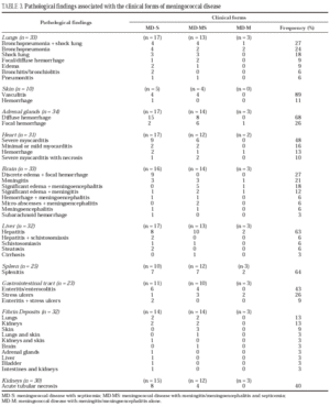
Adequate material for histopathological reassessment was available for all the cases. Of the 42 deaths involving clinically suspected meningococcal disease submitted to necropsy, 34 (81%) met the inclusion criteria; of these, 56% (19/34) were patients 14 years of age or younger and 44% (15/34) were over 14 years of age. The other 8 were investigated as differential diagnoses. Table 1 shows the distribution of cases according to inclusion criteria and clinical forms. Table 2 shows the clinical and pathological classification of the 34 cases of meningococcal disease. Among the total, 11 of the 17 MD-S cases, all 14 MD-MS cases, and 2 of 3 MD-M cases were confirmed pathologically. Thus, necropsy confirmed the clinical diagnosis in 79% of cases (Kappa = 0.64; P < .0001). The most marked histopathological alterations are shown in table 3. Diffuse vascular lesions, thrombosis and hemorrhages of varying intensity were seen in numerous organs or tissues. The lungs were most often affected by multiple processes, 100% (33/33); followed by skin alterations, 100% (9/9); adrenal hemorrhage, 94% (32/34); myocarditis, 71% (22/31); meningitis/meningoencephalitis, 70% (23/33); and hepatitis, 69% (22/32). Bacterial and reactive hepatitis was found in all clinical forms and hepatic schistosomiasis was seen in four cases. In the brain, significant edema was observed in 30% (10/33).
The clinicopathological correlations, based on the availability of corresponding clinical and pathological data, are shown in table 4. Pulmonary histopathological involvement was found in 100% of the necropsies, but manifestations of respiratory failure were recorded in 74% (11/15) of the MD-S patients, 77% (10/13) of the MD-MS, and all three MD-M cases. Adrenal hemorrhage and shock were highly correlated; table 3 shows that diffuse adrenal hemorrhage was more frequent in MD-S, 88% (15/17), as compared to MD-MS, 57% (8/14). In the clinicopathological correlation study, diffuse adrenal hemorrhage predominated in the MD-S form, 87% (13/15), with 92% (12/13) presenting shock; in the MD-MS form, there were eight deaths with diffuse adrenal hemorrhage and 88% (7/8) presented shock (table 4). Diffuse adrenal hemorrhage was not observed in MD-M. In the nine cases with focal adrenal hemorrhage, shock was documented in four (44%). Microthrombi were seen in 58% (18/31) of the necropsies, particularly in the skin, kidneys, and lungs (table 3). The incidence of microthrombi was identical in MD-S and MD-MS cases--64%--while clinically suggested disseminated intravascular coagulation detected by abnormal clotting tests was seen in 39%, among which 29% (4/14) were MD-S and 57% (8/14) MD-MS (table 4). Microthrombi were observed in 18 cases, in which 12 clinical manifestations of disseminated intravascular coagulation were detected, thus giving a clinical correlation of 67%. In the 31 clinical cases, meningitis or meningoencephalitis were observed in 22 deaths; of these, a correlation with meningeal signs was recorded in 52% (table 4). Evidence of meningismus was documented in four cases with clinical diagnoses of MD-S, in which cerebrospinal fluid showed fewer than 10 cells/mm3; however, two of these cases had pathological findings compatible with meningitis and meningoencephalitis. In addition, among the 14 MD-MS cases, five had no meningeal signs, but CSF showed more than 10 cells/ mm3. Severe necrotizing myocarditis was seen in MD-S (10/17) and MD-MS (8/12) (table 3), but clinical heart failure predominated in MD-MS (table 4), with a 27% correlation (7/26 cases) between clinical heart failure and cardiac involvement at necropsy. Among the 22 necropsies in which the digestive tract was reexamined and for which clinical data were obtained (table 4), enteritis or enterocolitis was seen in 55% (12/22), particularly in the MD-S form (8/10), and diarrhea was recorded in 25% (3/12) all MD-S cases. Stress ulcers were present in the three clinical forms, with an overall rate of 36% (8/22), but corresponding digestive hemorrhage was less common, 38% (3/8).
Multiple organ failure was recorded as the cause of death in the majority of necropsies--59% (20/34)--associated with shock in 65% (13/20), followed by cerebral edema from meningitis or meningoencephalitis--29% (10/34)--and myocarditis with left ventricular failure--12% (4/34).
Of the 8 cases that were excluded, other etiological agents were recovered in four (two cases of grampositive diplococci and one each of Escherichia coli and Klebsiella oxytoca) by way of microbiological tests or necropsy specimens. The other four cases involved, respectively, a pathological diagnosis of lymphoma, metabolic disorder due to gastroenteritis, idiopathic thrombocytopenic purpura, and hemorrhagic bronchopneumonia associated with purulent meningoencephalitis.
Discussion
Of the 42 deaths with clinically suspected meningococcal disease, 8 were excluded, thus giving a clinical diagnostic error rate of 19% (8/42). In an extensive study that also included hemorrhagic purpura and leukemia among the differential diagnoses, Daniel7 reported a clinical diagnostic error rate of 5%.
The clinicopathological evaluation of 34% of the meningococcal disease cases showed diffuse vascular lesions with fibrin thrombi and hemorrhage of varying intensity in several organs, especially the skin, lungs, and adrenal glands, frequently associated with sepsis, findings that have been documented by other authors3,4,11. There was high concordance (79%) between the clinical and pathological diagnoses, although it varied according to the clinical forms (table 2). Of the 7 cases in which the clinical diagnosis and necropsy findings were discordant, 6 were among the 17 cases classified clinically as MD-S, but in which central nervous system histological alterations were recorded at necropsy, thereby changing the clinical form to MD-MS. One of the three patients diagnosed clinically as MD-M and with septicemia by pathology, was hospitalized for 10 days and progressed with nosocomial pneumonia, severe respiratory failure, and shock, a condition possibly associated with secondary bacterial sepsis.
Clinical manifestations of respiratory failure presented a high correlation with pathological findings (77%), calling attention to the high rate of pulmonary involvement among patients who do not survive (table 4). Variable lower frequencies of respiratory failure have been recorded in other clinical studies, including 5.7% only in the MD-S and MD-MS forms in an overall case series of 562 patients12, and 53% of pulmonary edema in 35 cases of severe MD13.
The Waterhouse-Friderichsen syndrome, characterized by infection and shock, classically attributed to bilateral adrenal hemorrhage, is often seen in fulminating meningococcemia, although shock is not always associated with demonstrable hemorrhage2,3,7,8. In contrast to these reports, the present series showed a high correlation between distribution of hemorrhage in the adrenal cortex and manifestations of shock. Only 17% (4/23) of patients with shock showed focal adrenal hemorrhage at necropsy, as compared to 83% (19/23) with diffuse adrenal hemorrhage. Some authors have commented that shock associated with absence of adrenal hemorrhage suggests the possibility of acute cardiogenic shock related to myocarditis as an etiological factor in the syndrome3,14. A causal relationship between endotoxemia and shock in the MD-S and MD-MS forms has been debated for decades2,7,5,4 and may be partially related to the enormous increase in nitric oxide during endotoxemia, leading to vasoplegia and refractory hypotension15.
There was also a high clinicopathological correlation between disseminated intravascular coagulation and presence of fibrin deposits (67%); however clinical evidence of disseminated intravascular coagulation with corresponding pathological findings was more common in MD-MS than in MD-S. The reason for this may be that reports of fatal cases of MD-MS contain more clinical and laboratory information, since they progress less rapidly, thus facilitating diagnosis of the syndrome. This could also explain why there was no statistical difference in the occurrence of disseminated intravascular coagulation between the two forms in the clinical follow-up of surviving patients (unpublished data).
As expected, meningitis and meningoencephalitis were seen in all the necropsies classified clinically as MD-M and MD-MS, although surprisingly, as shown in table 3, they were also observed in some cases classified as MD-S--38% (6/16). Additionally, meningeal signs were not reported in 36% (5/14) of MD-MS (table 4), but CSF cellularity was increased in all these cases. The occurrence of meningeal signs may not be reported in very severe patients and those with major sensory depression16. On the other hand, meningeal signs were recorded in 4/14 cases of MD-S (29%) without CSF alterations (table 4), although two of them had histopathological evidence of meningitis or meningoencephalitis.
Enteritis and enterocolitis were not documented in the literature consulted, but this histopathological finding (table 3), especially associated with evidence of gastrointestinal hemorrhage, could explain the presence of abdominal pain and diarrhea, atypical manifestations of MD, with diarrhea reported as a sign of severity17.
Myocarditis was more severe and more frequent in patients with MD-MS (67%) and MD-S (59%), respectively, coinciding with findings by Hardman3. There was a high frequency of myocarditis (71%) with a low clinical correlation (24%). Although focal or diffuse myocarditis is not an uncommon finding in necropsies of patients who have died of meningococcal disease, clinical descriptions of it have been infrequent1,3,4. Of the 23 fatal cases with myocarditis examined by Neveling & Kashula4, only one presented clinical cardiac manifestations. These authors explain the lack of clinical diagnosis by the masking of signs and symptoms by septicemia and shock, while Böhm5 attributes it to the fulminating course of the disease. The 12% frequency of myocarditis as the cause of death in the present study, together with the data from the literature as a whole, speak for cardiac monitoring in all clinical forms of MD.
Bacterial and reactive hepatitis were evidenced in all the clinical forms, although clinical and laboratory alterations were not documented. Focal hepatitis in necropsies of meningococcal disease has been shown by another author18. Hepatic schistosomiasis was seen in 4 patients with MD-S; this parasitic disease can alter the reticuloendothelial system, the integrity of which is an important defense against N. meningitidis19.
The considerable frequency of cerebral edema (29%) and myocarditis (12%) in this study highlights the need to keep these diagnoses in mind, aiming at early treatment.
The fact that important findings such as adrenal hemorrhage and myocarditis showed similar frequencies in MD-S and MD-MS in this pathologic study suggests that these forms may represent variations in the severity of the septicemic form. There is an ongoing need for studies based on routine necropsies, if possible in association with other diagnostic techniques, aimed at providing a better understanding of the pathophysiogenesis of MD and supporting the respective clinical diagnostic and therapeutic approaches.
Acknowledgments
We are most grateful to Professor Jurema Paula Merêncio, medical pathologist, for her skillful assistance in the morphological study and to Dr. Christopher Peterson for revision of the text.


